Sarcoidosis: Muscle
|
History Early images General patterns Granulomas Typical Small (Early) Resolving Immune features Muscle fibers Perimysial pathology |
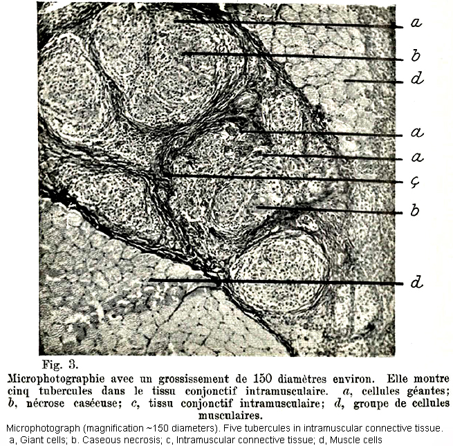
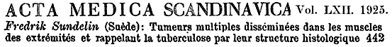
Sarcoidosis: Early images of nodular sarcoid involving muscle (Sundelin 1925)
 Sundelin 1 |
 |
 Sundelin |
Resolving Granuloma
Sarcoidosis: Patterns of sarcoid involving muscle
- Immune Myopathy with Perimysial Pathology & Granulomas
- Chronic Myopathy with Fatty replacement & Granulomas
- Sarcoid + IBM
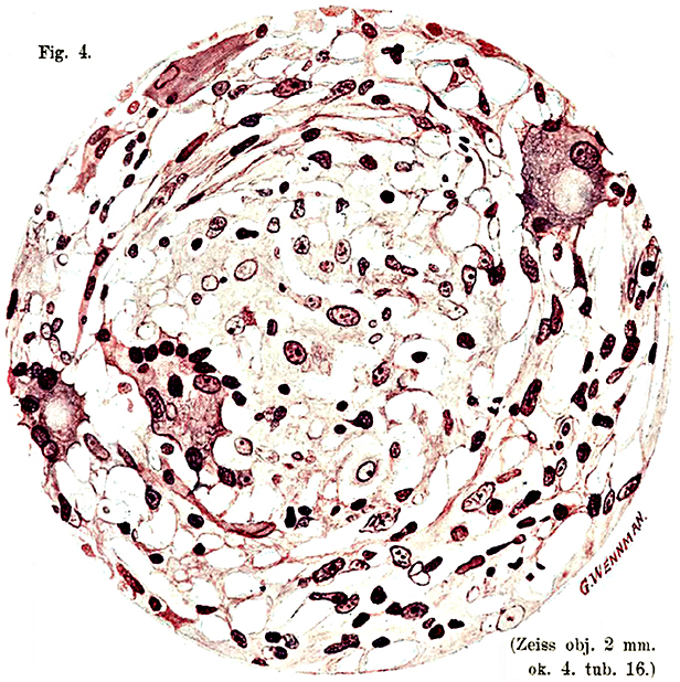
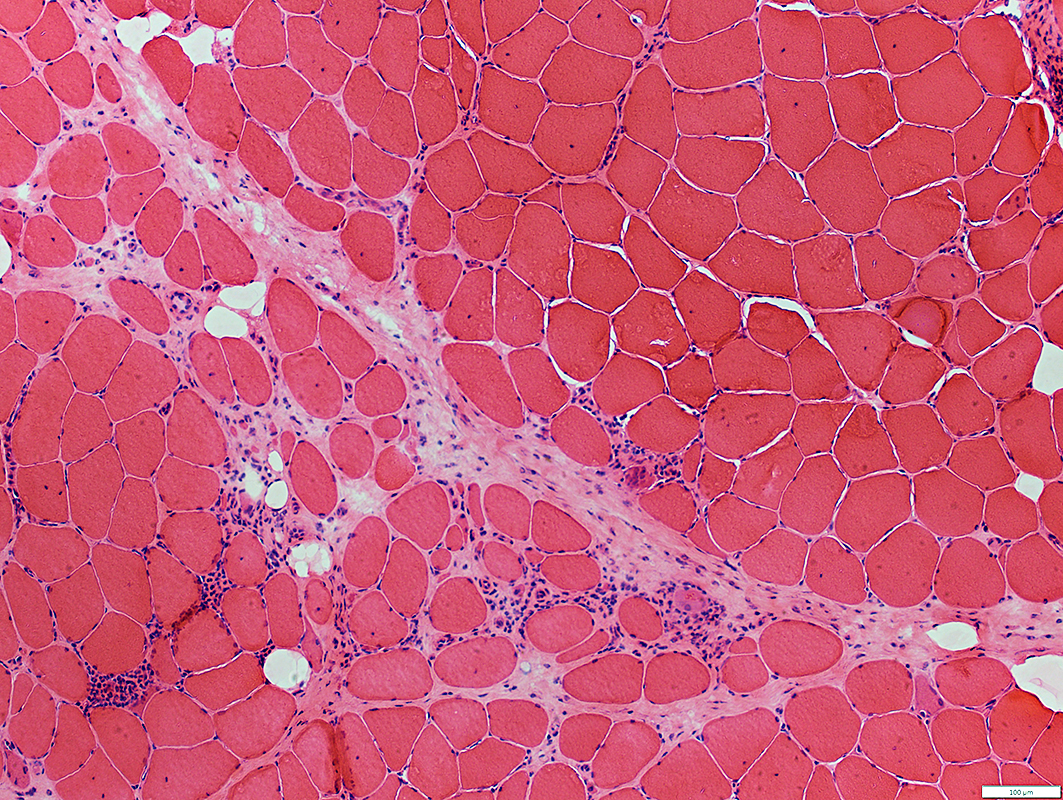
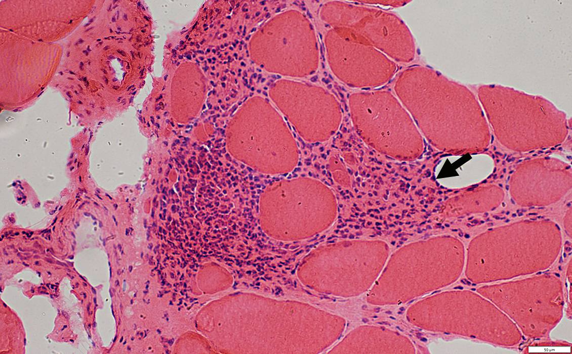
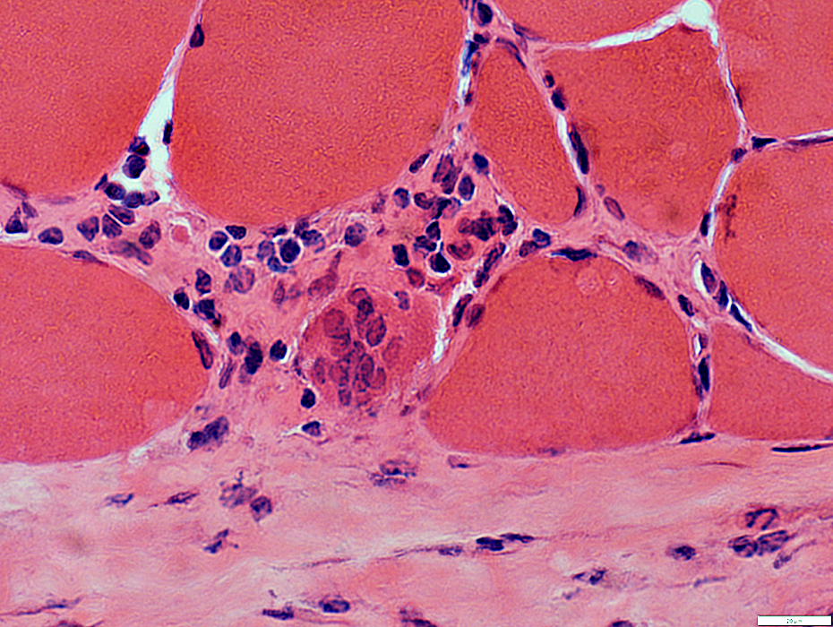
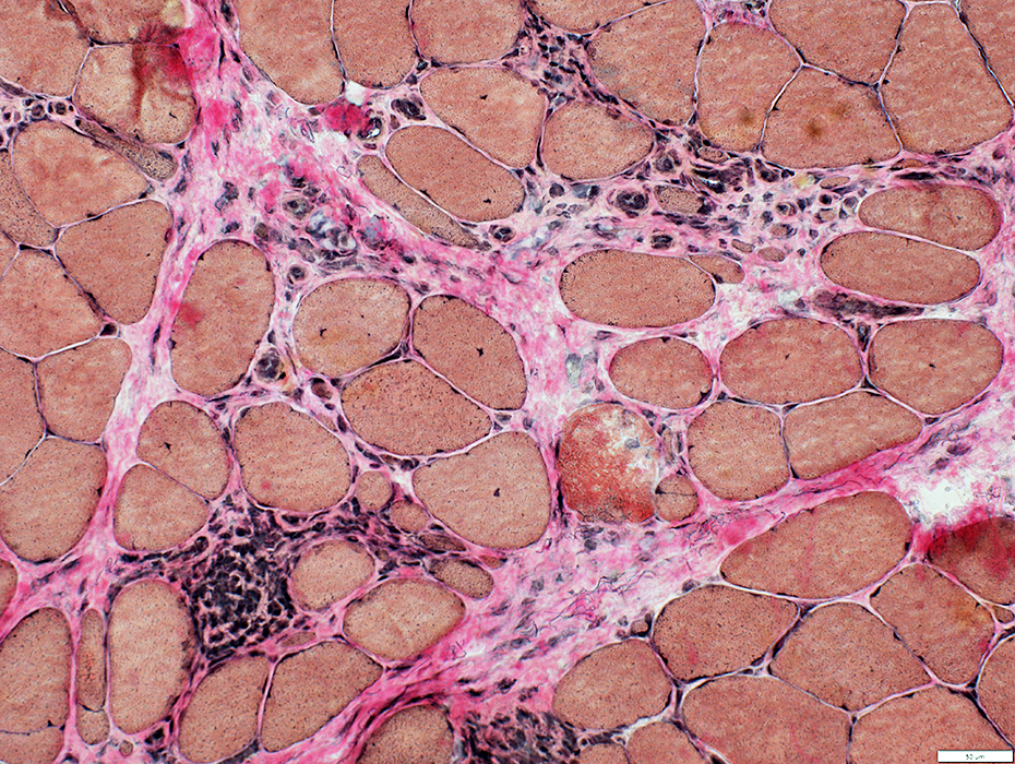
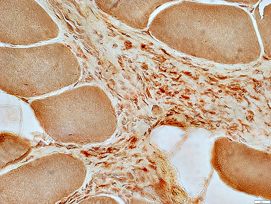
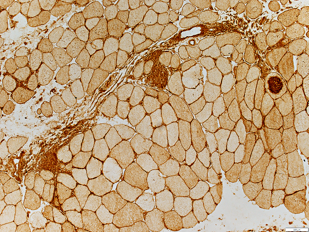
Immune myopathies with Perimysial connective tissue pathology (IMPP) & Granulomas
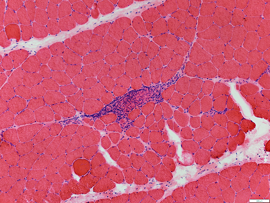 H&E stain |
Muscle fibers: Scattered small fibers in areas remote from granuloma
Perimysium: Fragmented structure
Myopathy: Multiple focal regions with
Small muscle Fibers
Increased endomysial connective tissue
Inflammation: Small cluster of cells, possibly replacing a muscle fiber
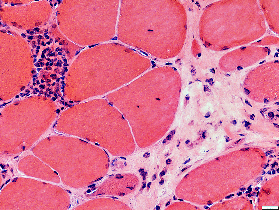
 H&E stain |
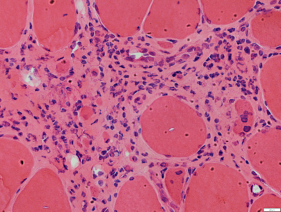
Histiocytic inflammation
Clusters of histiocytes
In granulomas
Associated with necrotic muscle fibers
Scattered histiocytes: in perimysial connective tissue
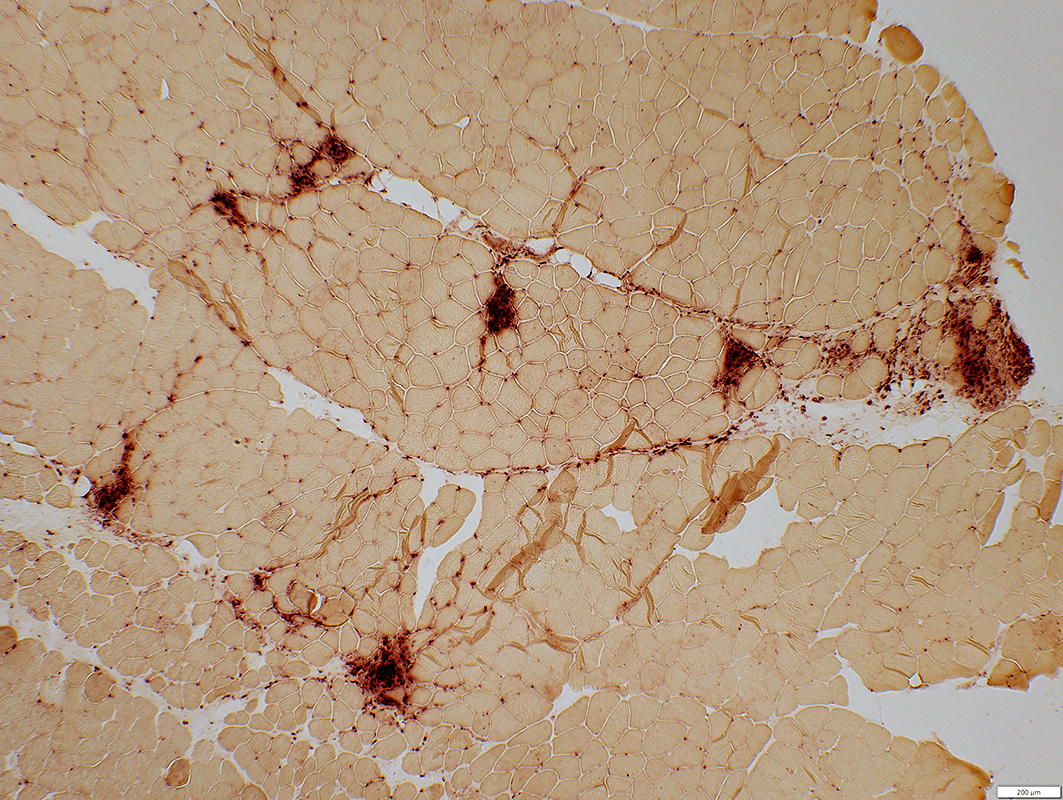 Acid phosphatase stain |
 Congo red stain |
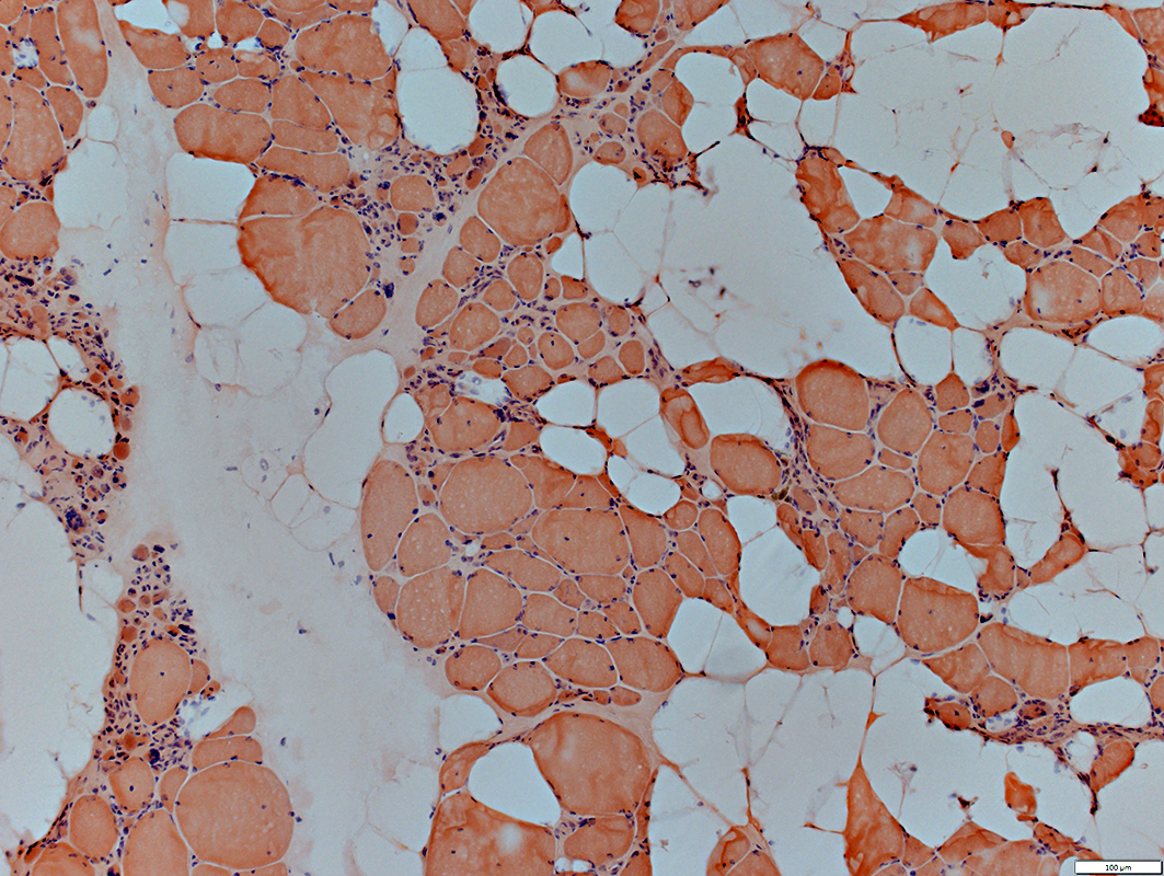
Perimysium & Endomysium: Replaced by Fat
Granulomas with giant cells in endomysium or perimysium
 Congo red stain |
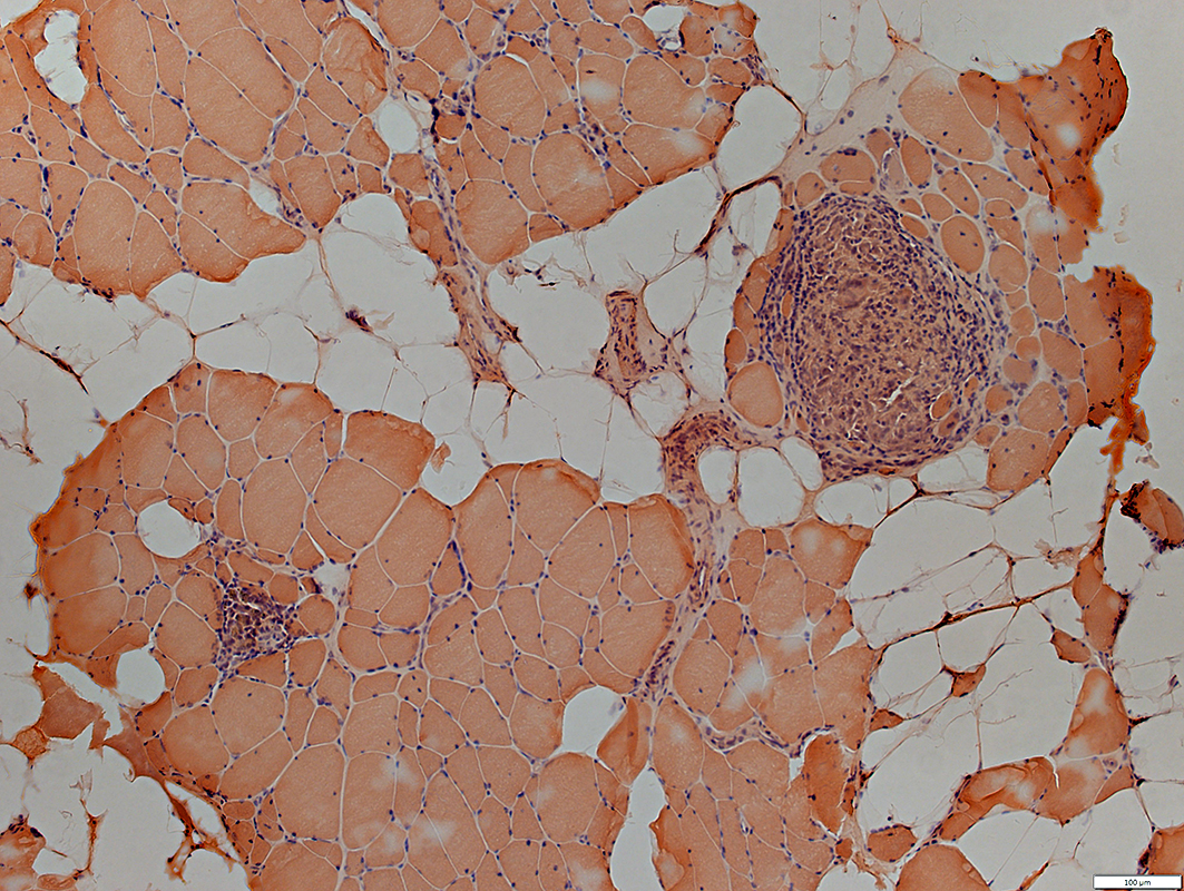
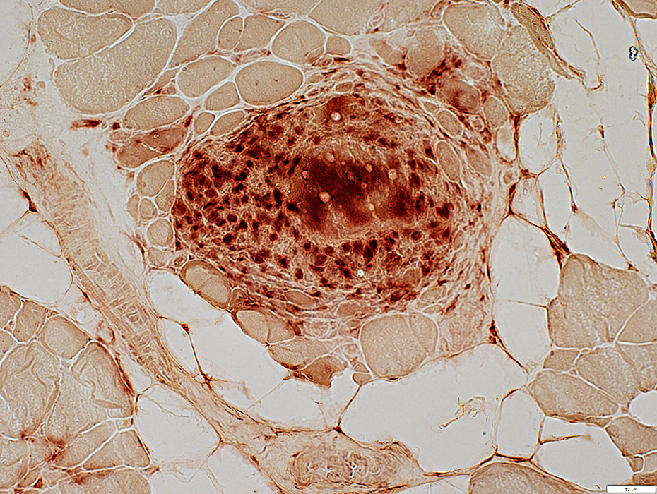
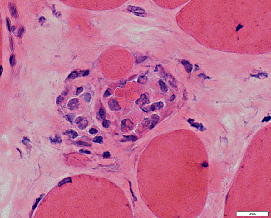
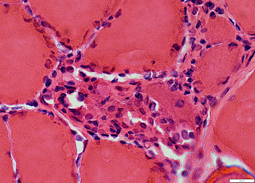
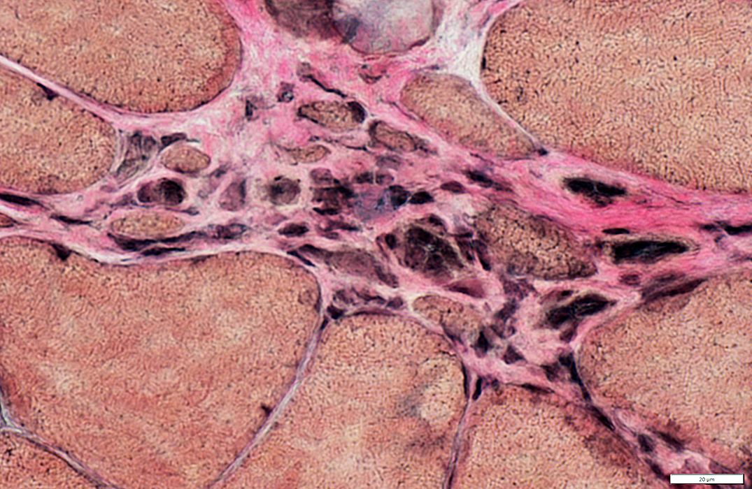
Sarcoid: Granulomas
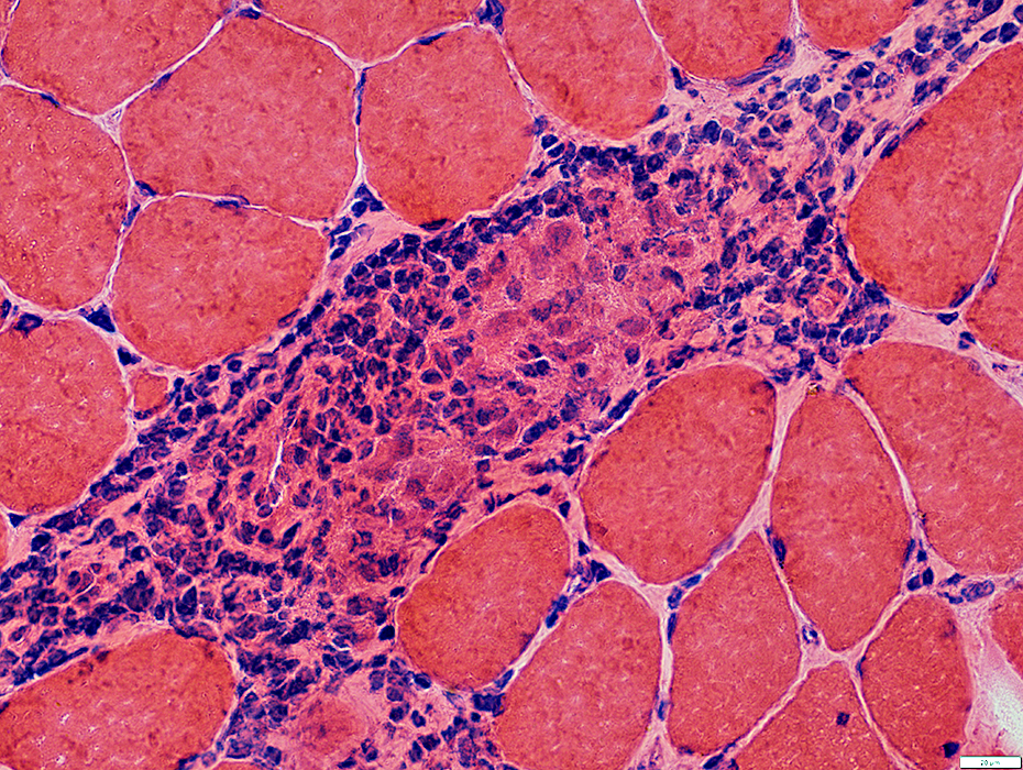 H&E stain |
 H&E stain |
Large cells & multinucleated cells in central regions
Smaller cells (Lymphocytes) in peripheral regions
Small muscle fibers often neighbor granuloma in some areas
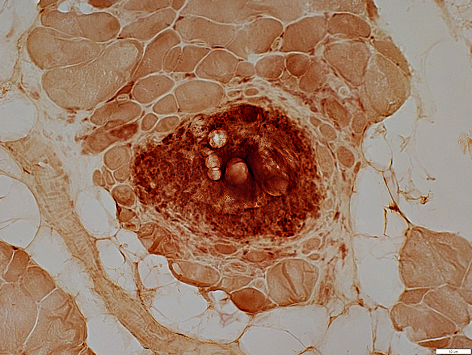
 Congo red stain |
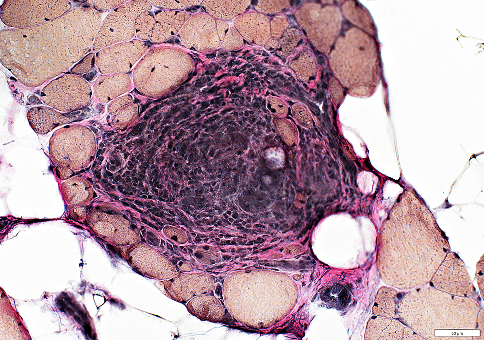 VvG stain |
Small muscle fibers in periphery of granuloma
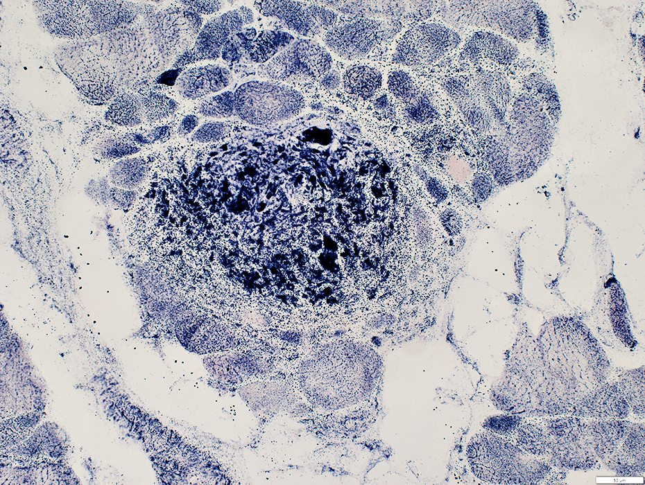
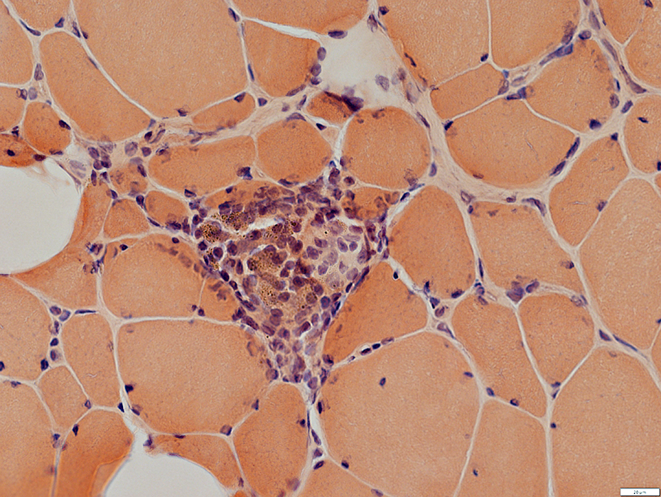
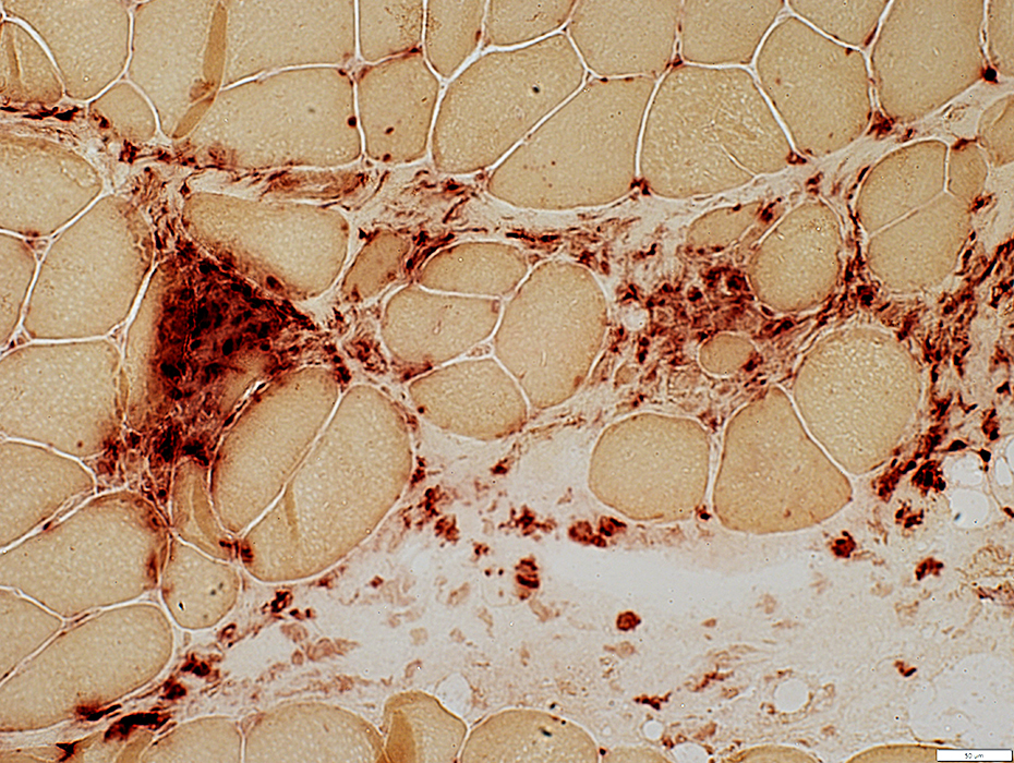
 Acid phosphatase stain |
Acid phosphatase (Above) stains
Around nuclei of individual histiocytes
Diffusely in Cytoplasm of giant cell
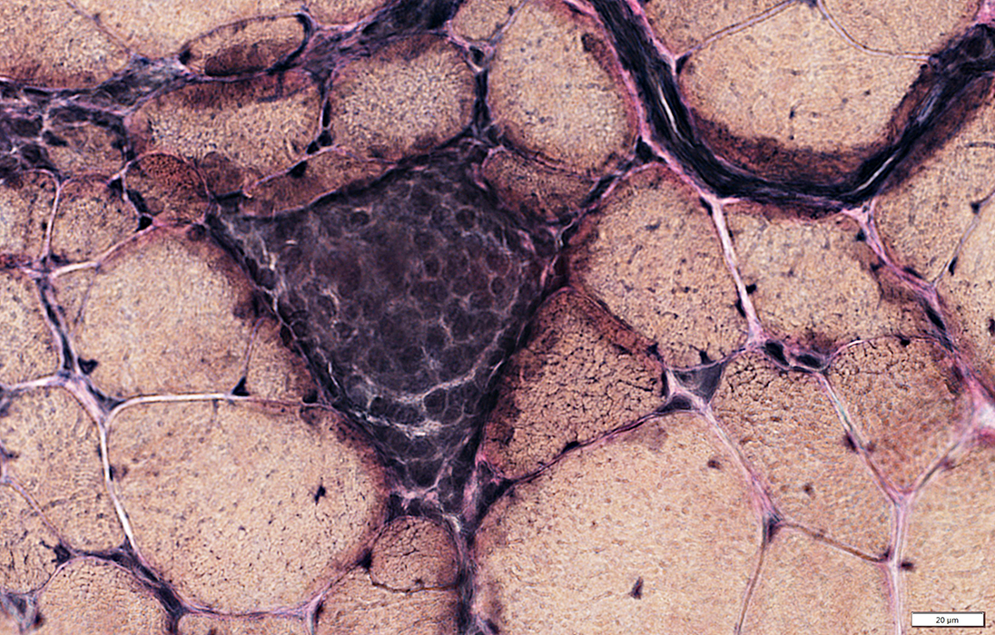
Esterase (Below) stains
Cells within granuloma in confluent pattern
 Esterase stain |
Granuloma
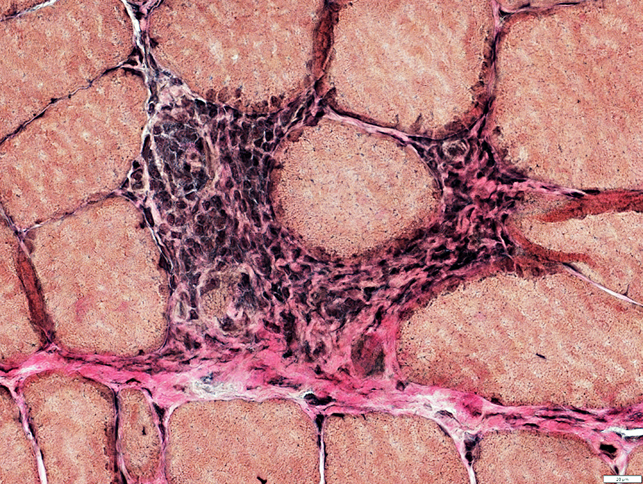
Cells in granuloma have abundant mitochondria
 SDH stain |
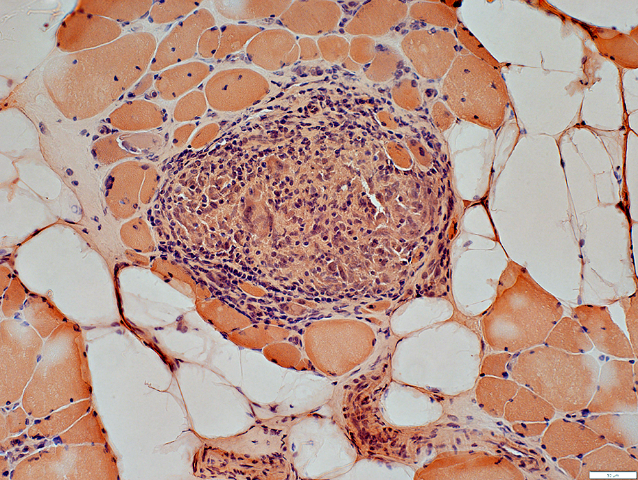
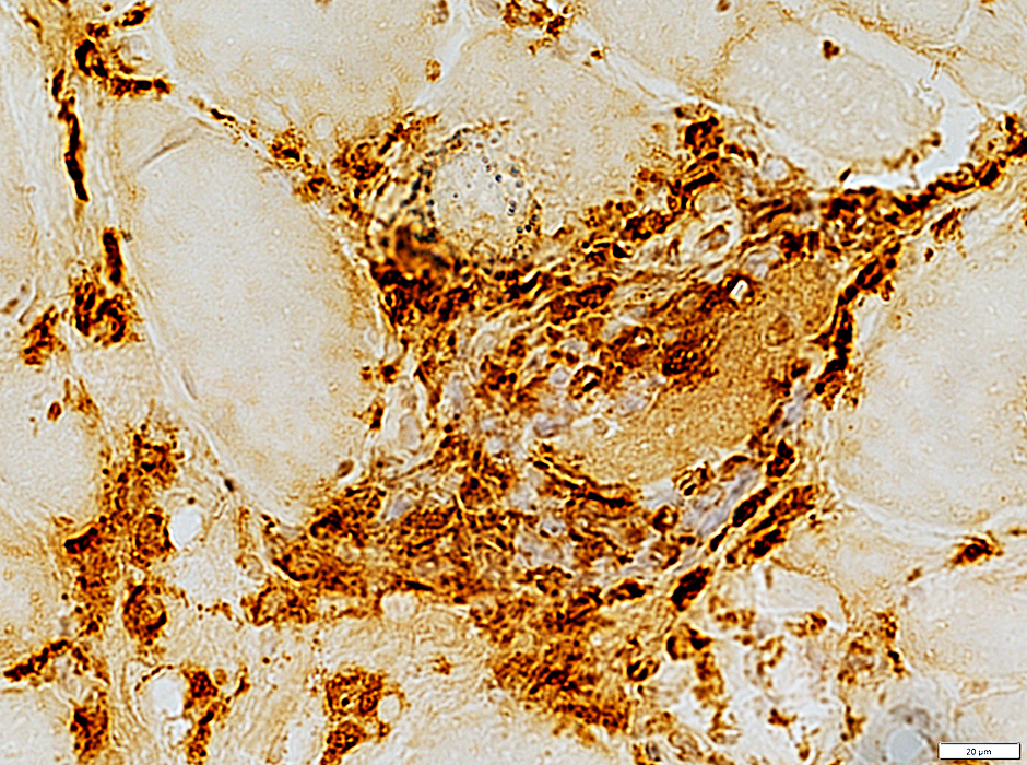
Granuloma
Cells in granuloma stain strongly for histiocyte marker CD68
Activated histiocytes (CD68+) are also scattered in endomysium, possibly near capillaries
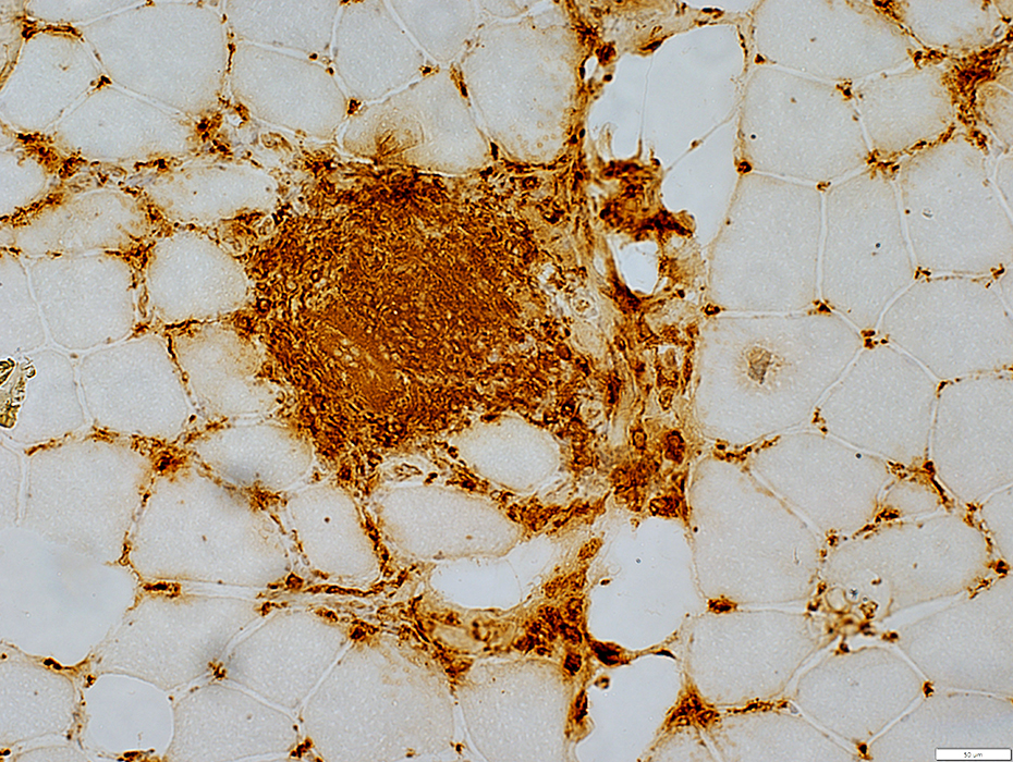 CD68 stain |
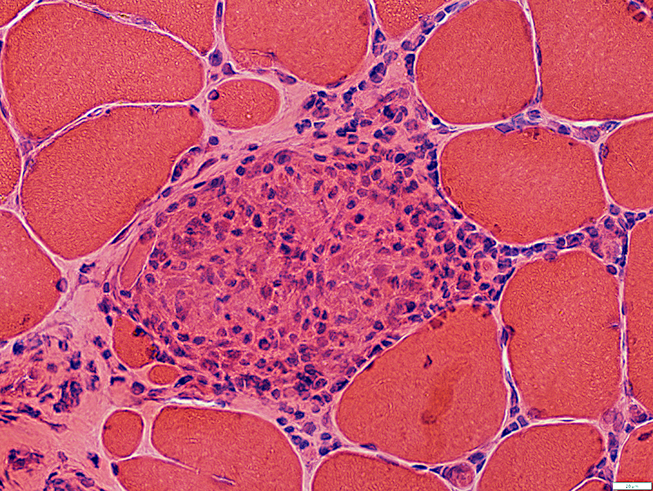
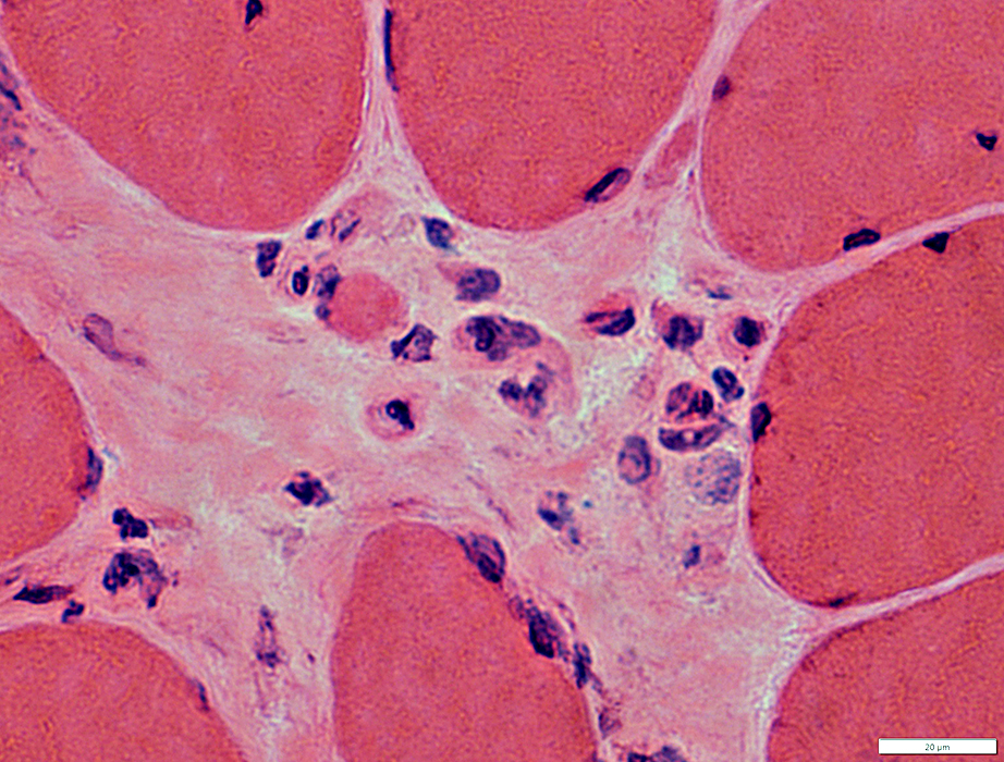
Sarcoid: Small Granulomas
 H&E stain |
Histiocytes replace muscle fibers & remain in place
 Congo red stain |
 VvG stain |
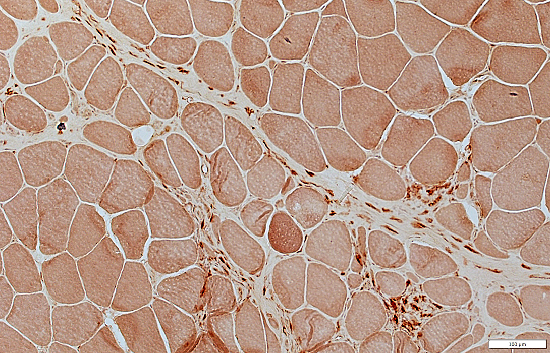
Sarcoidosis, Chronic: Muscle fiber pathology
 VvG stain |
Immune cells surround muscle fibers (Above)
Muscle fiber damage: Muscle fibers focally invade & progressively replace muscle fibers (Below; Arrow)
 Gomori trichrome stain |
 H&E stain |
Muscle fiber damage: Muscle fibers focally invade & replace muscle fibers
 H&E stain |
 CD68 stain |
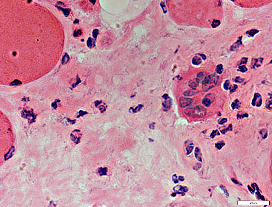
Sarcoidosis: Muscle fiber pathology
Muscle fiber damage: Fragments of muscle fibers (Arrow) remain in some areas of a granuloma
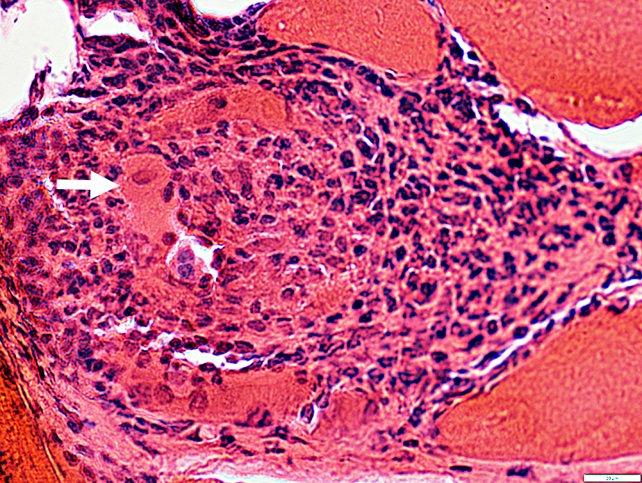 H&E stain |
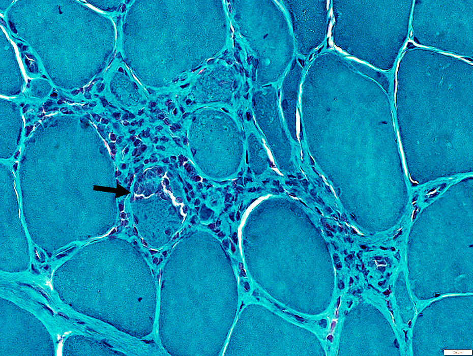
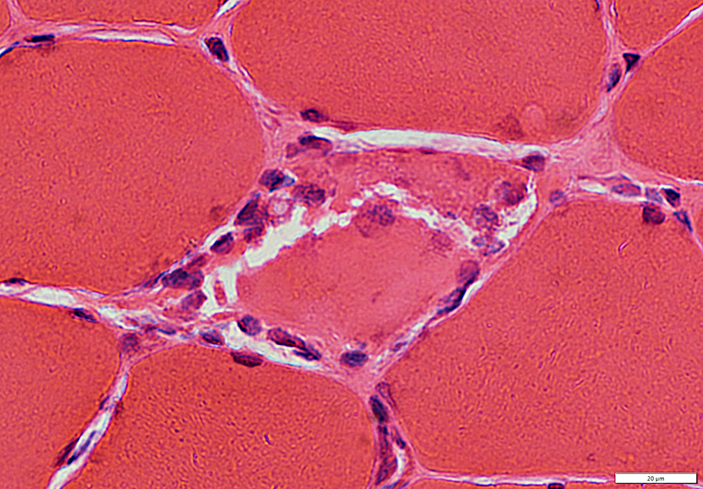
Sarcoidosis: Resolving granulomas
 H&E stain |
Granulomas may resolve leaving focal regions (Arrow) with
Increased, or damaged, connective tissue
Scattered
Cells, Some multinucleated
Small muscle fibers
 H&E stain |
 Congo red stain |
Cellular connective tissue
Small muscle fibers
 VvG stain |
 H&E stain |
 H&E stain |
 H&E stain |
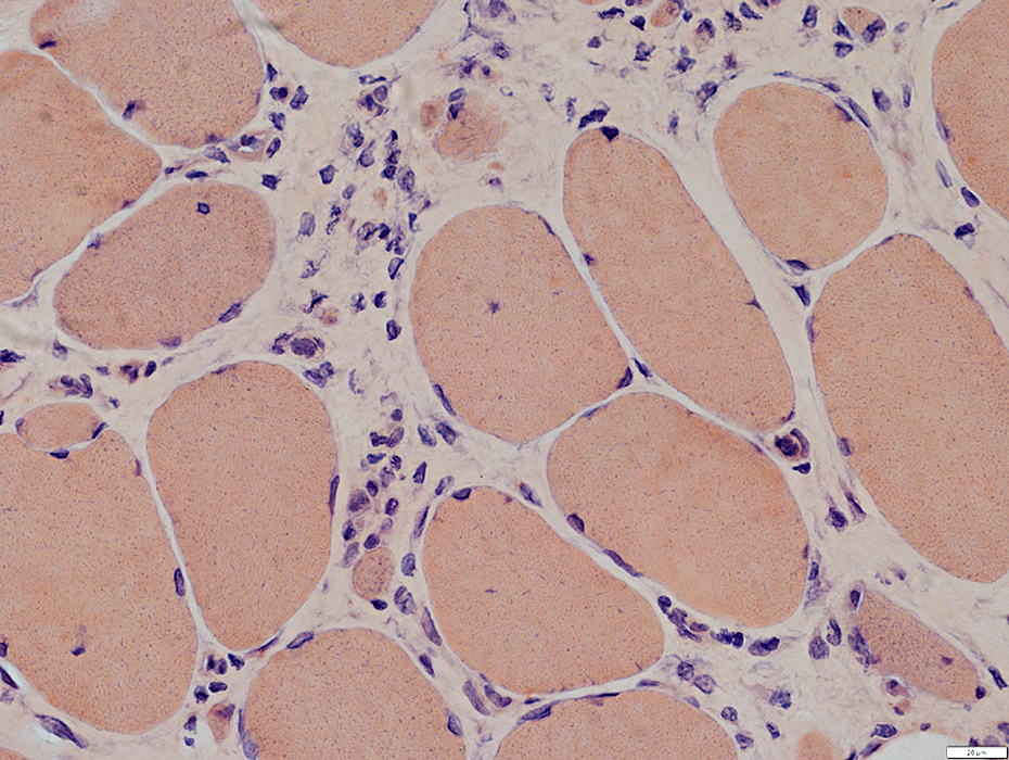
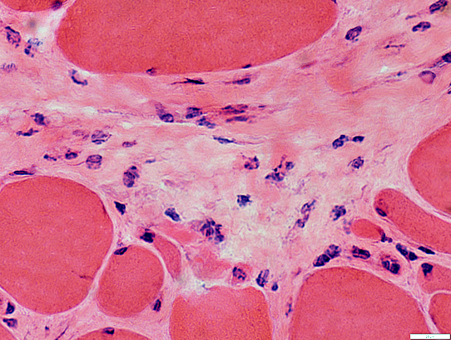
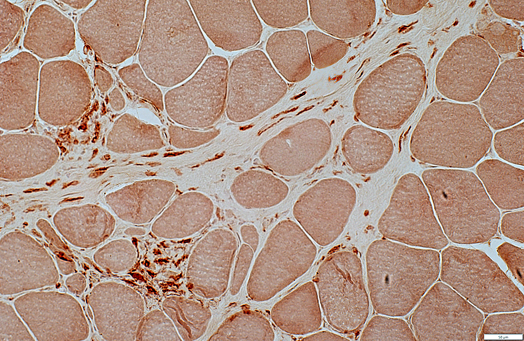
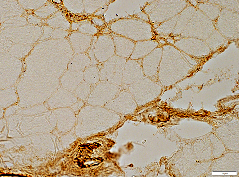
Sarcoidosis: Perimysial pathology
 VvG stain |
Structure: Fragmented
Contents: Scattered histiocytes
 VvG stain |
 H&E stain |
Structure: Fragmented
Contents: Scattered histiocytes
 H&E stain |
 Acid phosphatase stain |
Contains scattered histiocytes (Acid phosphatase + (Red)) in areas with normal & fragmented structure
 Acid phosphatase stain |
 Esterase stain |
Contains scattered histiocytes (Esterase + (Red)) in areas with normal & fragmented structure
 Esterase stain |
Perimysial Connective Tissue
Regions in some patients have abnormal alkaline phosphatase staining
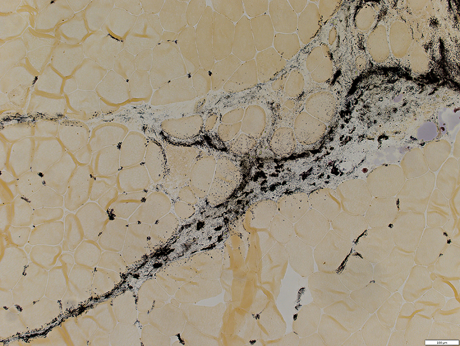 Alkaline phosphatase stain |
Sarcoid muscle: Immune features
C5b-9 deposition: Endomysial & Perimysial connective tissue
 C5b-9 stain |
MHC Class I upregulation by muscle fibers
 MHC Class I stain |
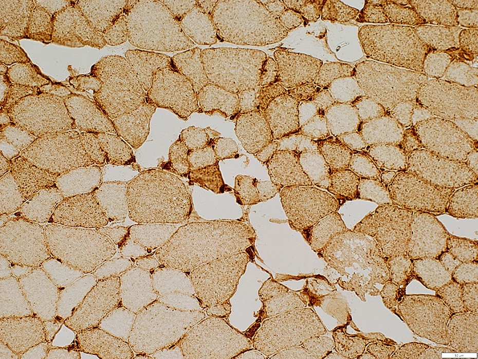 MHC Class I stain |
Return to: Neuromuscular Home Page
12/16/2021