Local Hypertrophic Neuropathy: Neurofibroma/Schwannoma Hybrid Nerve Sheath Tumors (N-S HNST)
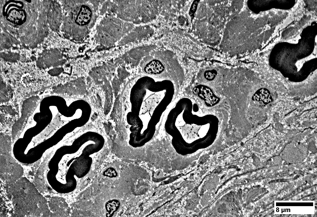
Nerve: Varied Structure
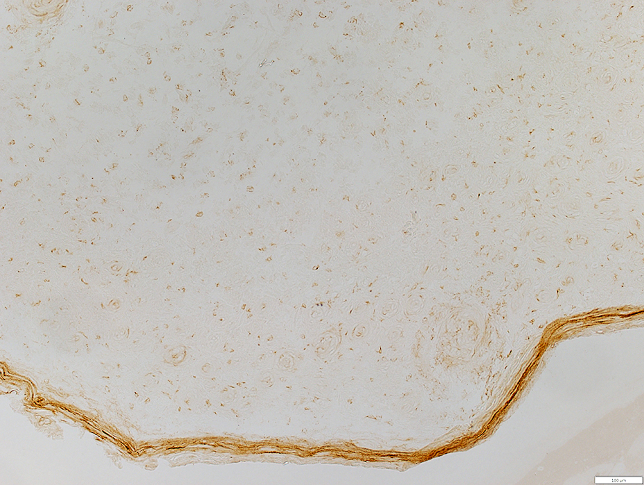
Large & Small rounded regions separated by varied amounts of endoneurial connective tissue
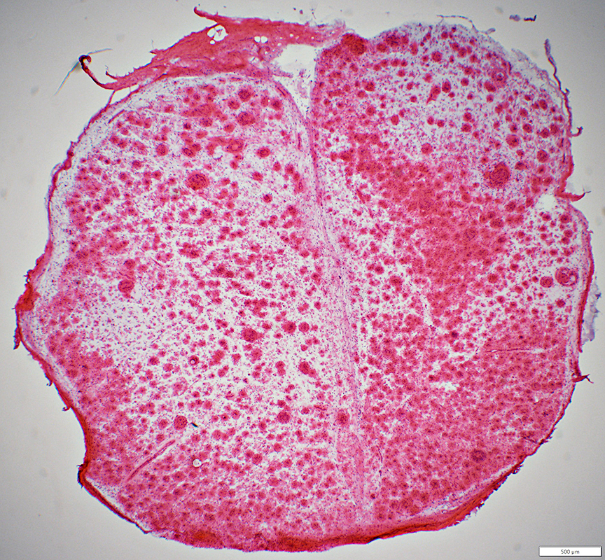 H&E stain |
N-S HNST Nerve organization
 H&E stain |
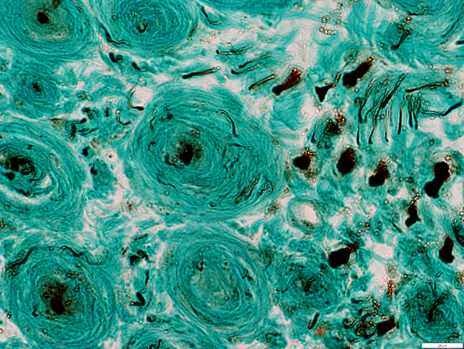
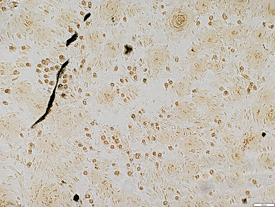
Varied sizes
Scattered in pale endoneurium
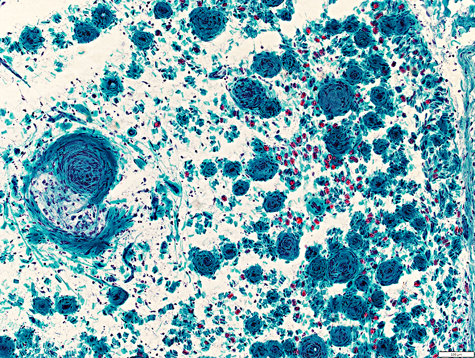 Gomori trichrome stain |
Scattered in pale endoneurium
Myelinated axons (Red) are present in some areas
 Gomori Trichrome stain |
 H&E stain |
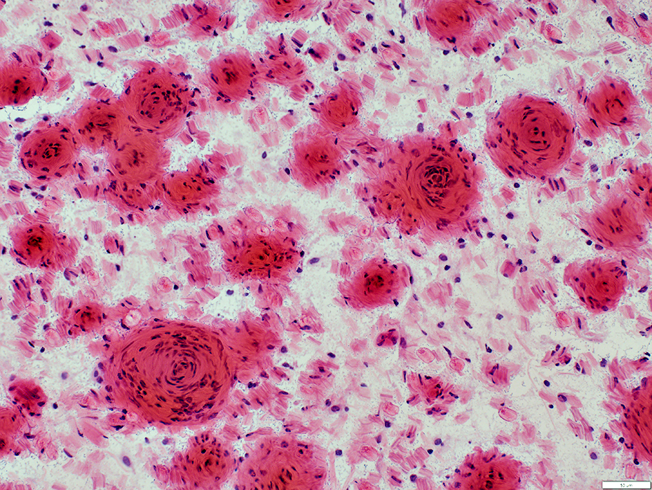
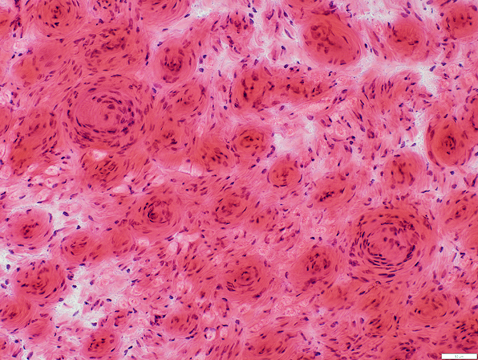
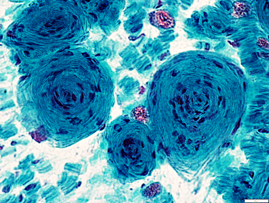
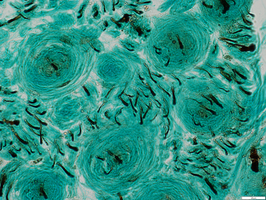
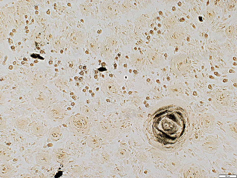
Varied sizes
Clustered in endoneurium
 Gomori trichrome stain |
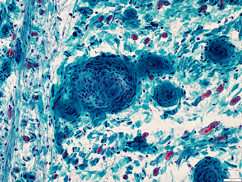
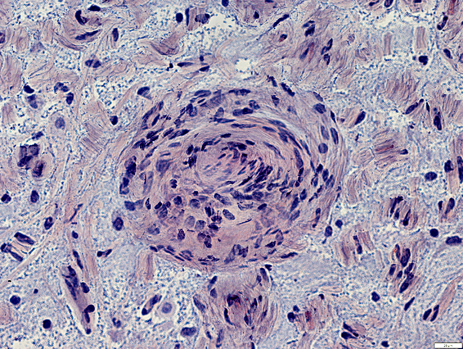
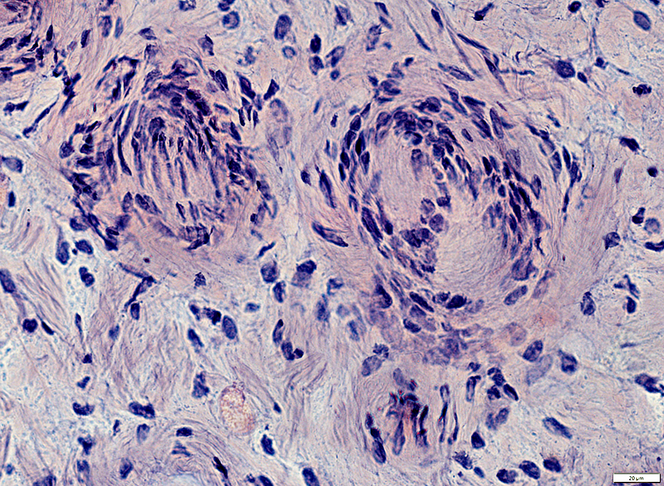
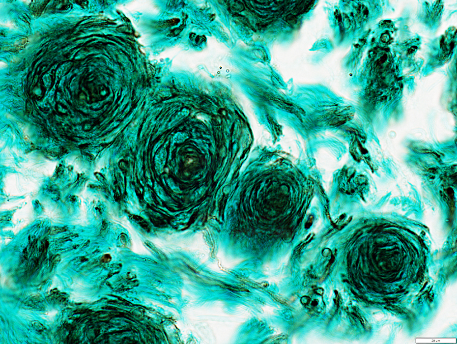
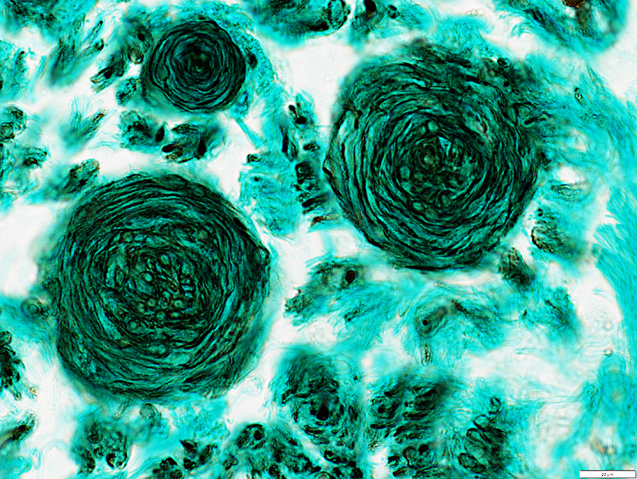
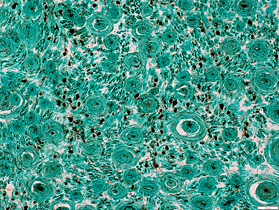
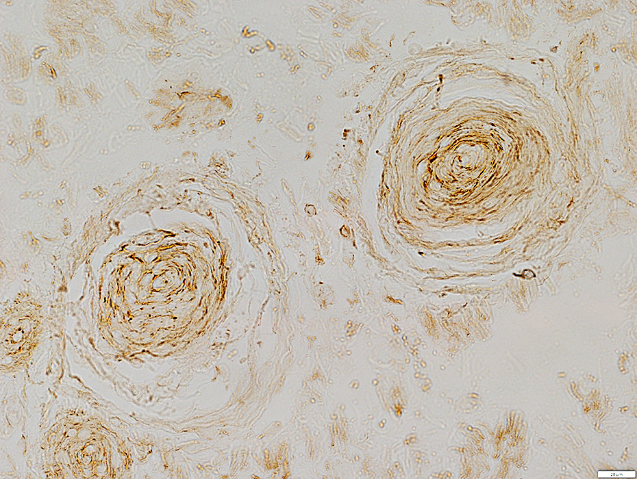
Onion Bulb-like Structures
ATPase staining around periphery
 ATPase ph 4.3 stain |
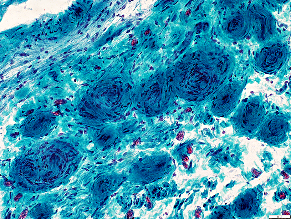
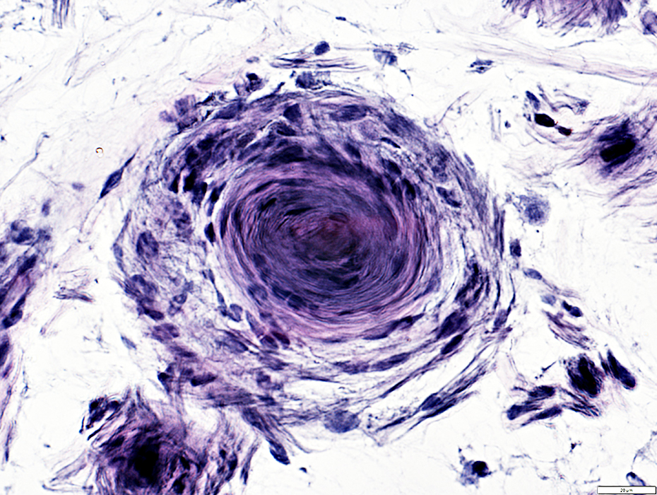
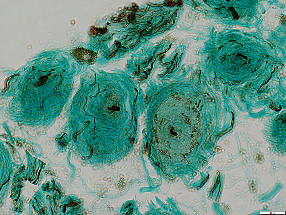
 H&E stain |
Concentric layers of cells & connective tissue
 Gomori trichrome stain |
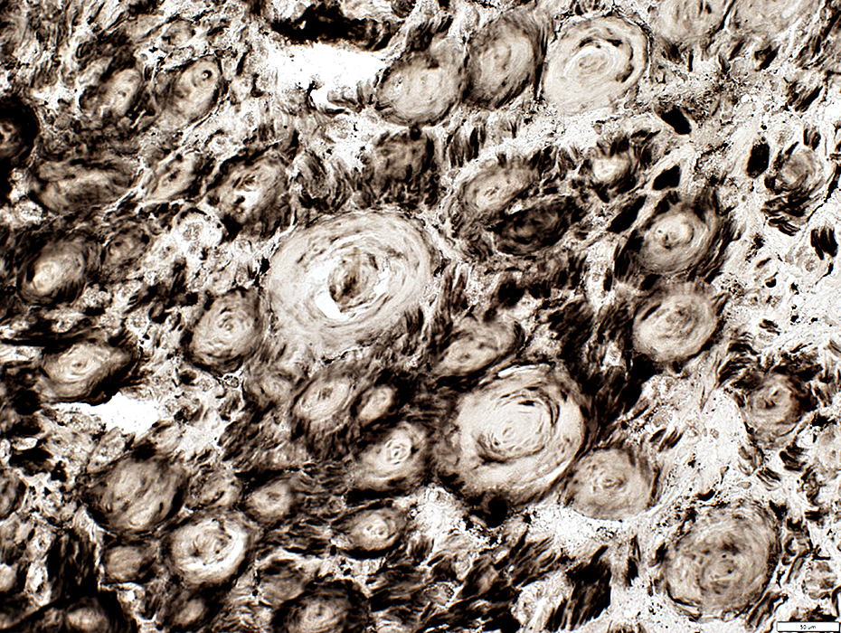
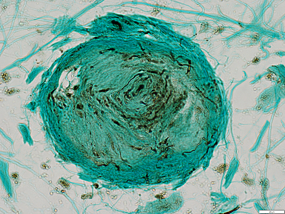
 VvG stain |
Concentric cell layers
Connective tissue: Looser organization in outer layers
 VvG stain |
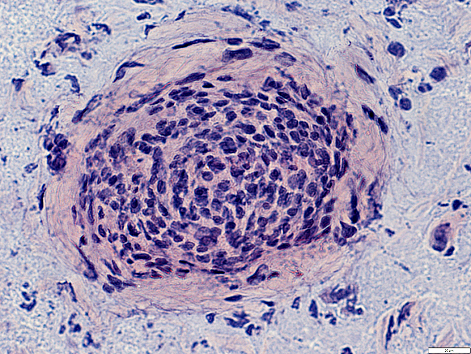
Onion Bulb-like Structures: Nuclei Patterns
 Congo red stain |
Surrounded by pale-stained endoneurial regions
 Congo red stain |
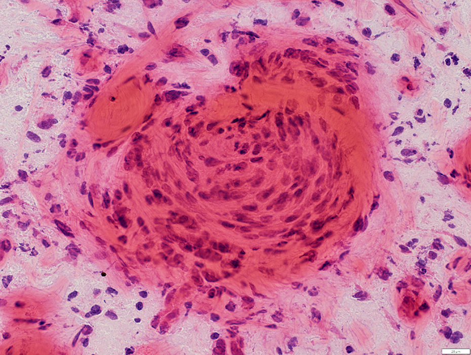
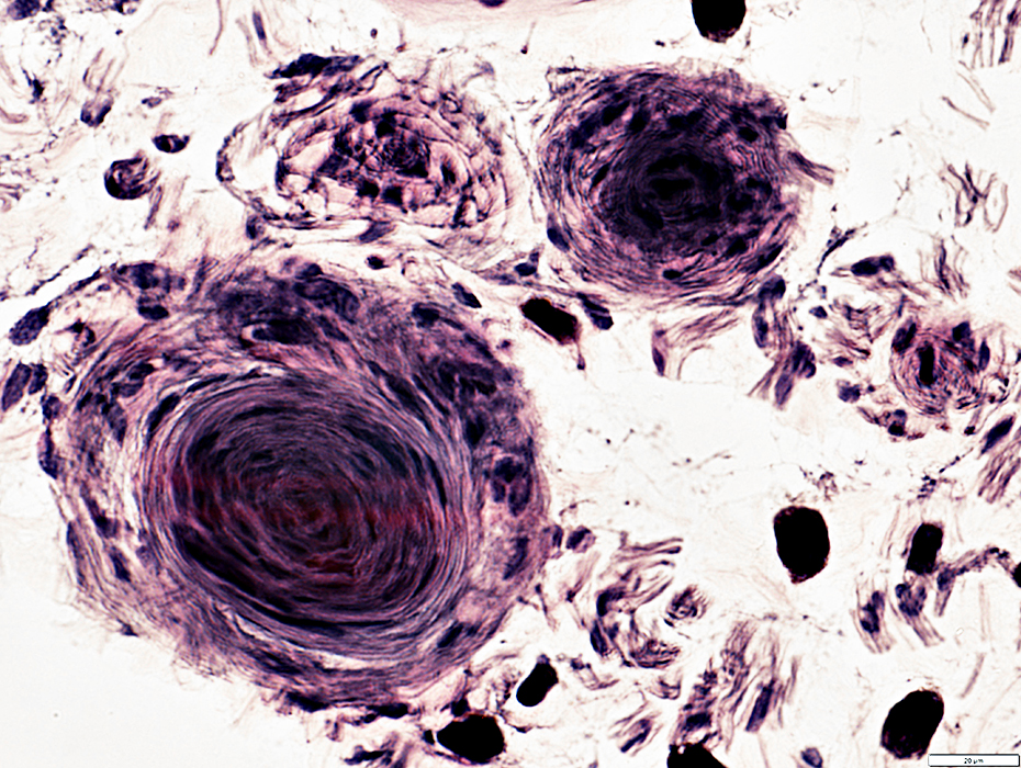
Verocay body-like organization in some areas
Palisades of nuclei separated by fibrillary connective tissue
 Congo red stain |
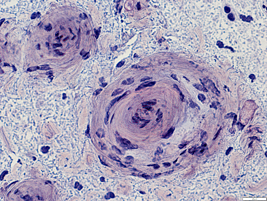
Some structures have many nuclei
 Congo red stain |
Onion Bulb-like Structures: Schwann cells (NCAM stain)
 NCAM/Gomori stain |
Cells & concentric processes stain for NCAM: Similar to non-myelinating Schwann cells
 NCAM/Gomori stain |
Onion Bulb-like Structures: Molecular features
Cells & concentric processes stain for NCAM
Some cells co-stain for NCAM & P0 (Yellow): Similar to Büngner band cells
Scattered myelinated axons (Red) are present in surrounding endoneurial regions
P0(r)_07ap.jpg) NCAM (Green)/P0 (Red) stain |
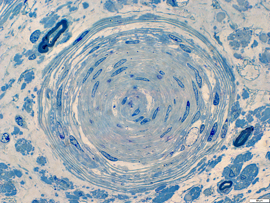
Onion Bulb-like Structures: Structural Patterns
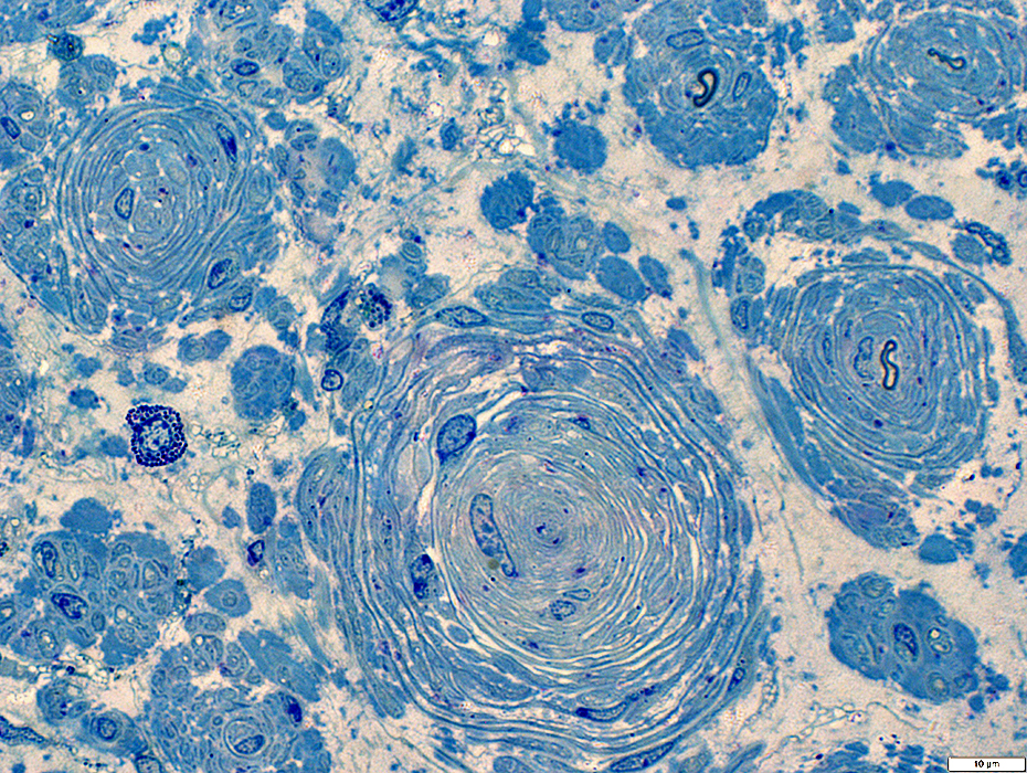 Toluidine blue stain |
Concentric layers of cells & basal lamina
Basal lamina: Looser organization in outer layers
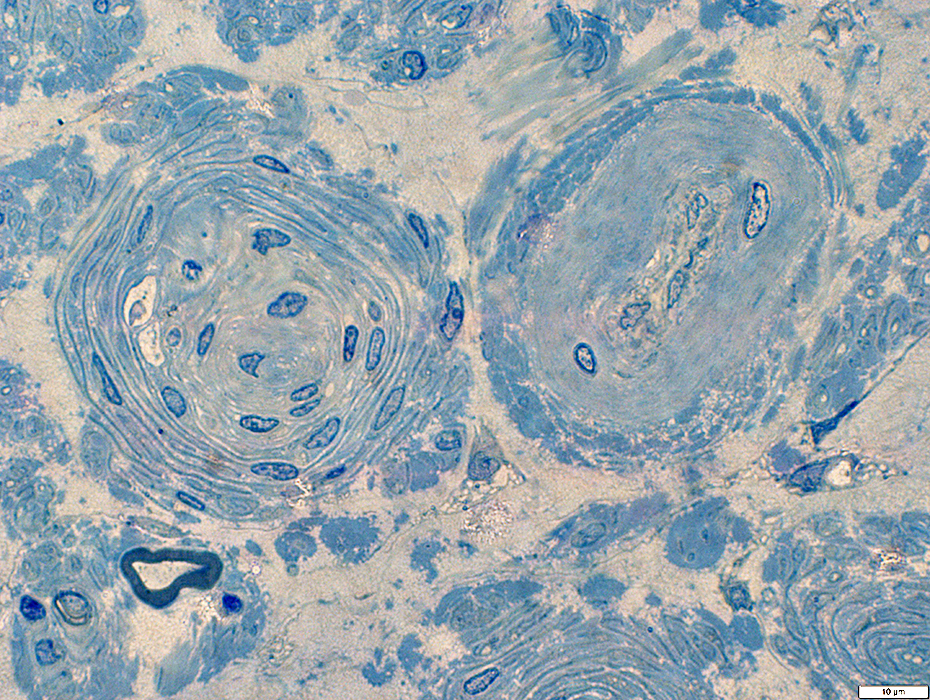 Toluidine blue stain |
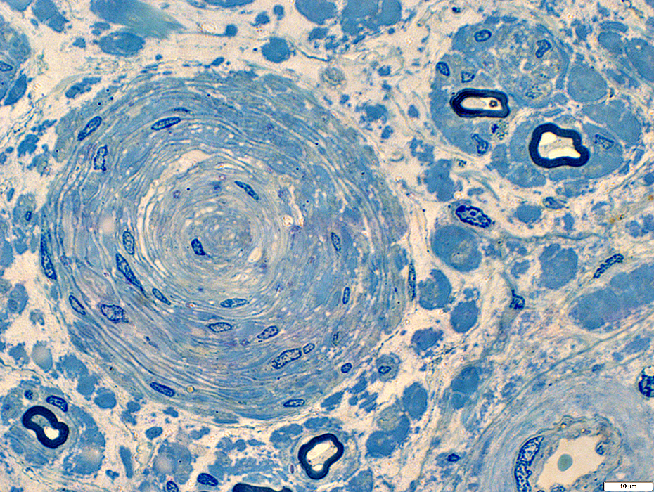 Toluidine blue stain |
Concentric layers of cells & basal lamina
Basal lamina: Looser organization in outer layers
 Toluidine blue stain |
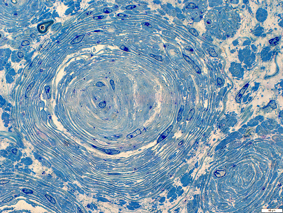 Toluidine blue stain |
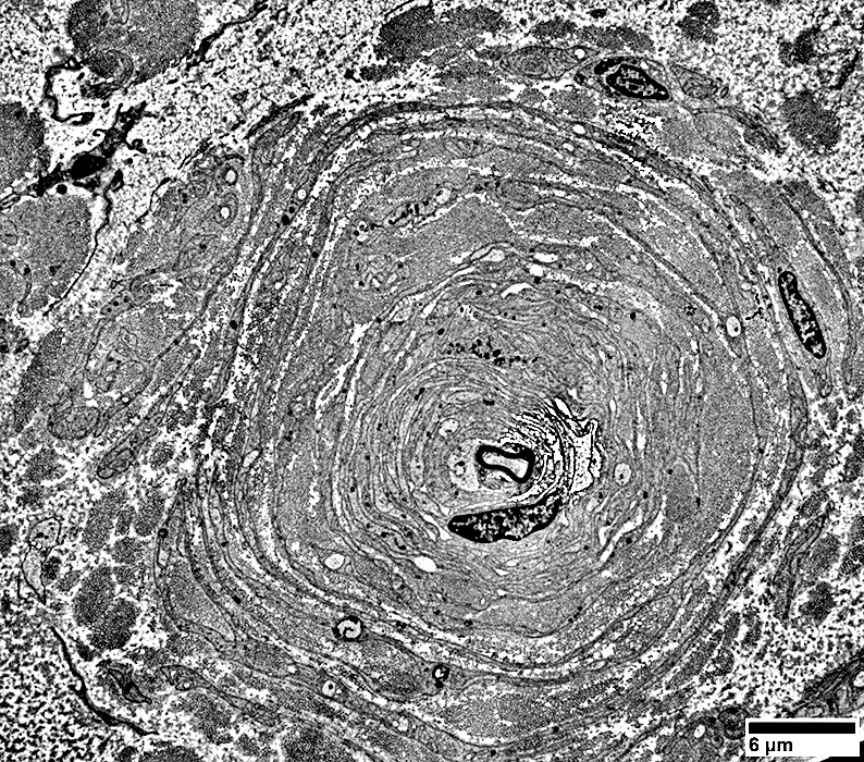
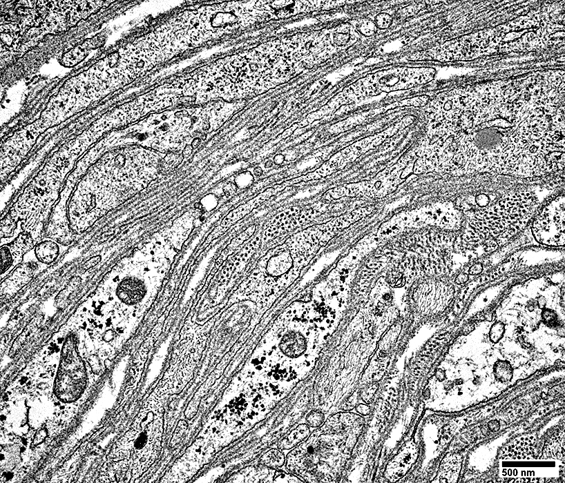
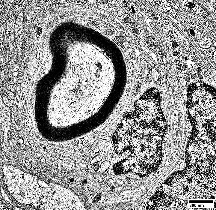
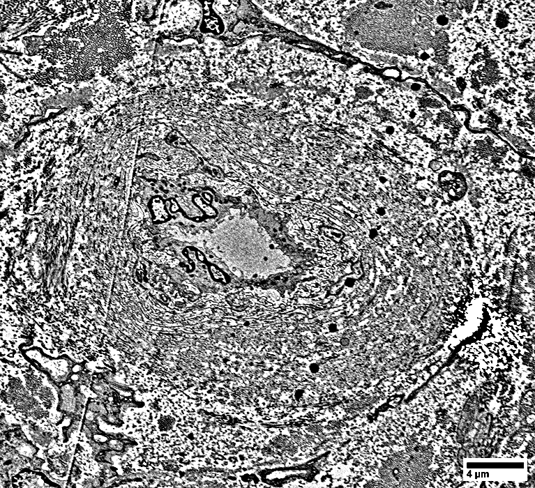
Onion Bulb-like Structures: Ultrastructure
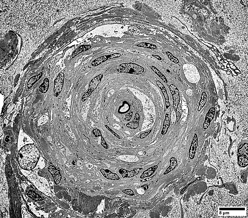 From: R Schmidt |
Basal lamina layers: Abundant
Schwann cell processes: Circumferential
Axons: Central (thin or no myelin) & Few peripheral
Collagen: Less common than in onion bulbs related to demyelination
 From: R Schmidt |
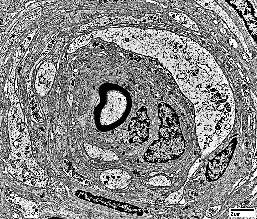
 From: R Schmidt |
Basal lamina layers: Abundant
Schwann cell processes: Circumferential
Axons: Central (thin or no myelin) & Few peripheral
Collagen: Less common than in onion bulbs related to demyelination
 From: R Schmidt |
 From: R Schmidt |
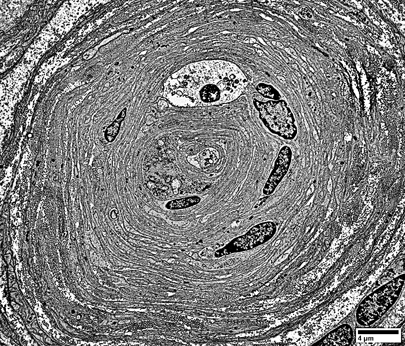
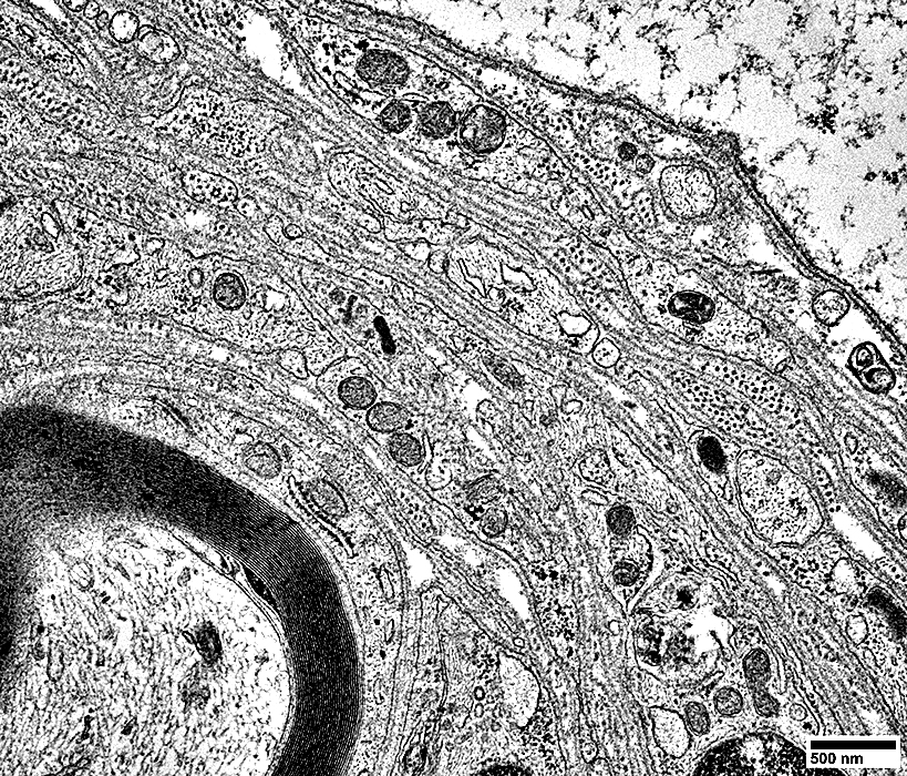
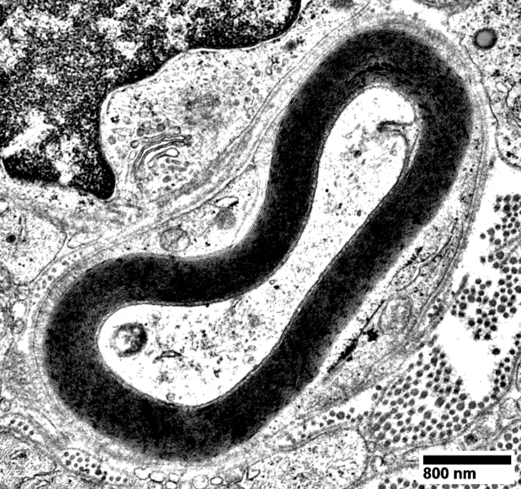
Onion Bulb-like Structures: Variant
Circumferential large processes
Central axon: Thinly myelinated
 From: R Schmidt |
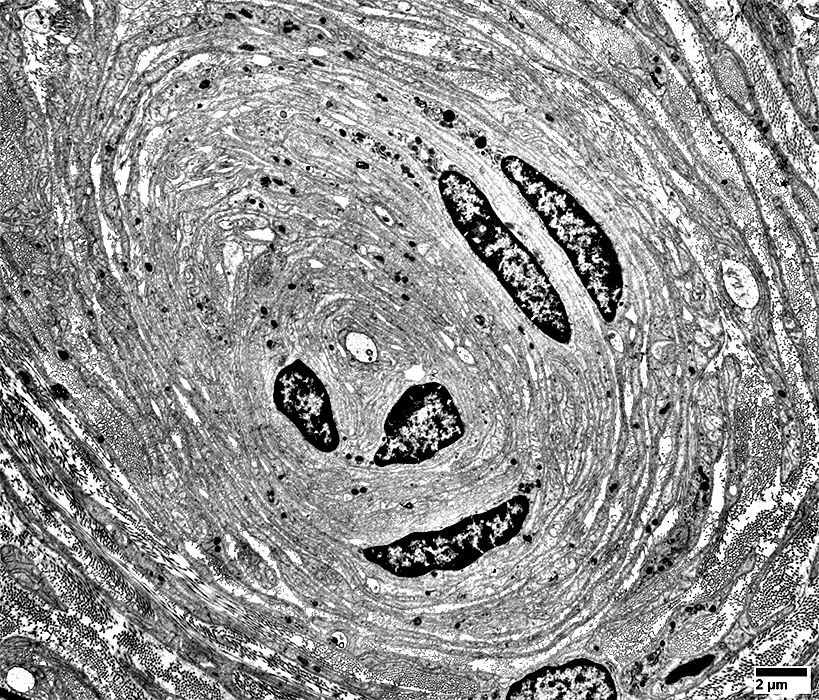
Onion Bulb-like Structures
Abundant parallel basal lamina
Schwann cell processes
Little collagen
 From: R Schmidt |
 From: R Schmidt |
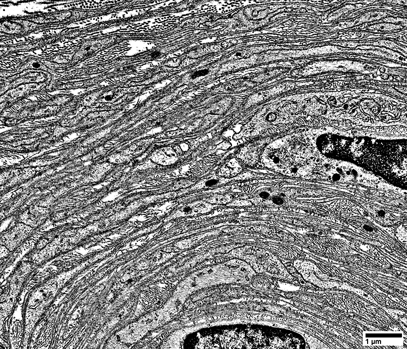 From: R Schmidt |
Abundant parallel basal lamina
Schwann cell processes
Little collagen
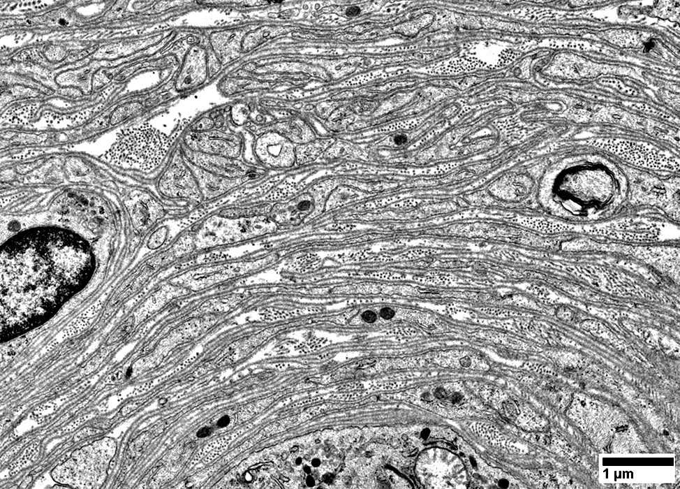 From: R Schmidt |
 From: R Schmidt |
 From: R Schmidt |
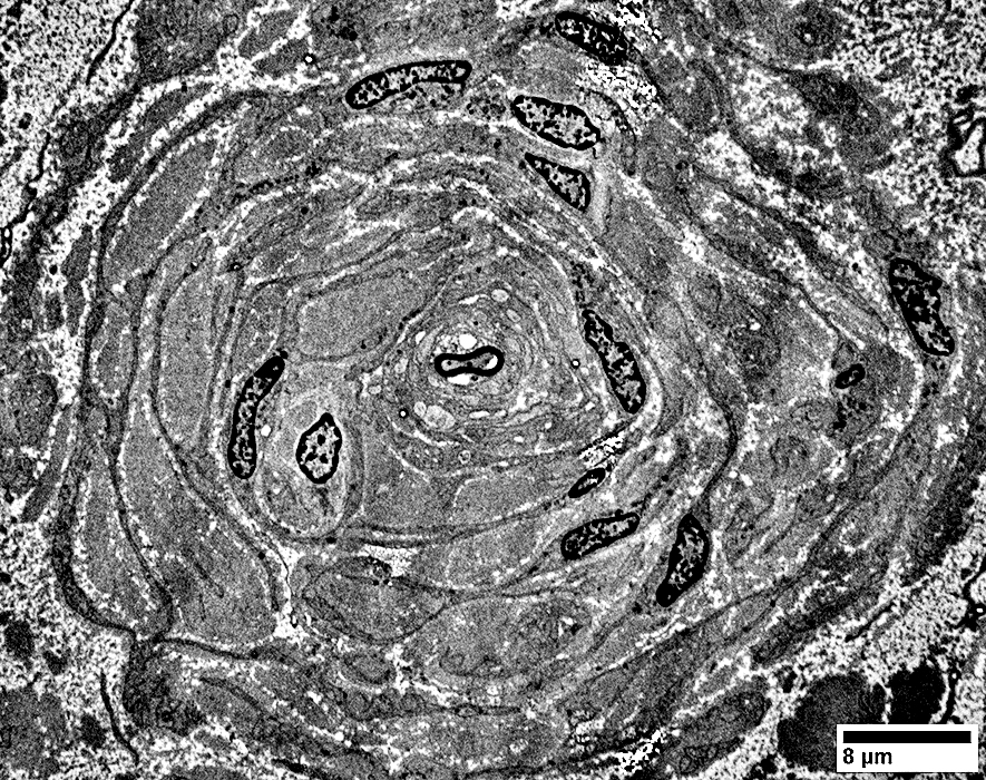
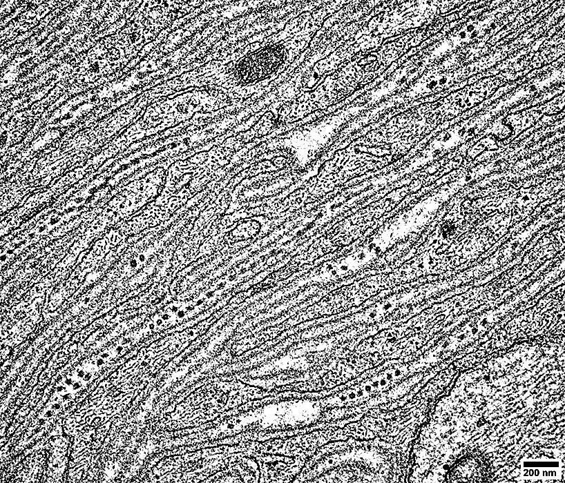
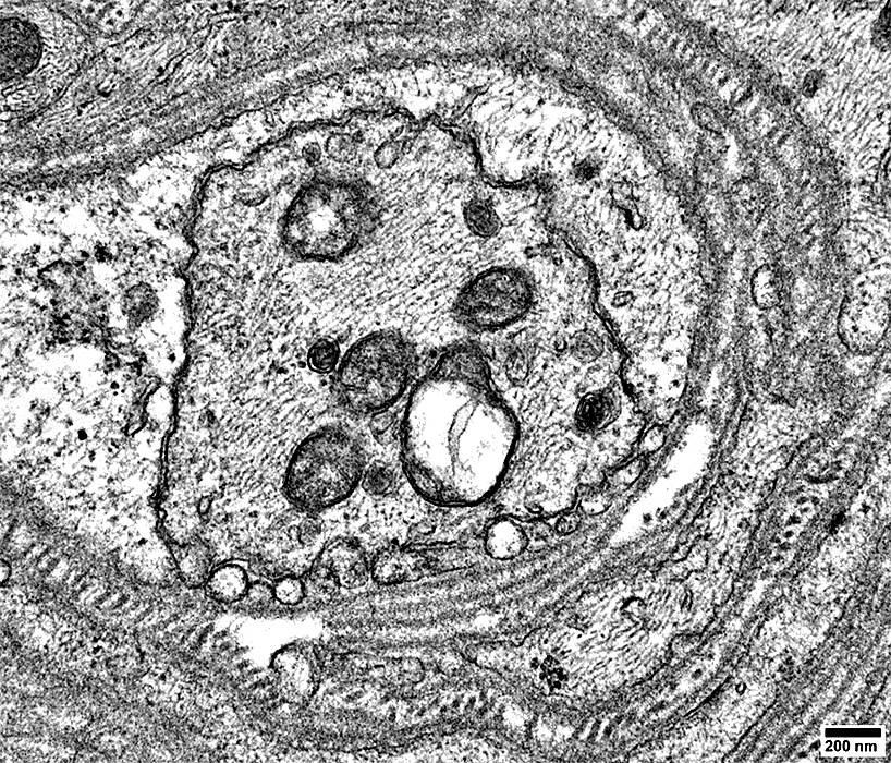
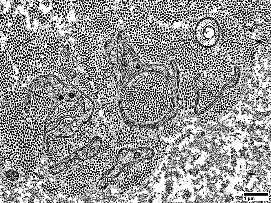
Onion Bulb-like Structures: Axons, Central & Other
 From: R Schmidt |
Thin, or No, Myelin
Some structures have no central axons
 Neurofilament/Gomori stain |
Larger axon: At center of some structures
Smaller axons: Present in circumferential layers
Some structures have no central axons
See: Control nerve
 Neurofilament/Gomori stain |
 Neurofilament/Gomori stain |
Larger axon: At center of some structures
Smaller axons: Present in circumferential layers
Some structures have no central axons
 Neurofilament/Gomori stain |
 Neurofilament/Gomori stain |
P0(r)lp1.jpg) Neurofilament(Green)/P0 (Red) stain |
P0 (Red) stains some cells in onion bulb-like structures
Axons: More loss of large than small axons
Varied degrees of pathology: More axon loss & larger onion bulb like structures in some areas
P0(r)lp2.jpg) Neurofilament(Green)/P0 (Red) stain |
NCAM(r)lp1.jpg) Neurofilament(Green)/NCAM (Red) stain |
NCAM (Red)
Stains most cells in all onion bulb-like structures
Loss of most non-myelinating Schwann cells
Small axons
Are present in layers of many onion bulb-like structures
In other areas have no associated NCAM stain
Axons: More loss of large than small axons
NCAM(r)mp.jpg) Neurofilament (Green)/NCAM (Red) stain |
NCAM(r)mp2.jpg) Neurofilament (Green)/NCAM (Red) stain |
NCAM (Red) stains most cells in all onion bulb-like structures
Loss of most non-myelinating (NCAM) Schwann cells
Small axons are presnt in layers of many onion bulb-like structures
Axons: More loss of small than large axons
P0(r)mp.jpg) Neurofilament(Green)/P0 (Red) stain |
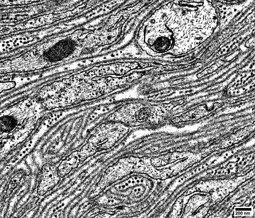
 From: R Schmidt |
Large axons have thin myelin for size
 From: R Schmidt |
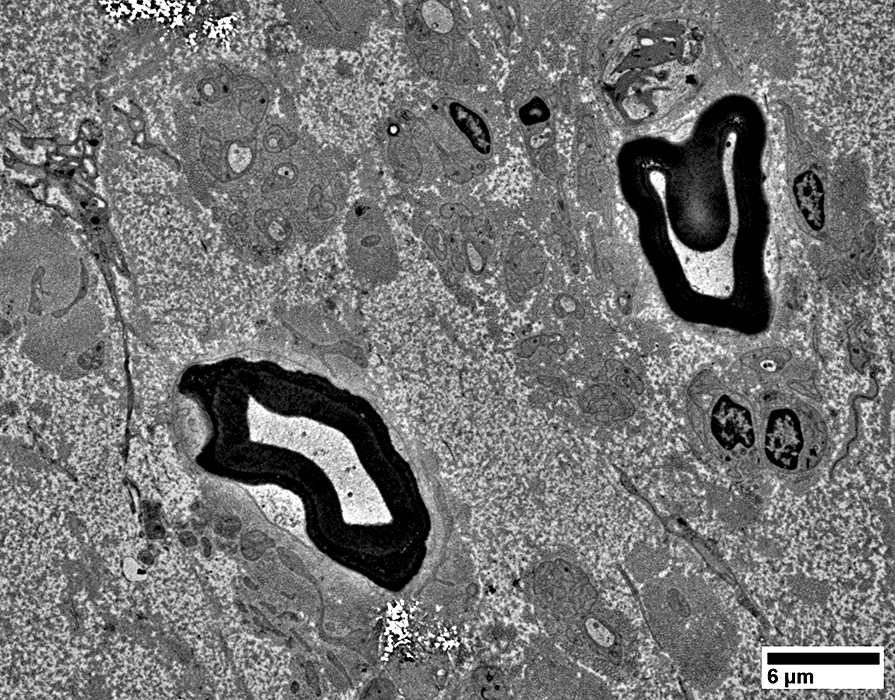
Local Hypertrophic Neuropathy/N-S HNST: Central axons, Ultrastructure
Larger axon with no myelin has irregular structure
 From: R Schmidt |
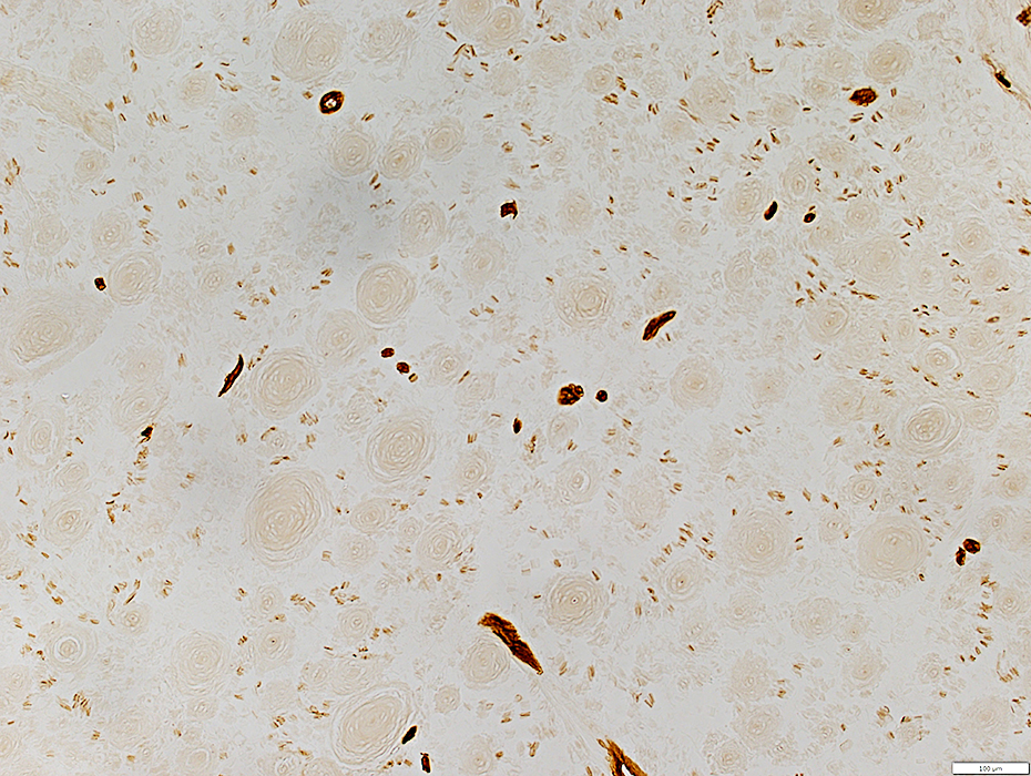
Local Hypertrophic Neuropathy/N-S HNST: Axons in regions other than onion bulb-like structures
 From: R Schmidt |
Small Axons: Present individually surrounded by Schwann cells (Below)
 From: R Schmidt |
MBP(r).jpg) Neurofilament(Green)/MBP (Red/Yellow) stain |
Small axons: Co-stain (Yellow) for neurofilament & Myelin basic protein (MBP) in absence of myelin
Large axons (Above): Have MBP (Red) in surrounding myelin
MBP(r)2.jpg) Neurofilament(Green)/MBP (Red/Yellow) stain |
 Alkaline phosphatase stain |
Endoneurial microvessels: Not increased in number
This differs markedly from perineuriomas
 Alkaline phosphatase stain |
 Ulex stain |
Endoneurial Microvessel: Thick, multilayered wall
 From: R Schmidt |
Local Hypertrophic Neuropathy/N-S HNST
Epithelial Membrane Antigen (EMA)
Stains Perineurium
No, or little, endoneurial EMA staining
See: Perineuriomas
 EMA stain |
Other Pathology
 CD163 stain |
Some histiocytic cells are present
 From: R Schmidt |
Local Hypertrophic Neuropathy/N-S HNST
Büngner band cells associated with collagen pockets
 From: R Schmidt |
Return to:
Return to: Neuromuscular Home Page
2/19/2025