PERINEURIOMA
|
Child Adult Muscle Ultrastructure |
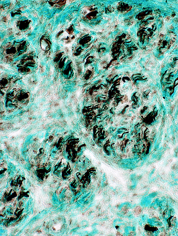
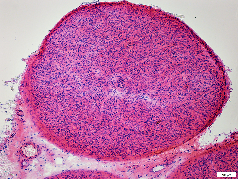
Perineurioma: Adult
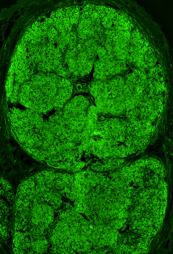
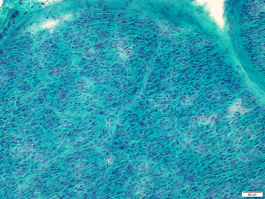
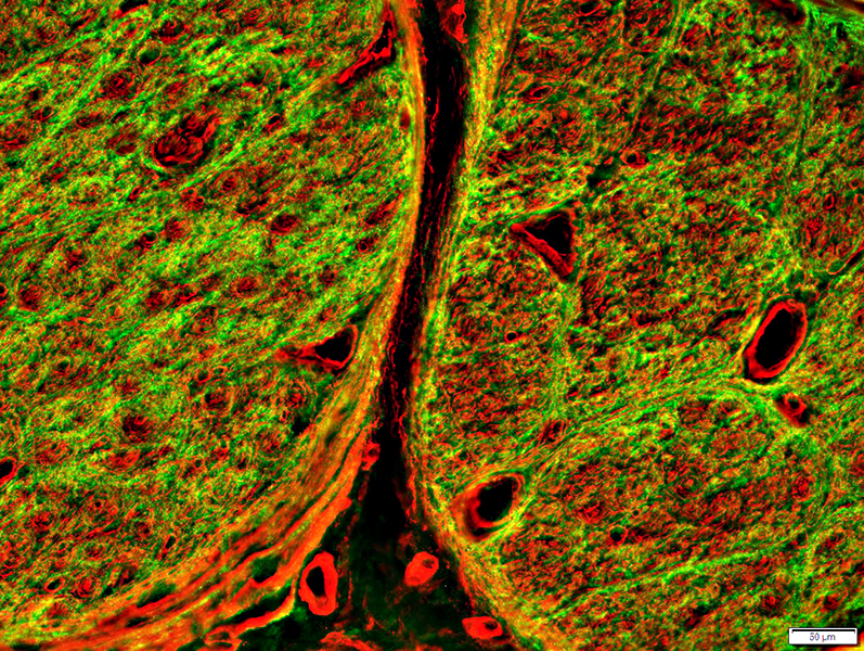
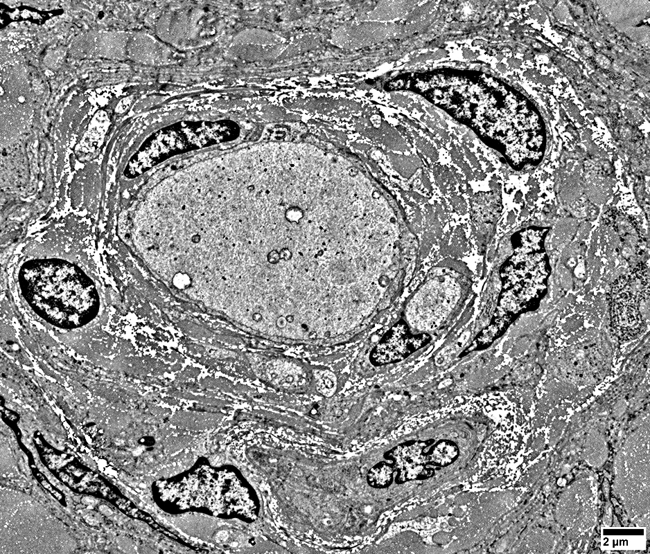
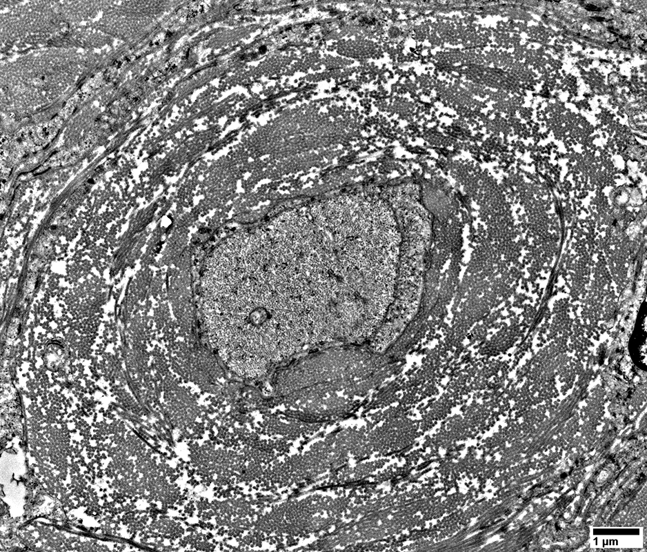
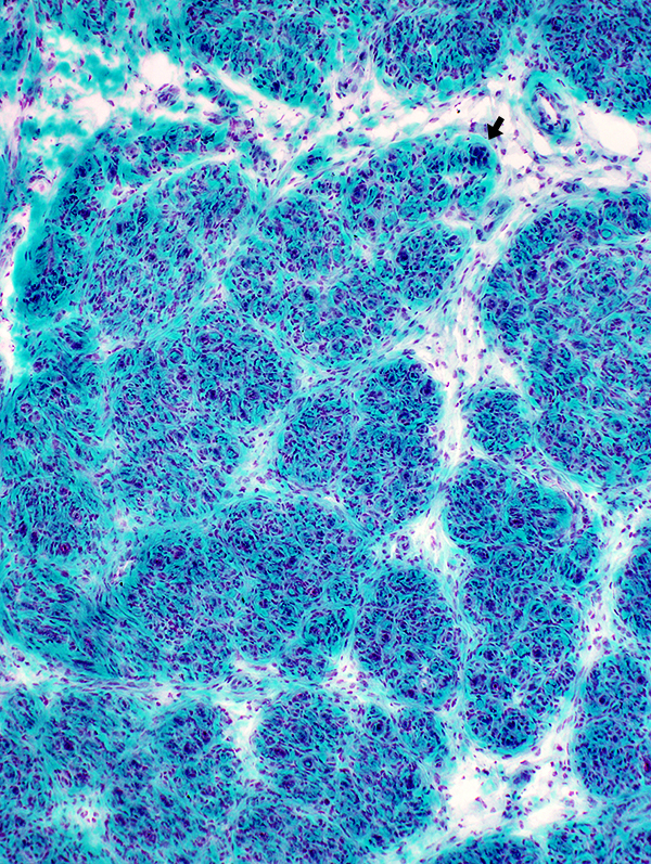 Gomori trichrome stain Perineurioma: Tissue organization Enlarged nerve with many, variably-sized, sub-fascicles Many capillaries (Arrow) Higher magnification (Right) shows many whorled pseudo-onion bulbs |
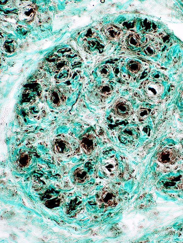
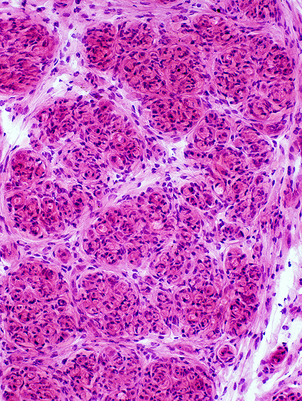 H&E stain |
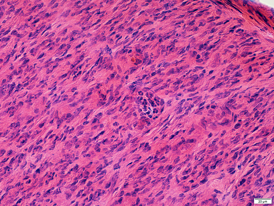
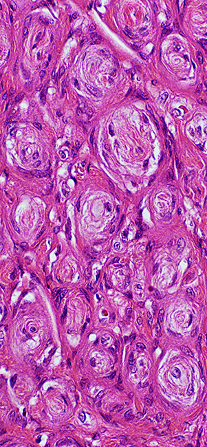 Whorled pseudo-onion bulbs
|
|
|
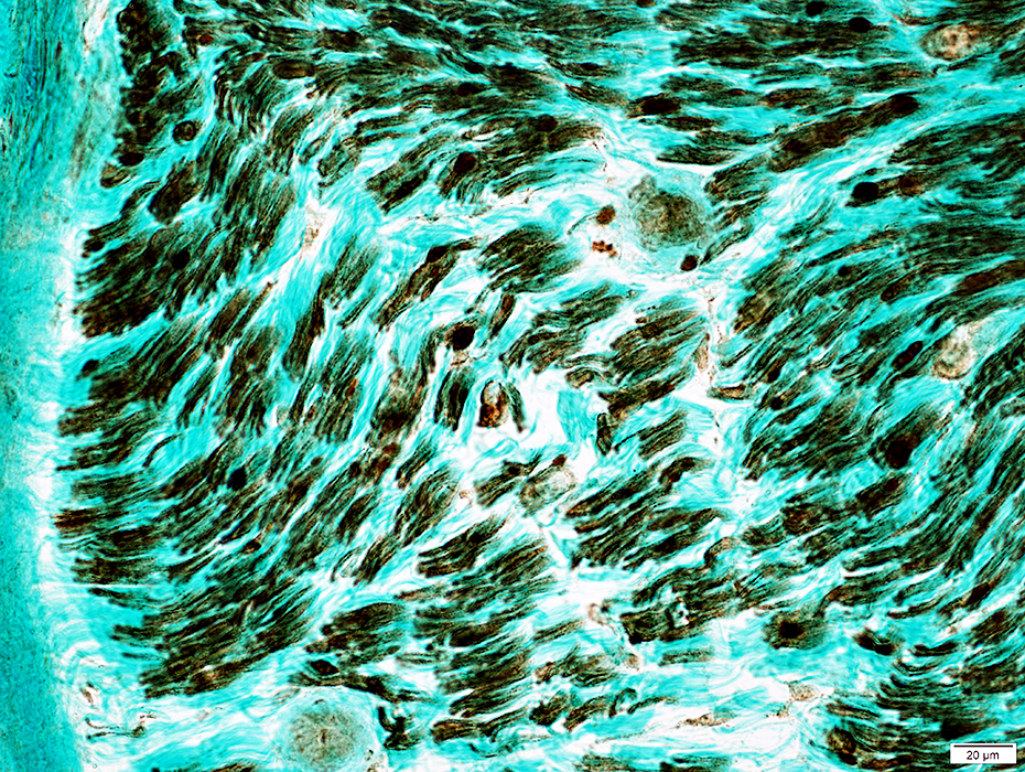
Variable types of axons in whorls: Some whorls have multiple small axons (Left); Other whorls contain large axons (Right) | |
 Neurofilament stain |
 Neurofilament stain |
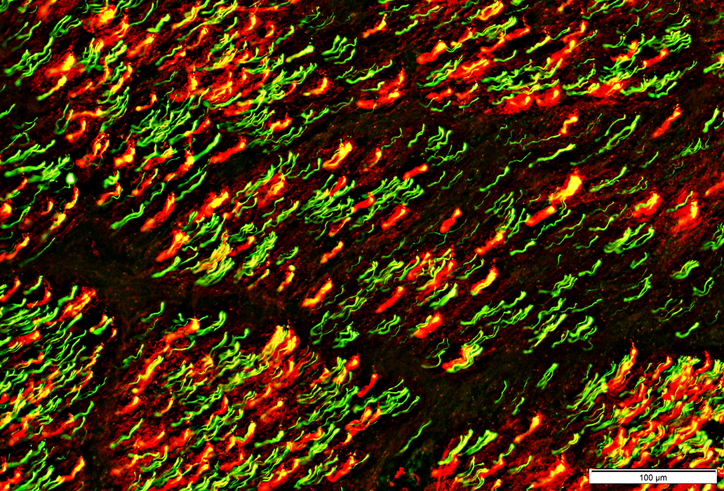
Differential fascicular involvement: More axons in some fascicles than others Neurofilament stain |
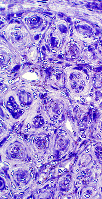
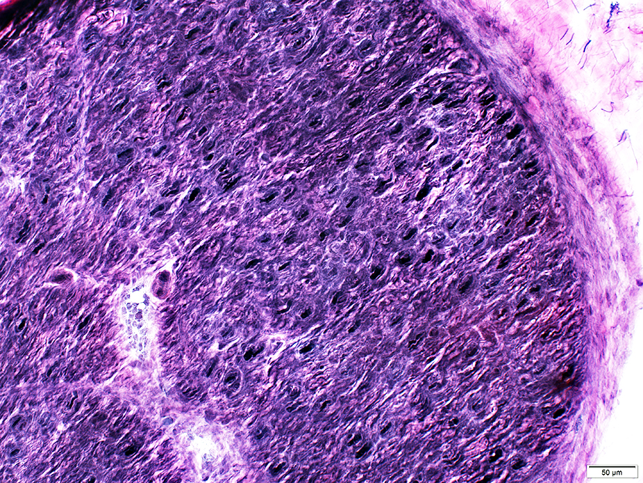
Some whorls contain myelinated axons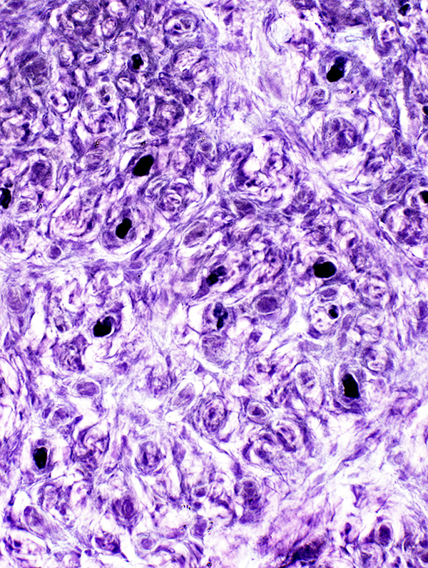 VvG stain |
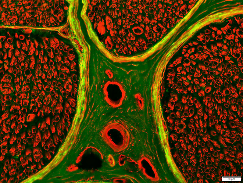
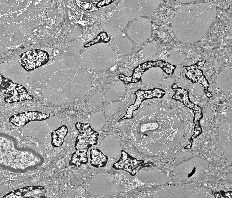
Perineurioma: Many endoneurial vessels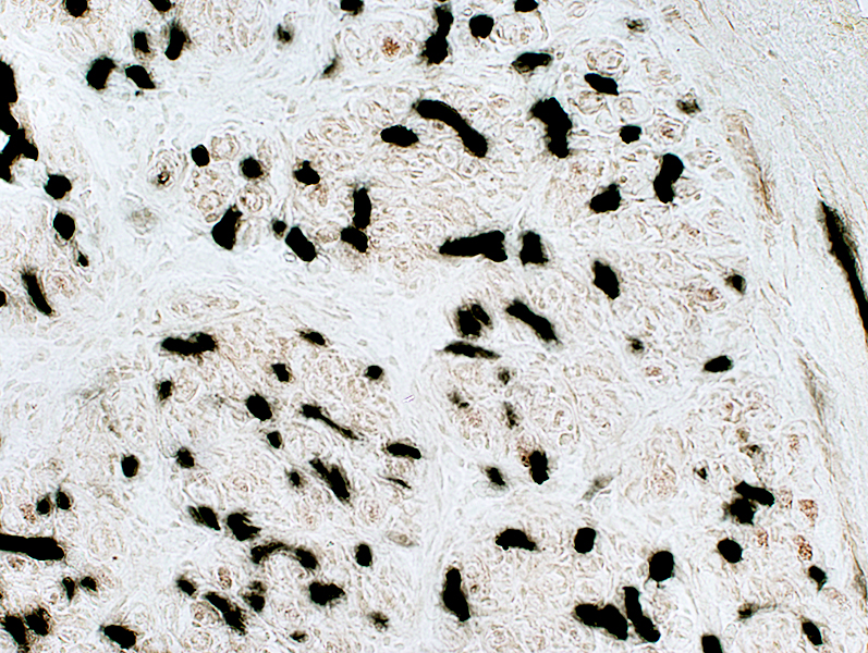 Alkaline phosphatase stain |
|
Epithelial Membrane Antigen (EMA) Perineurioma (Right): Staining of cells diffusely in endoneurium of enlarged nerve fascicle Normal nerve (Below): Staining in perineurium, but not endoneurium 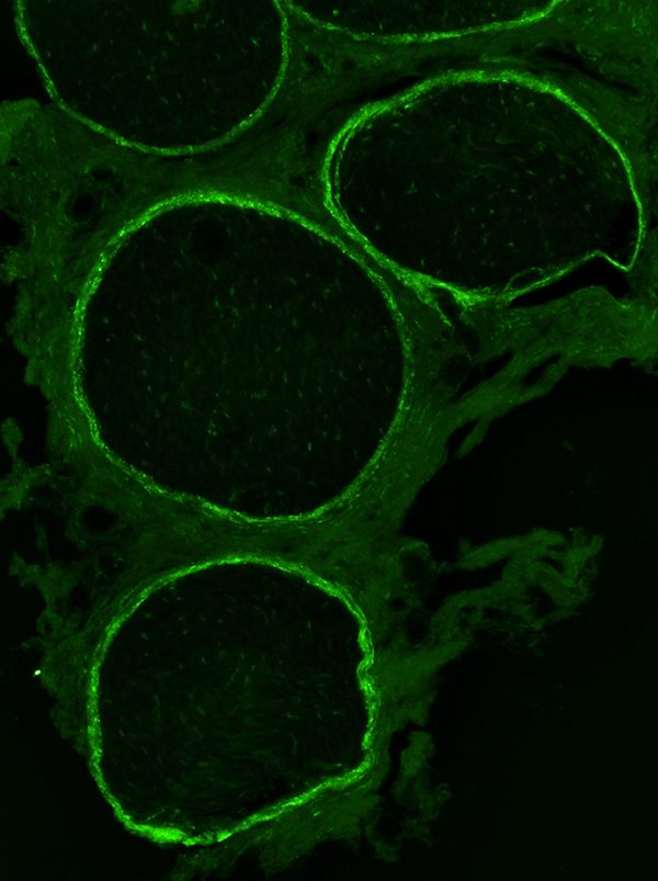
|
|
Perineurioma: EMA stains cells in whorls around axons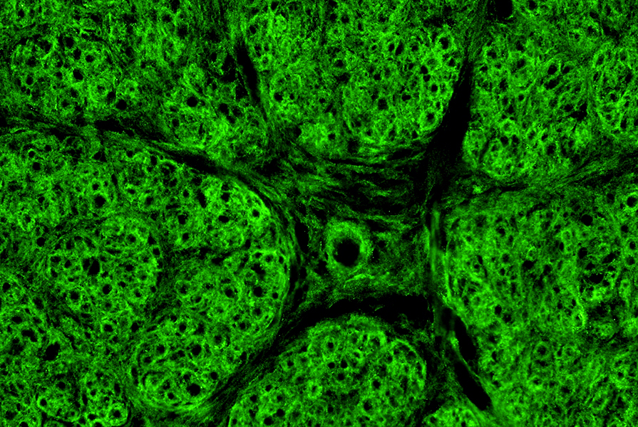
|
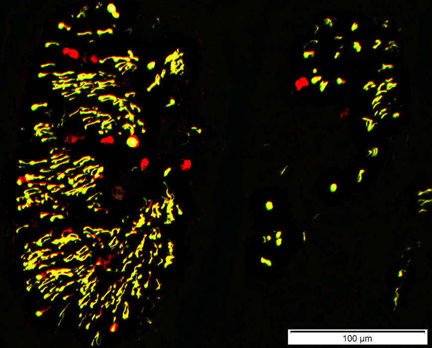
Perineurioma with axon loss: Collagen IV & Neurofilament stains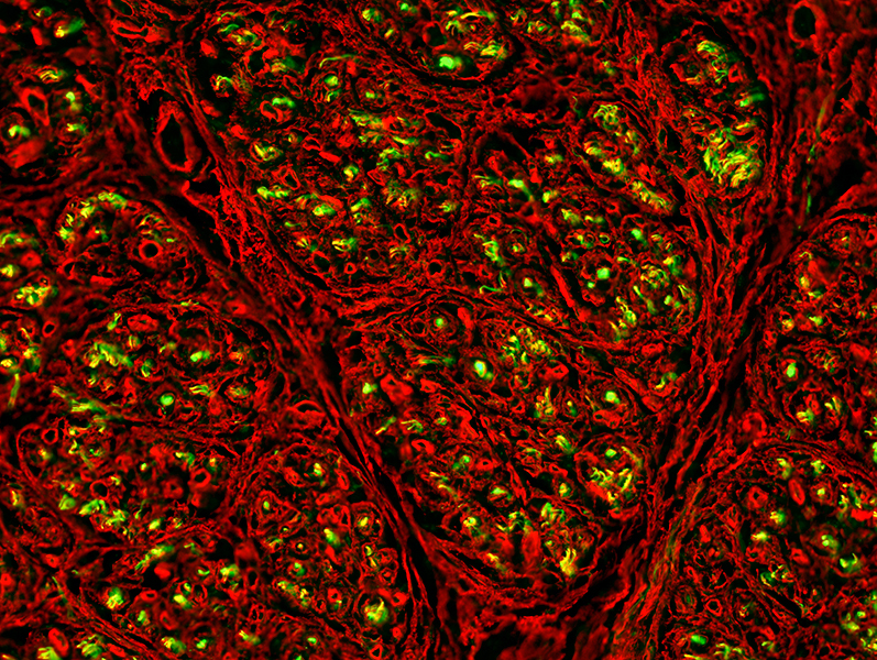 Neurofilament Green; Collagen IV Red |
Perineurioma: Chronic denervation & reinnervation of muscle
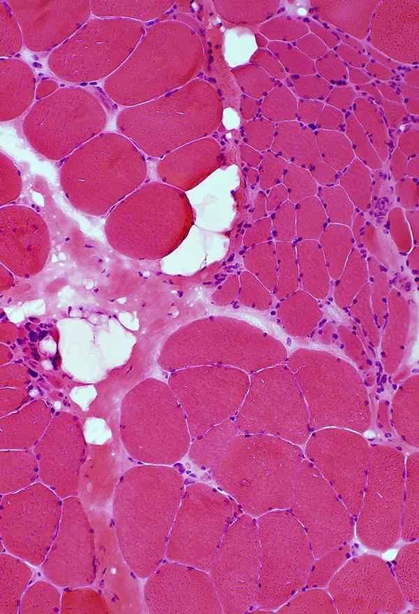 H&E stain |
Grouped Atrophy & Fiber type grouping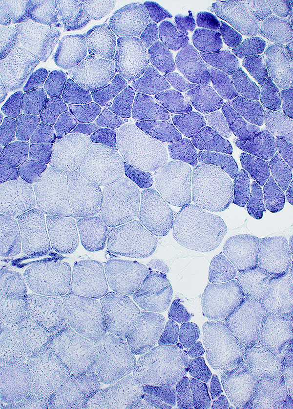 NADH stain |
Fiber type grouping: Large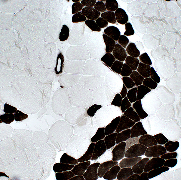 ATPase pH 4.3 stain |
Small angular fibers with targets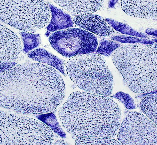 NADH stain |
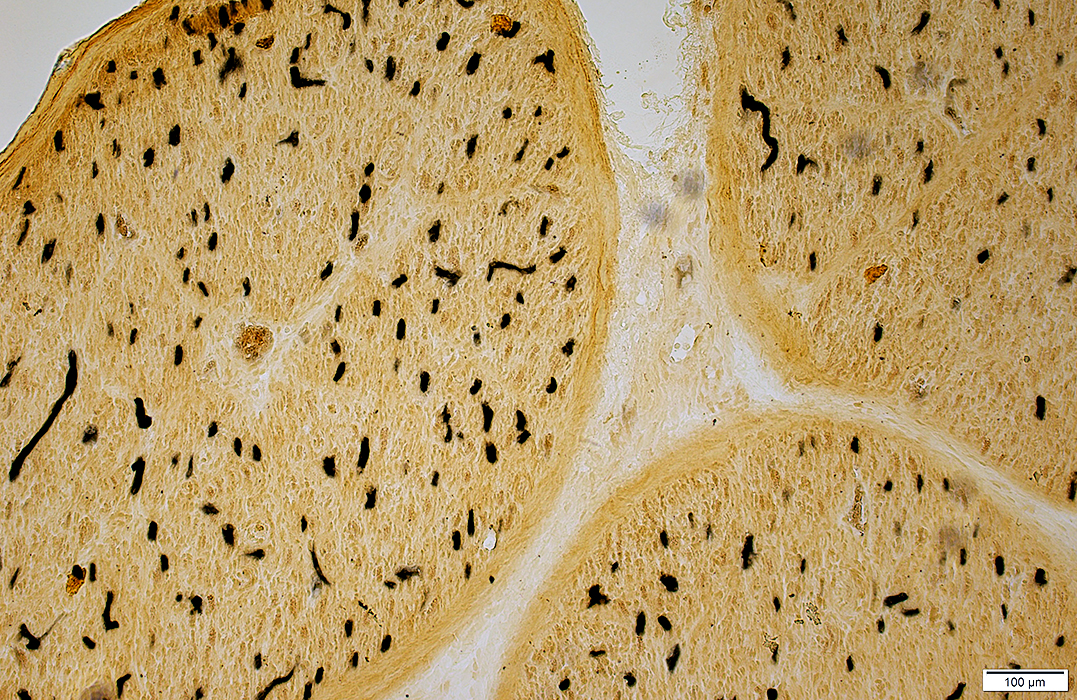
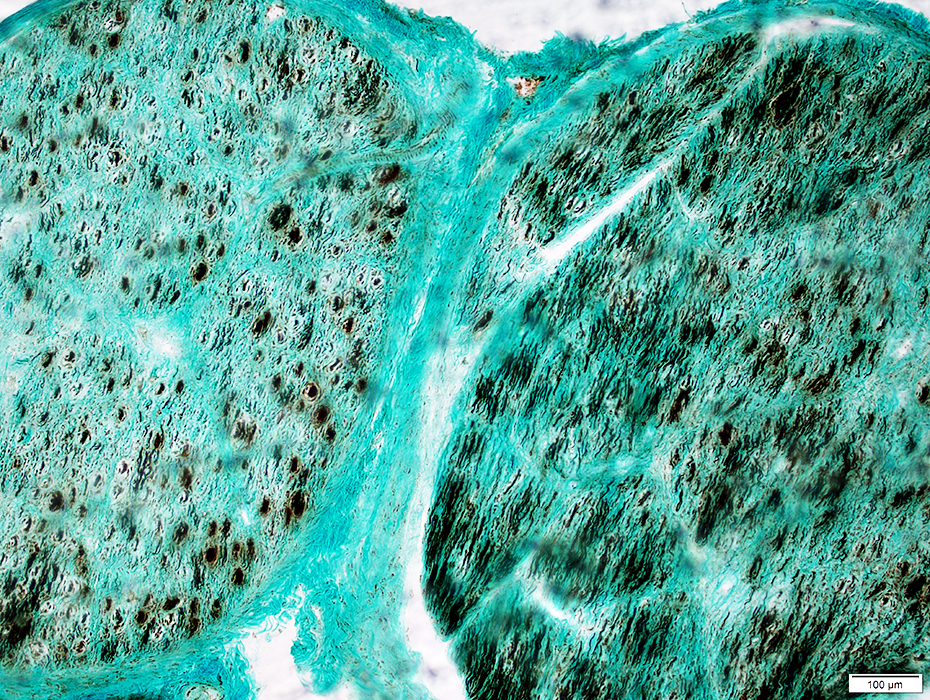
Perineurioma: Child
 H&E stain |
 H&E stain |
 Gomori trichrome stain |
Endoneurium: Cellular; Loss of myelinated axons
 VvG stain |
Perineurioma: Many endoneurial vessels (Alkaline phosphatase positive)
 Alkaline phosphatase stain |
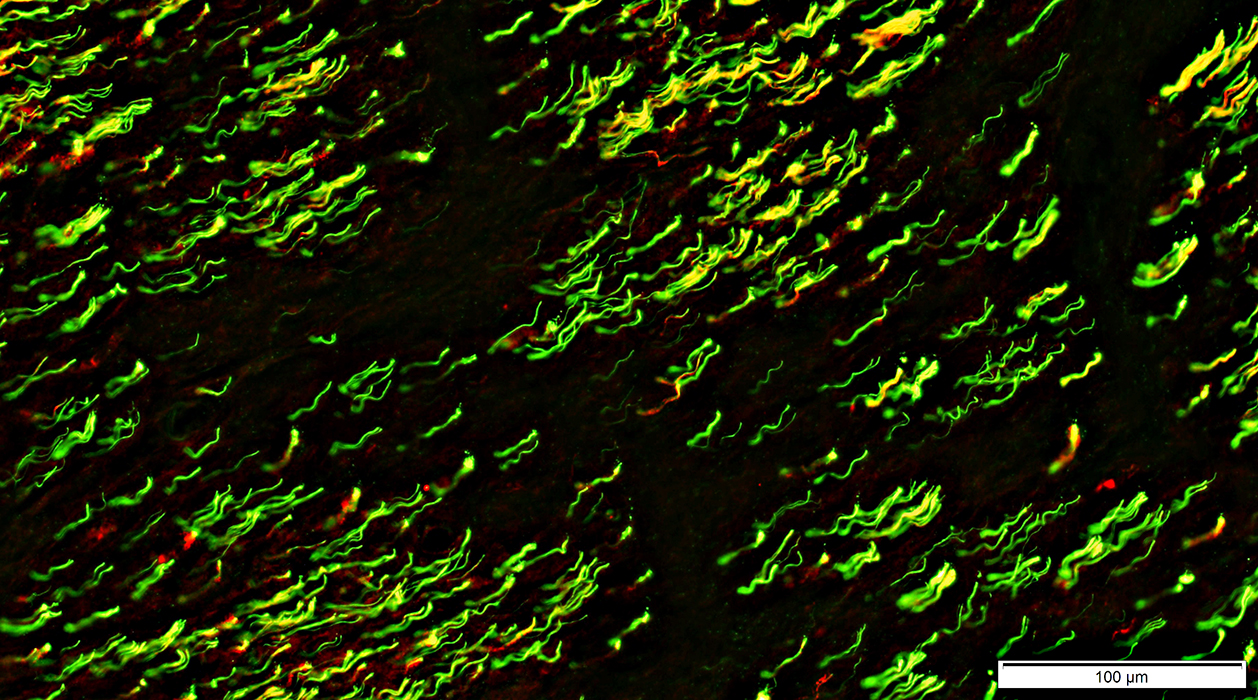
Perineurioma: Axon loss is varied among fascicles
 Neurofilament stain |
Perineurioma: ATPase positive small round structures
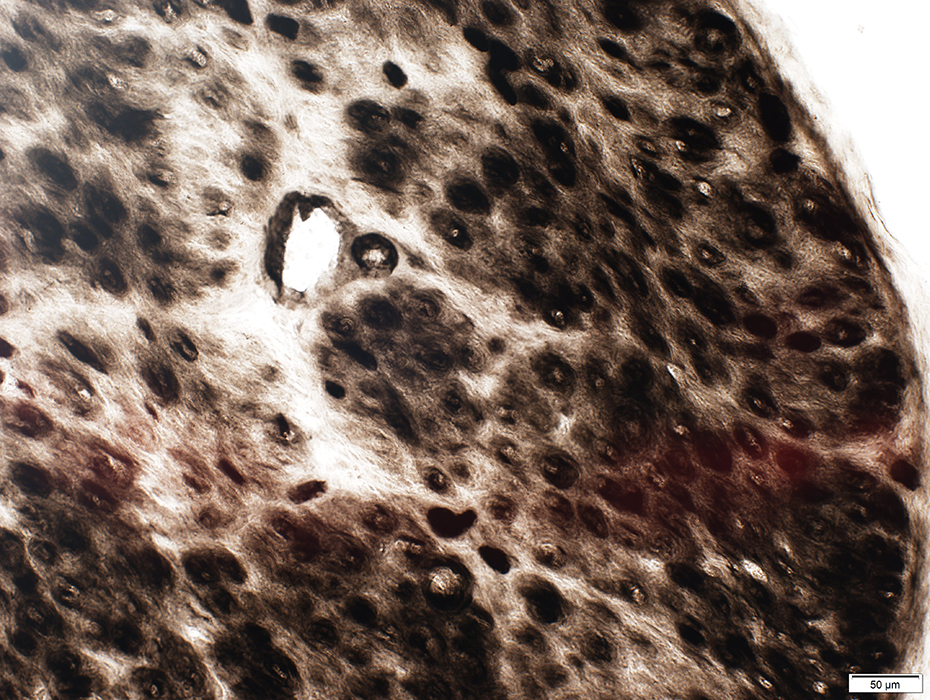 ATPase pH 4.3 stain |
Perineurioma: Cells contain Epithelial Membrane Antigen (EMA)
 Epithelial Membrane Antigen (EMA) (Green); Collagen IV (Red) |
EMA staining
Abnormally present in endoneurial regions of 2 large fascicles
Lost, or reduced, in usual perineurial region
Small normal fascicle (Arrow at bottom): EMA only stains perineurium
Perineurioma
EMA staining in endoneurial regions
 Epithelial Membrane Antigen (EMA) (Green); Collagen IV (Red) |
Control nerve
Epithelial Membrane Antigen (EMA): Present in perineurium but not endoneurium (Green-Yellow)
Collagen IV: Present in perineurium & endoneurium (Red-Yellow)
 Epithelial Membrane Antigen (EMA) (Green); Collagen IV (Red) |
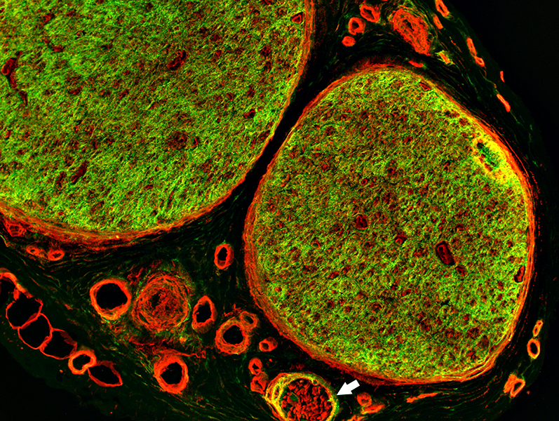
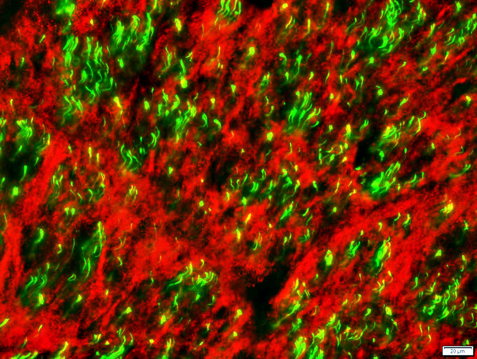
Perineurioma: Endoneurial Cell Population
 Neurofilaments (Green); NCAM (Red) |
Large axons (Green; Neurofilaments): Most are lost
Small axons (Green; Neurofilaments): Many may remain within perineurioma
Non-myelinating Schwann cells (Red; NCAM stain)
No NCAM staining: Small axons appear "Naked"
Present on ultrastructure
Stain for MBP
|
Control nerve Large axons (Green; Neurofilaments): Many are present; No associated NCAM stain Small axons (Yellow; Neurofilaments): Associated with non-myelinating Schwann cells (Yellow) Non-myelinating Schwann cells (Red & Yellow; NCAM stain): Associated with small axons, normally present in clusters 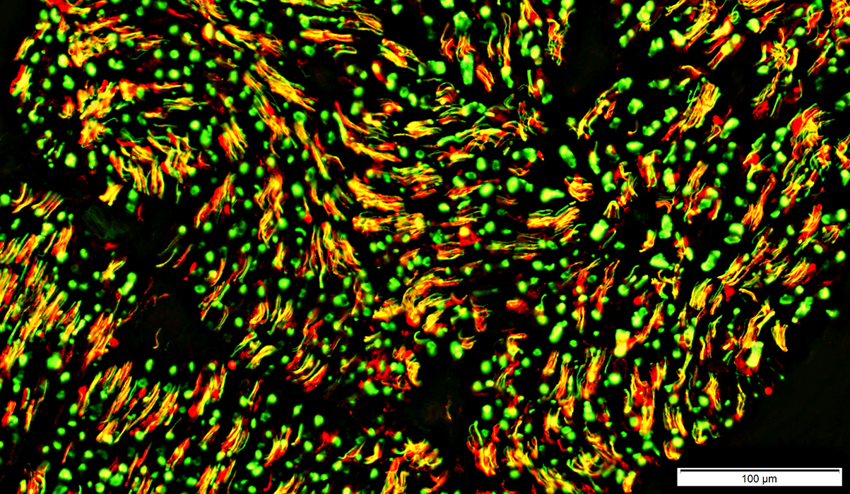 Neurofilaments (Green); NCAM (Red) |
|
Perineurioma Loss of NCAM-positive Non-myelinating Schwann cells in endoneurium 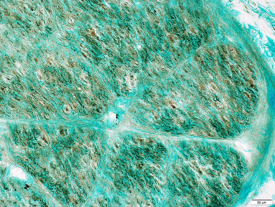 NCAM stain |
Control nerve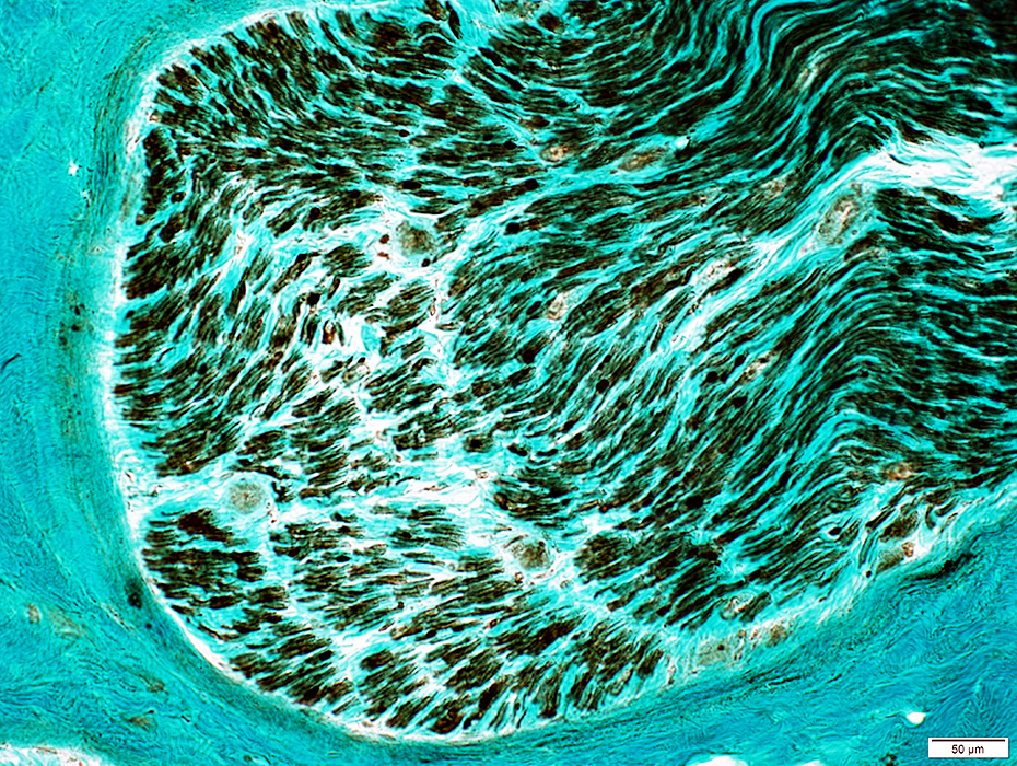 NCAM stain |
 NCAM stain |
Perineurioma
Most large axons (Yellow) have associated myelin (P0 stain (Red)) surrounding them
 Neurofilament (Green) & P0 (Red) stain |
Perineurioma
Immature Schwann cells: Surround remaining small, unmyelinated axons
Stain (Yellow) for MBP without myelin
Little or no NCAM
 Neurofilaments (Green); MBP (Red); |
Perineurioma: Axons surrounded by regions of Perineurial tumor cells (EMA staining)
 Neurofilament (Green) & EMA (Red) stain |
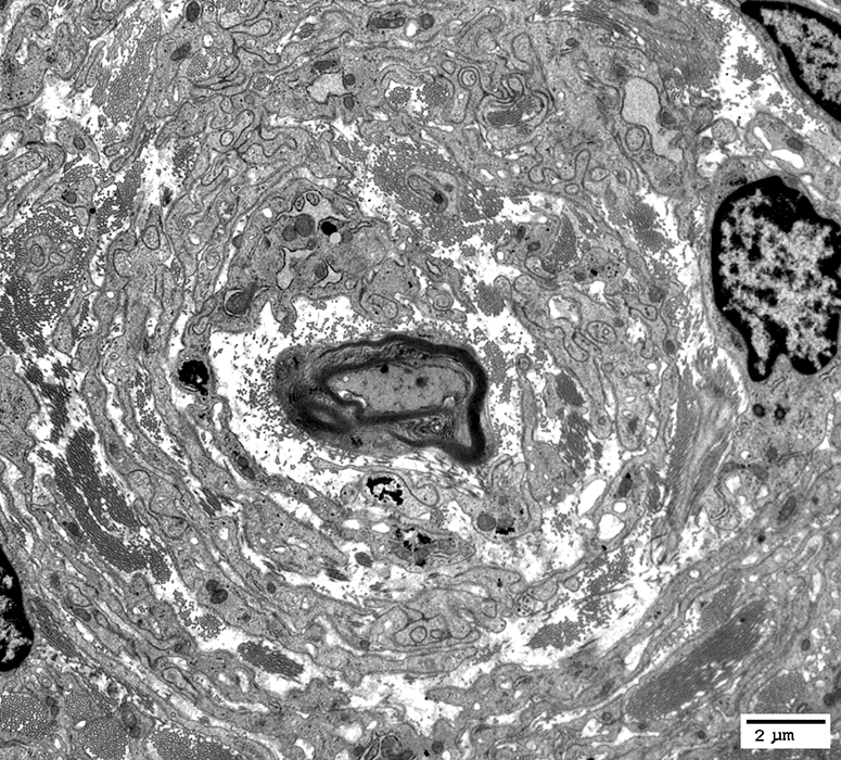
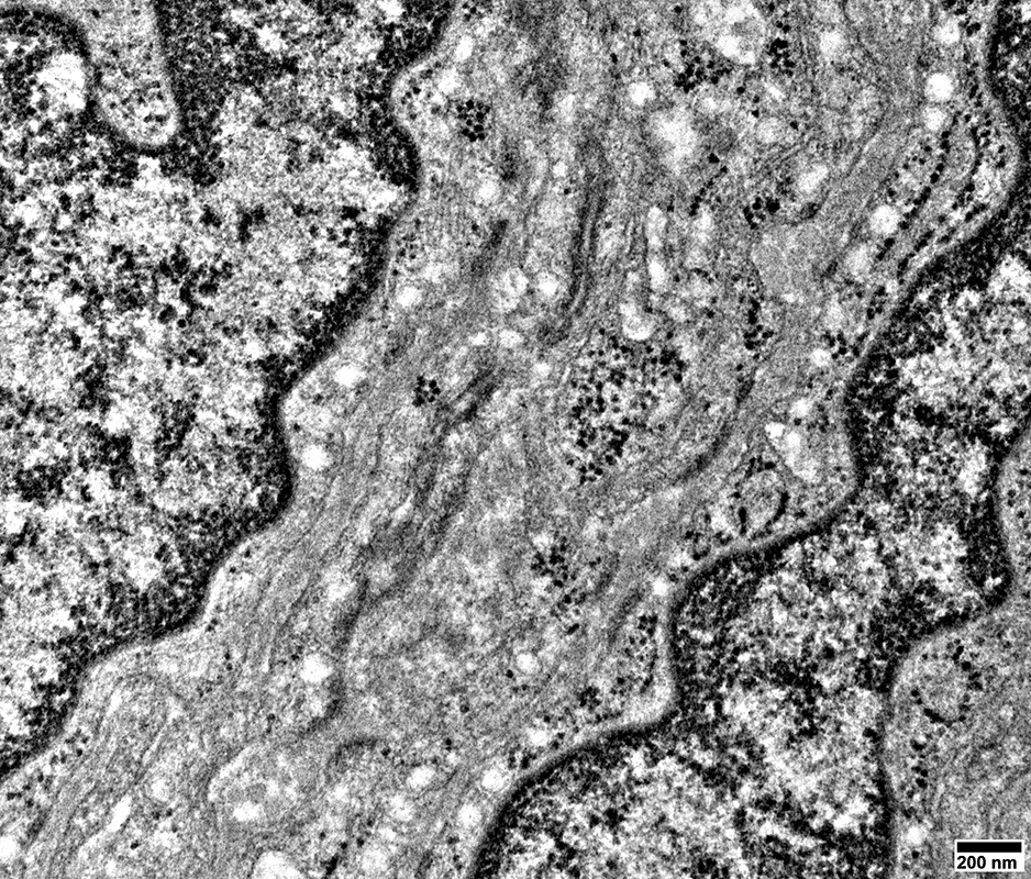
Perineurioma: Ultrastructure
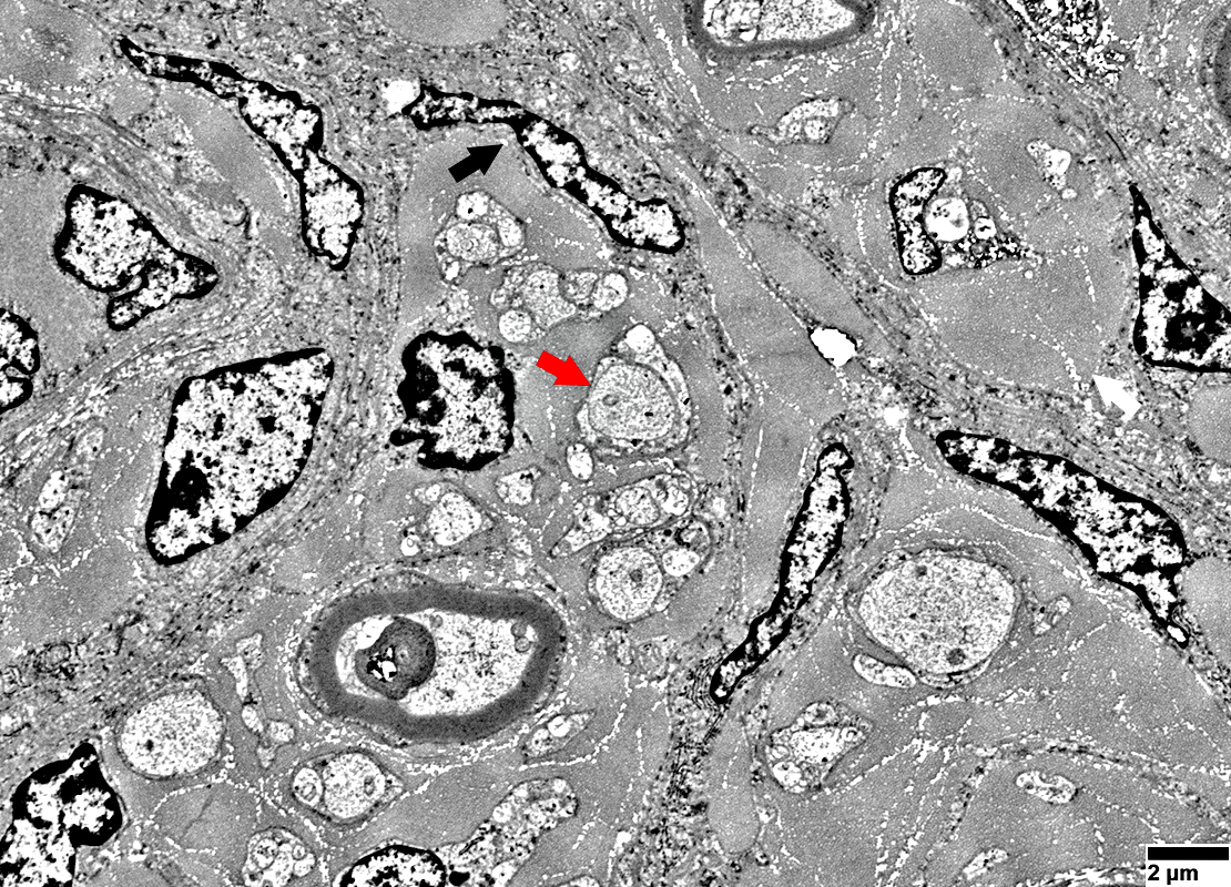 From: R Schmidt |
Perineurial tumor cells in endoneurium (Black arrow)
Long processes containing small round vesicles
Unmyelinated axons
Surrounded by thin Schwann cell processes (Red arrow)
Processes contain MBP but not NCAM
Large collagen pockets are present (White arrow)
 From: R Schmidt |
 From: R Schmidt |
Layered whorls of cells & processes around, but not immediately neighboring, unmyelinated axon
Central unmyelinated axon: Surrounded by non-myelinating Schwann cell & Collagen
 From: R Schmidt |
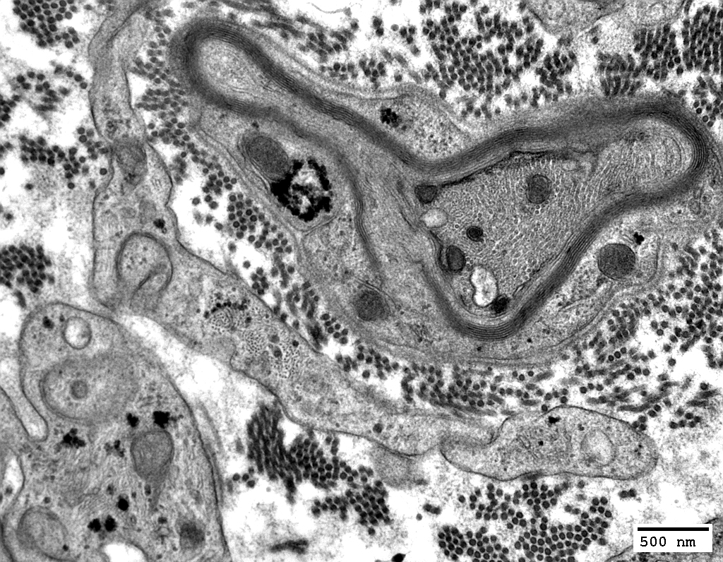
Layered whorls of processes around thinly myelinated axon
Central thinly myelinated axon: Surrounded by Myelin & Collagen
 From: R Schmidt |
Center: Axon, Myelinated or non-myelinated
Around axon: Myelin with Schwann cell, or Schwann cell alone
Surround: Collagen
Periphery: Perineurial cell processes
Interdigitated
Basal lamina: Varied thickness
 From: R Schmidt |
Surrounds only single axon
Stained by MBP
Not stained by NCAM
 From: R Schmidt |
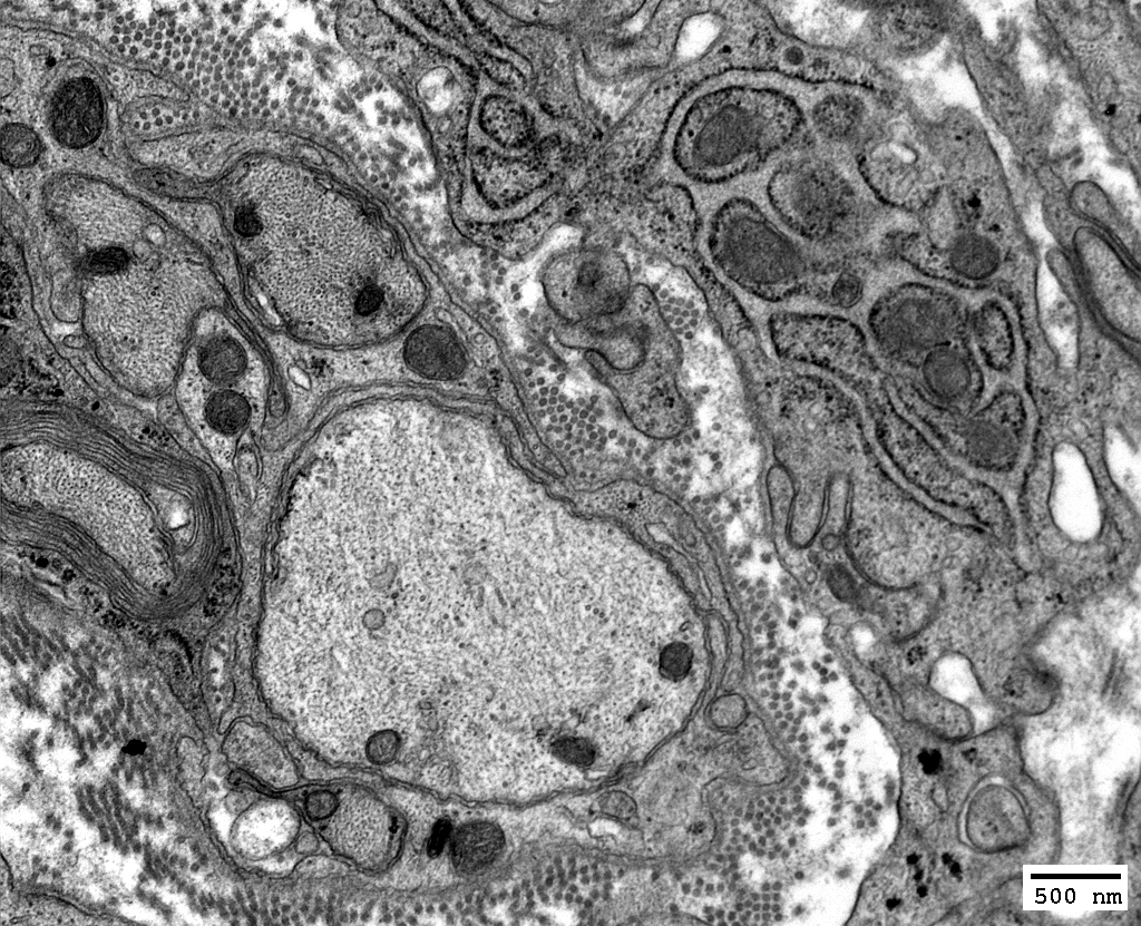
Perineurioma
 From: R Schmidt |
Fusion of regions of apposed membranes (Red Arrow)
 From: R Schmidt |
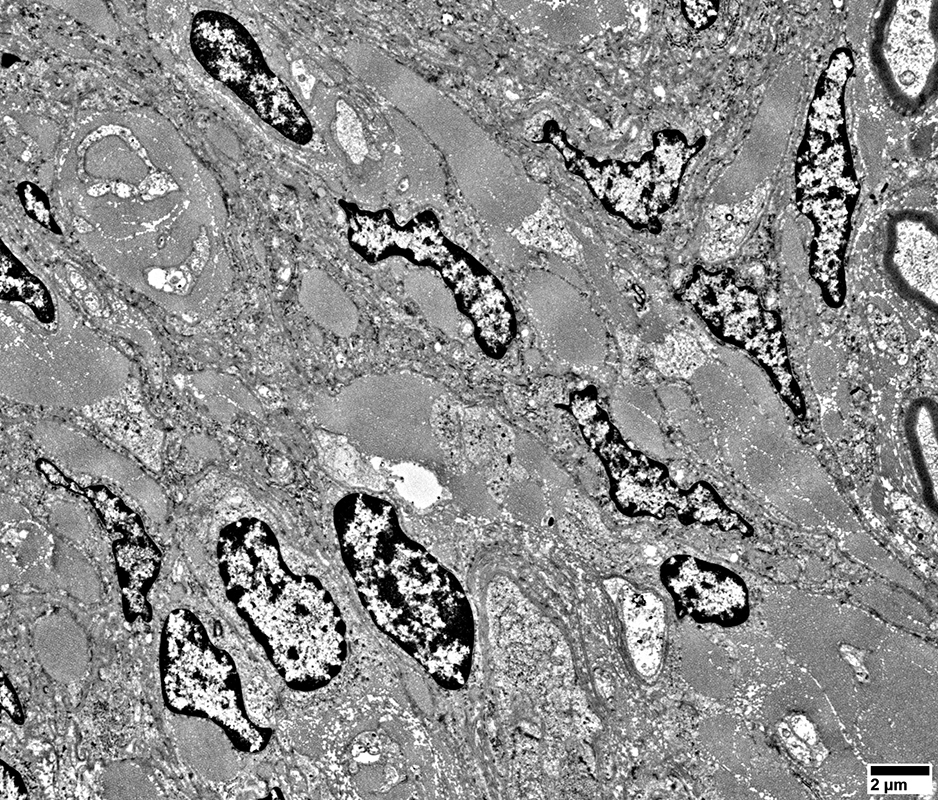
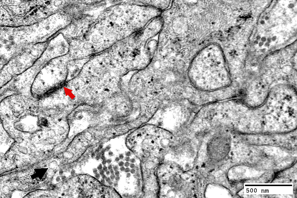
Perineurioma: Collagen Bulbs
From: R Schmidt |
Irregular elongated nuclei in perimysium, often near collagen pockets
Myelinated axon
Surrounded by region containing mainly collagen
Collagen pockets
Many in endoneurium
 From: R Schmidt |
Immediately surrounded by thin Schwann cell process: Stains for MBP, not NCAM
Within regions containing mainly collagen
 From: R Schmidt |
Return to Perineurioma
Return to Biopsy illustrations
Return to Neuromuscular Home Page
Return to Nerve biopsy
3/2/2026