Immune Checkpoint Inhibitors
|
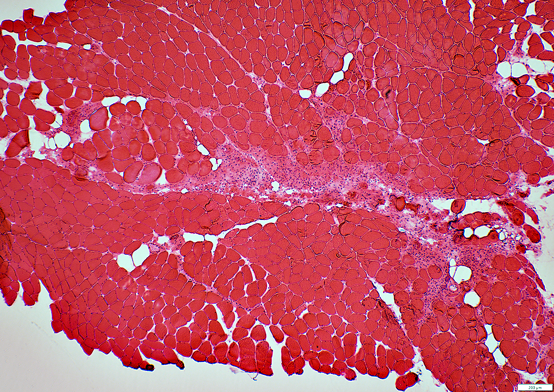
Immune Neuropathies Endoneurial Microvessel Inflammation Perineuritis/Epineuritis Immune Myopathies, Multifocal Cell foci, Mild fiber necrosis Cell foci, Clustered fiber necrosis |
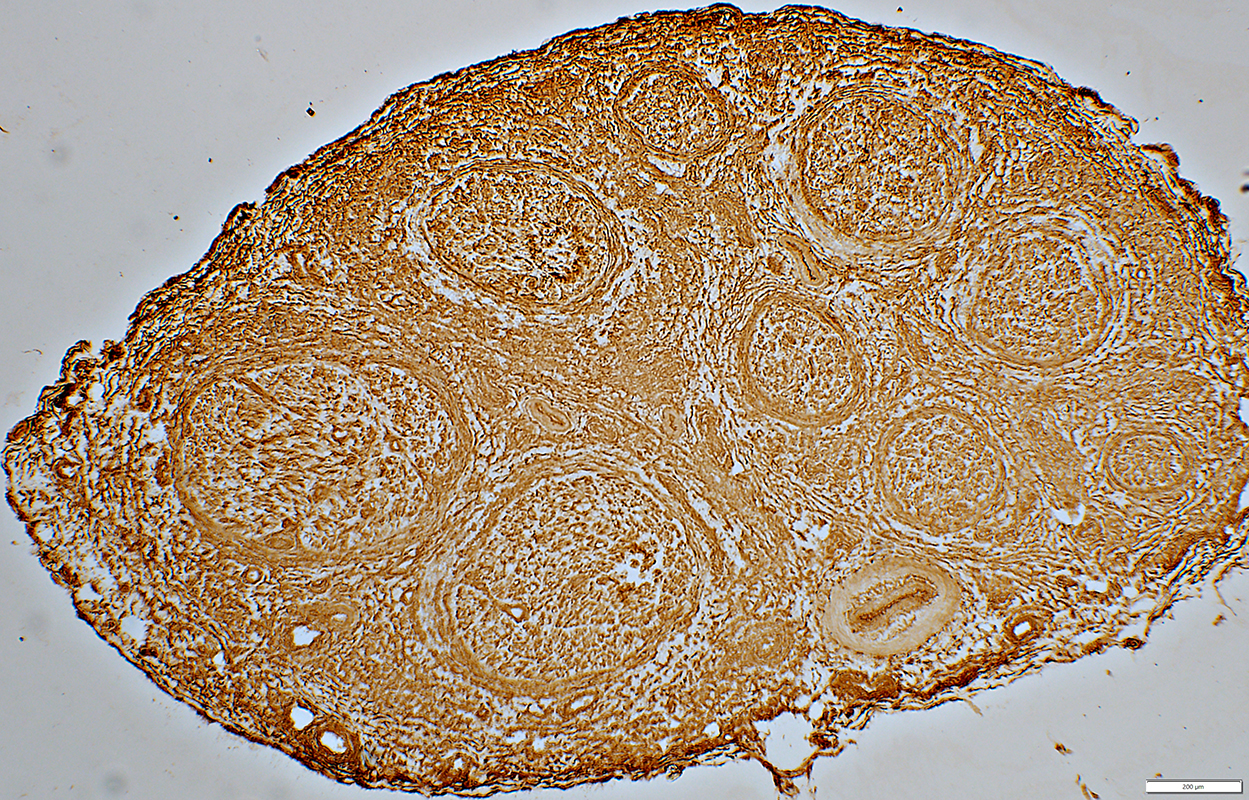
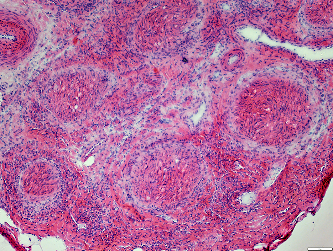
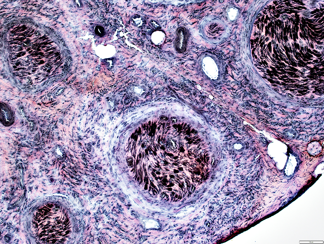
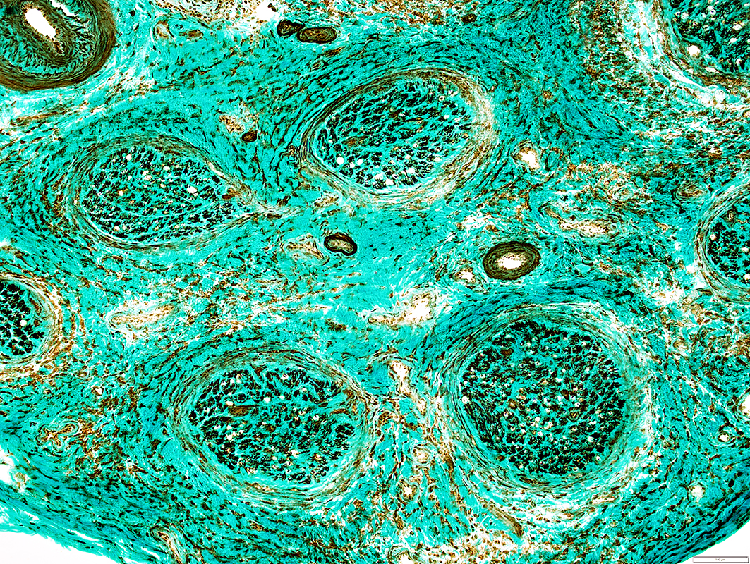
Polyradiculoneuropathy + Endoneurial Inflammatory Vasculopathy (Ipilimumab)
MRI: Root enhancement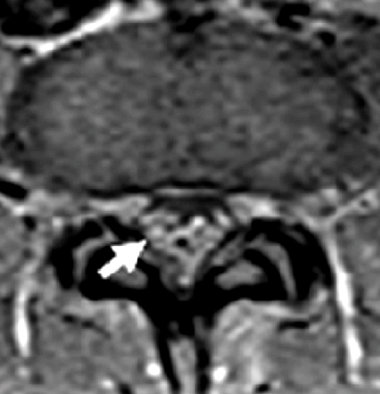
|
MRI: Meningeal enhancement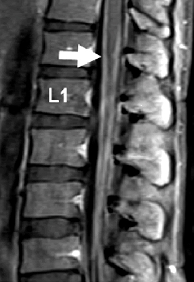
|
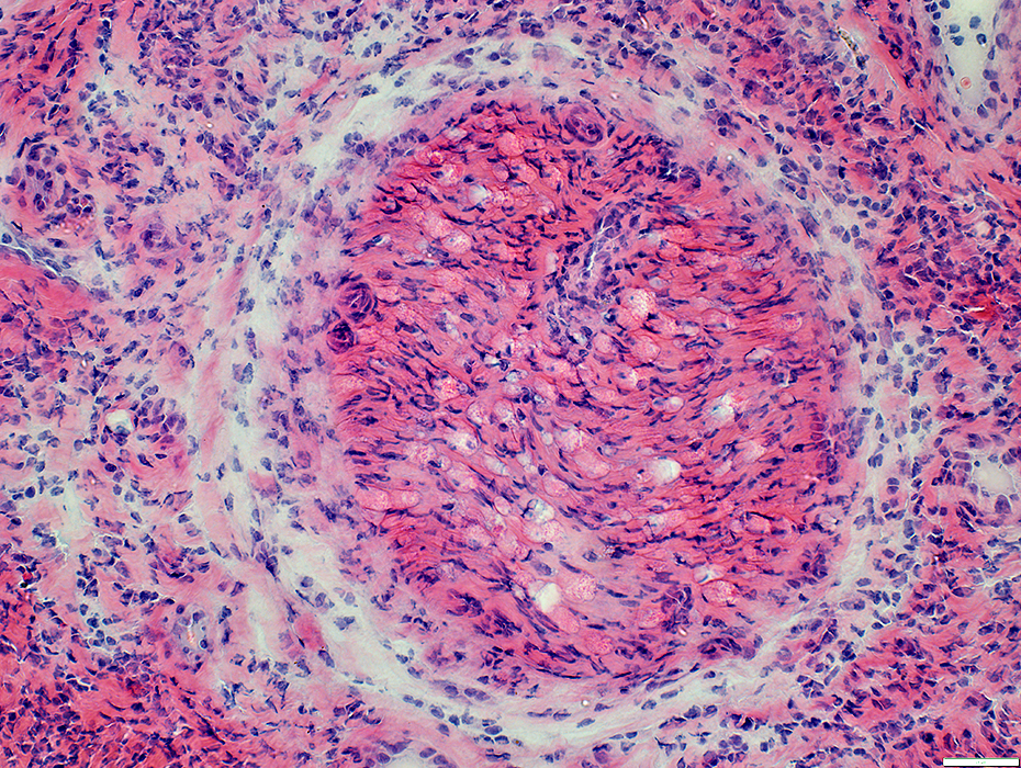
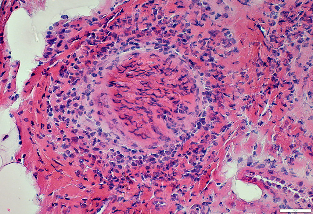
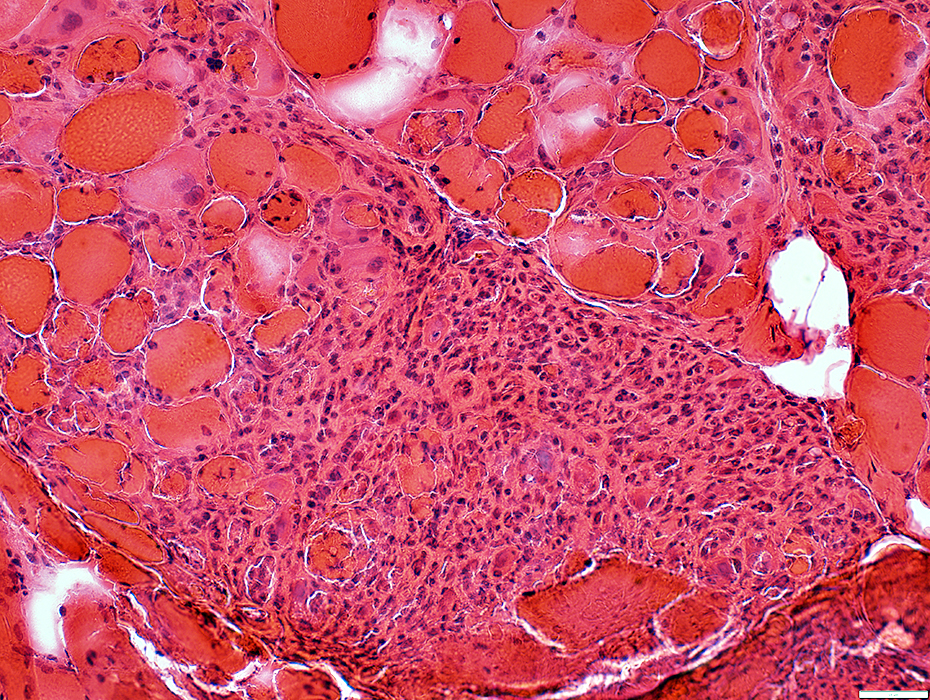
Endoneurial Microvessels: Perivascular inflammation
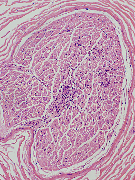 H&E stain |
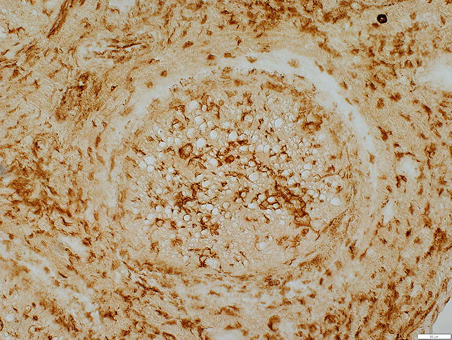
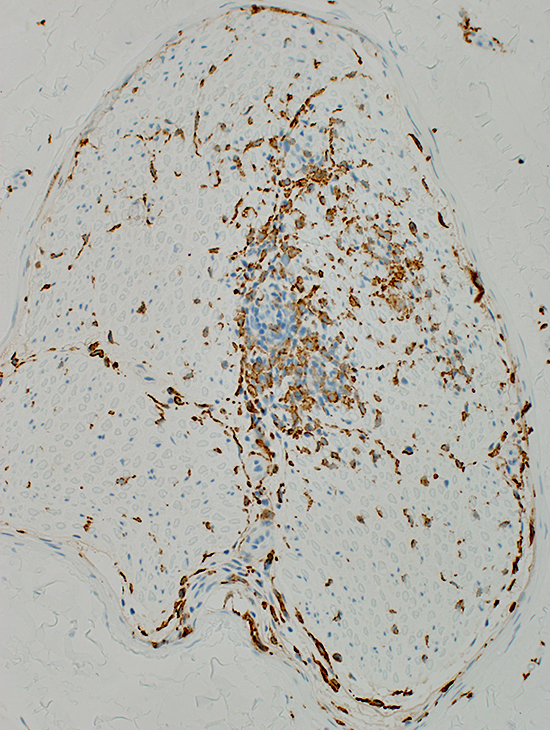 CD163 stain |
 H&E stain |
 CD3 stain |
 CD163 stain |
 Acid phosphatase stain |
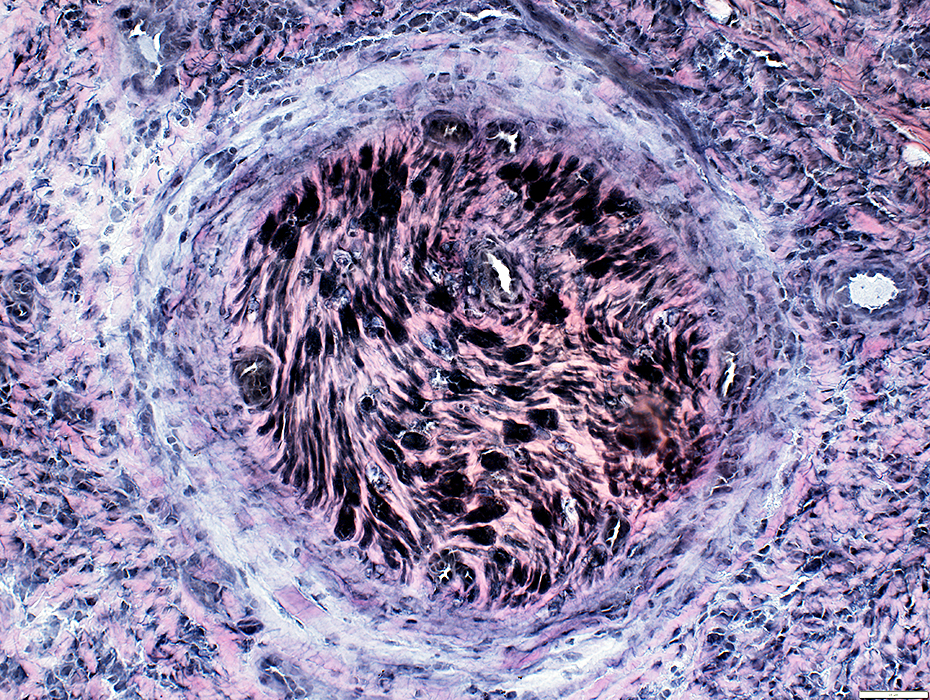
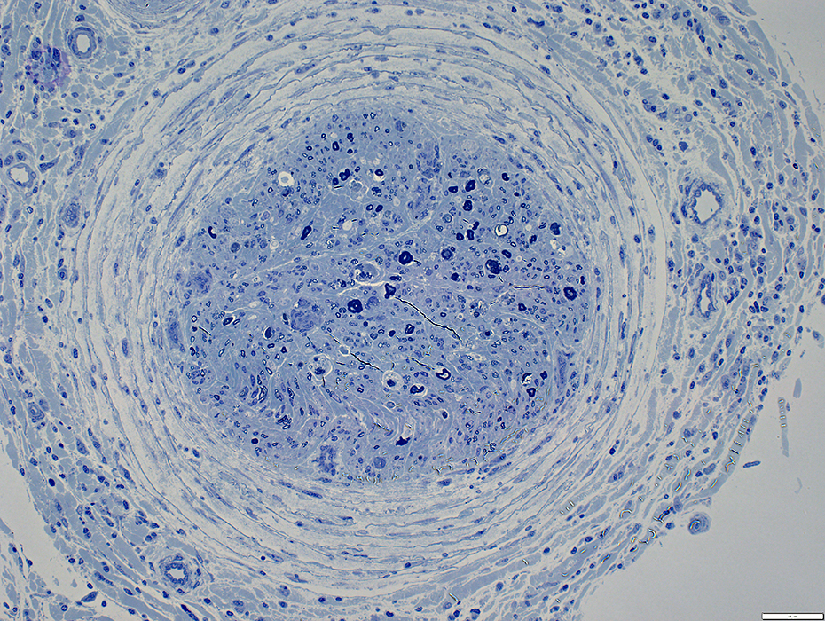
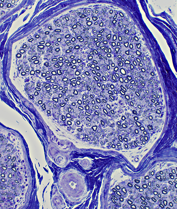 Toluidine blue stain |
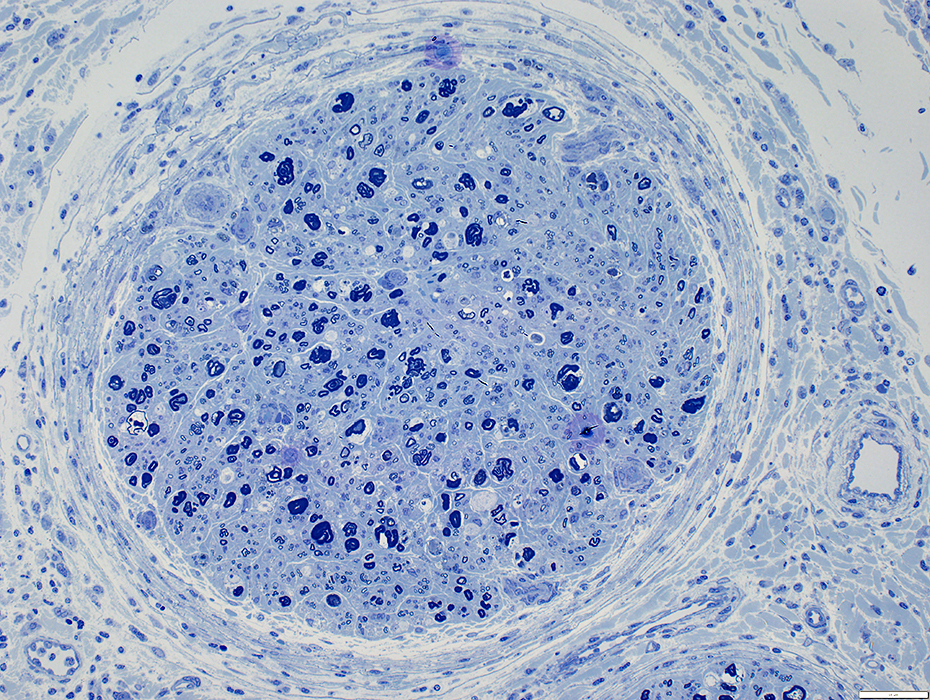
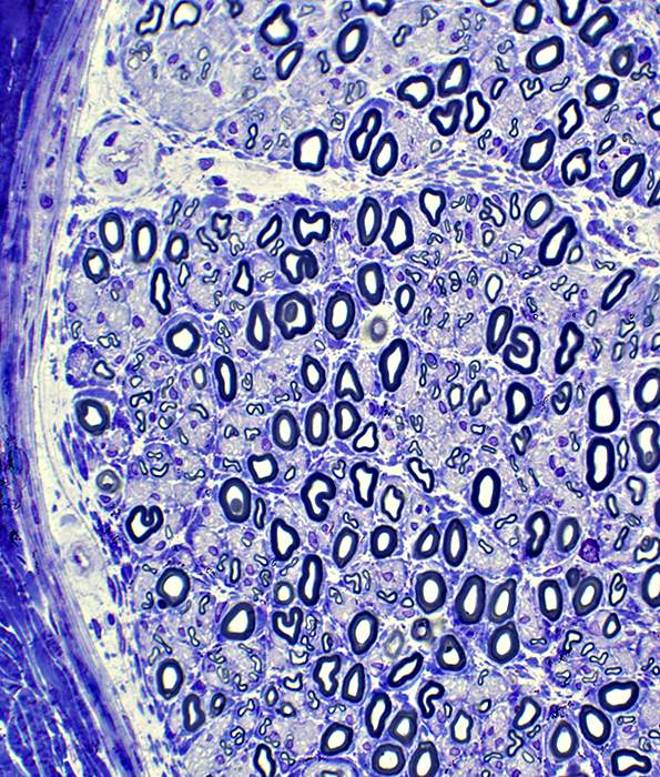 Toluidine blue stain |
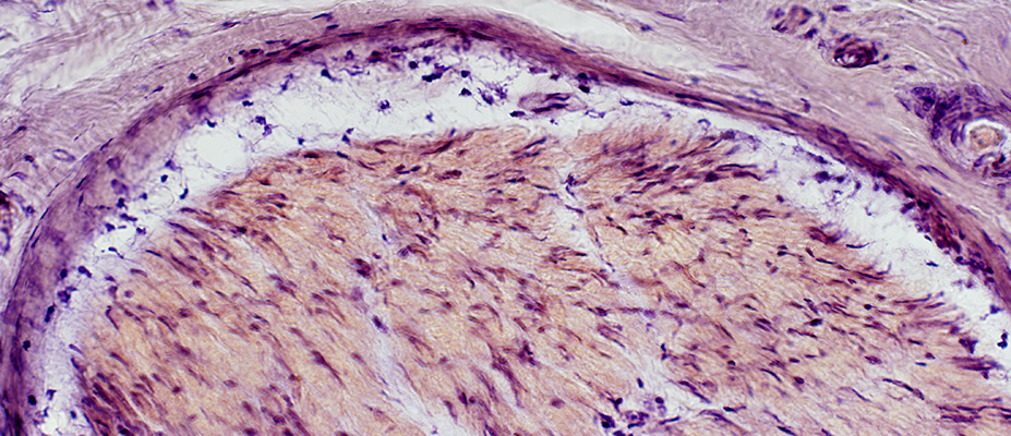 VvG stain |
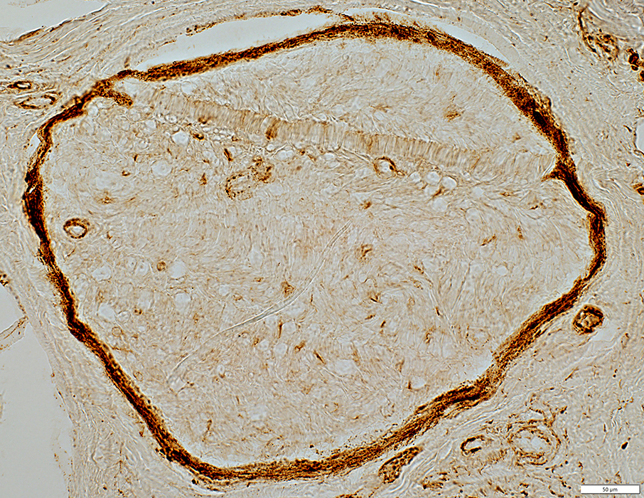
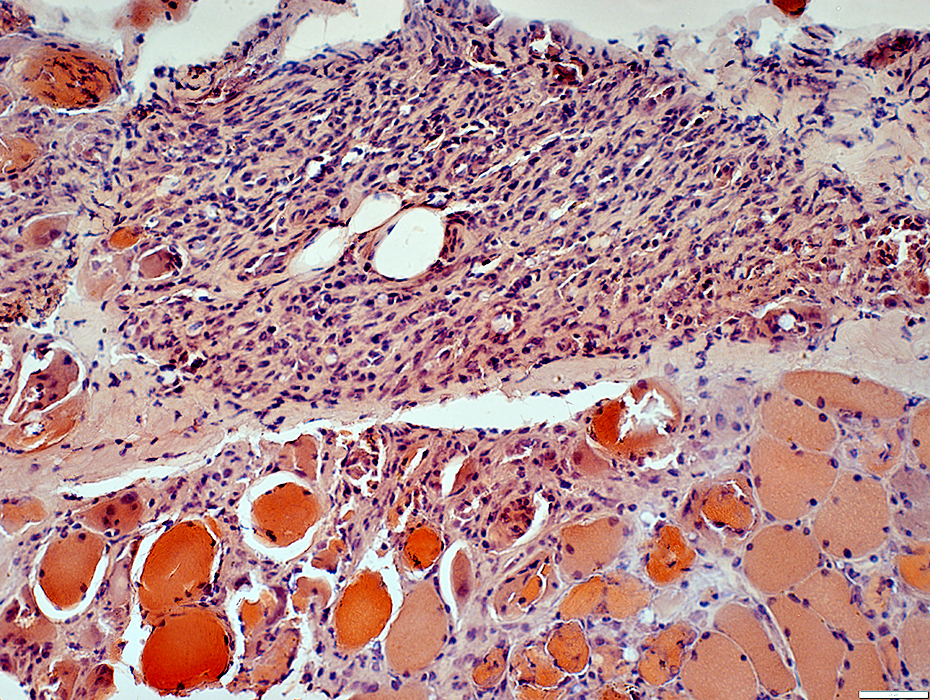
Perineuritis/Epineuritis/Vasculopathy: ICI-related
 H&E stain |
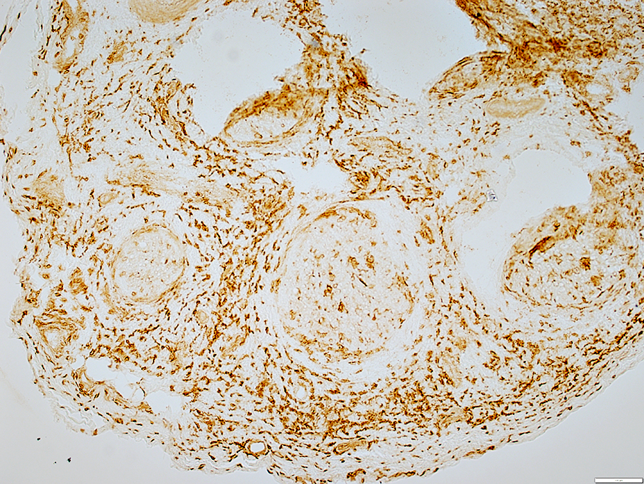
Pale & Wide around most fascicles
Epineurium
Structure: Irregular & Pale
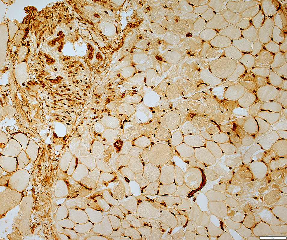
 MHC Class I stain |
Abnormally & Diffusely expressed in Epineurial connective tissue
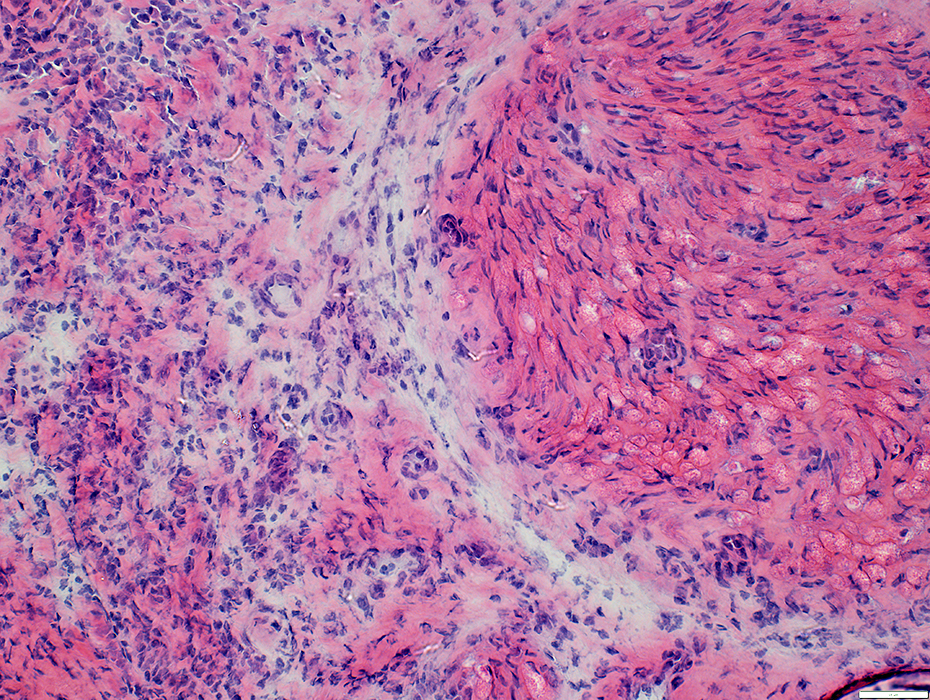
 H&E stain |
Pale & Wide around most fascicles
Epineurium
Structure: Irregular & Pale
Endoneurium
Wallerian Degeneration: Myelin irregularly stained, may be pale
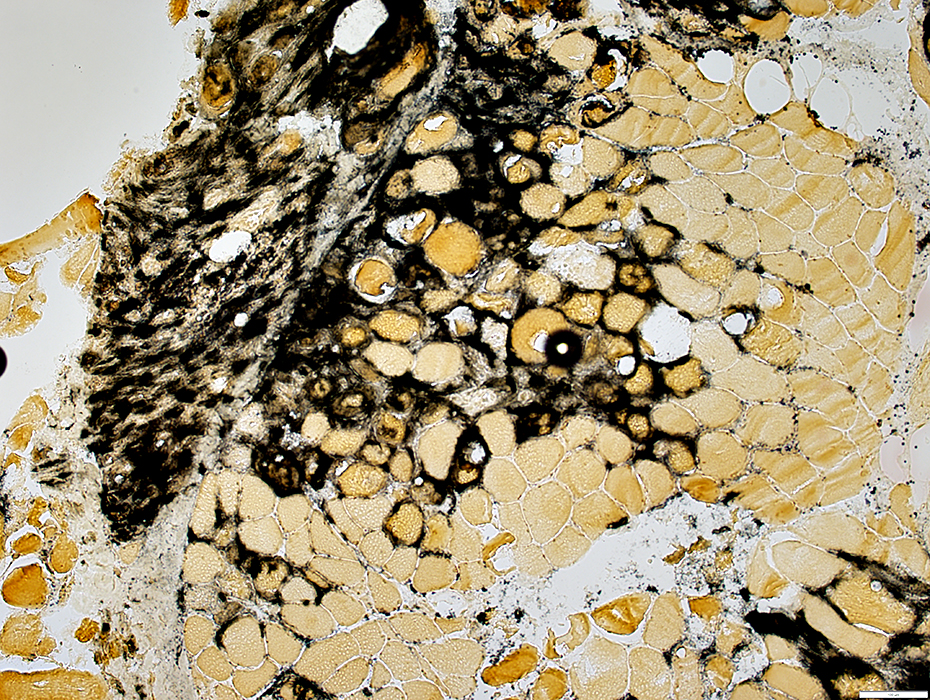
 VvG stain |
Loss of large axons
 Neurofilament stain |
Perineurial Pathology
 H&E stain |
Pale & Wide
Some areas have increased cells
 H&E stain |
Perineurium
Pale & Wide
Endoneurium
Wallerian Degeneration: Myelin irregularly stained & may be pale
 VvG stain |
ICI Perineuriopathy: C5b-9 stain
Perineurium: Loss of C5b-9 stain
Endoneurium: C5b-9 stains cells associated with Wallerian degeneration
Epineurium: C5b-9 stains scattered cells
 C5b-9 stain |
Control Nerve
Perineurium: Strong C5b-9 stain
 C5b-9 stain |
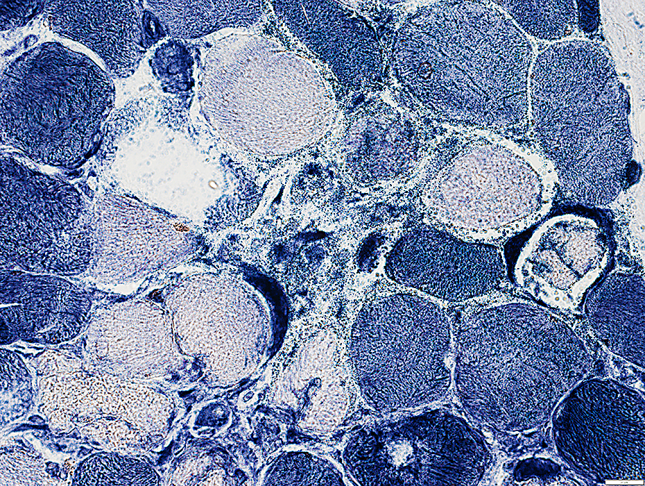
 Toluidine blue stain |
Pale & Wide
Endoneurium
Loss of most myelinated axons: Varied between fascicles
 Toluidine blue stain |
Epineurium
Structure: Irregular & Pale
 H&E stain |
 CD4 stain |
Scattered
Stain for CD4 & CD8
Perineurial Cells
Mostly CD8
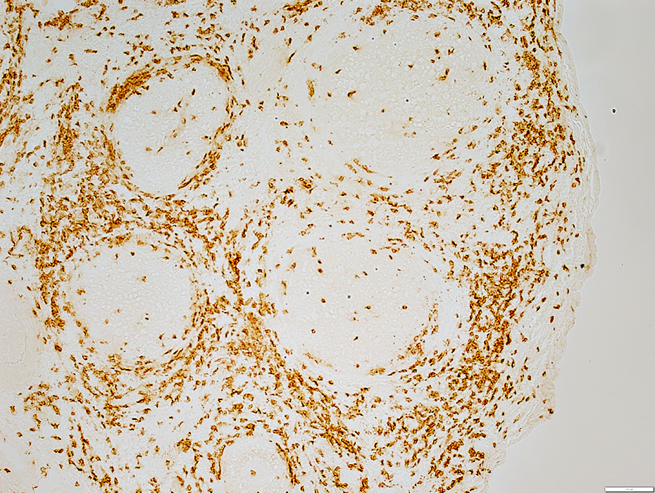 CD8 stain |
Epineurial & Endoneurial Cells
Many Histiocytes
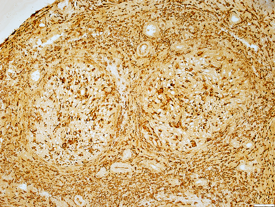 HAM56 stain |
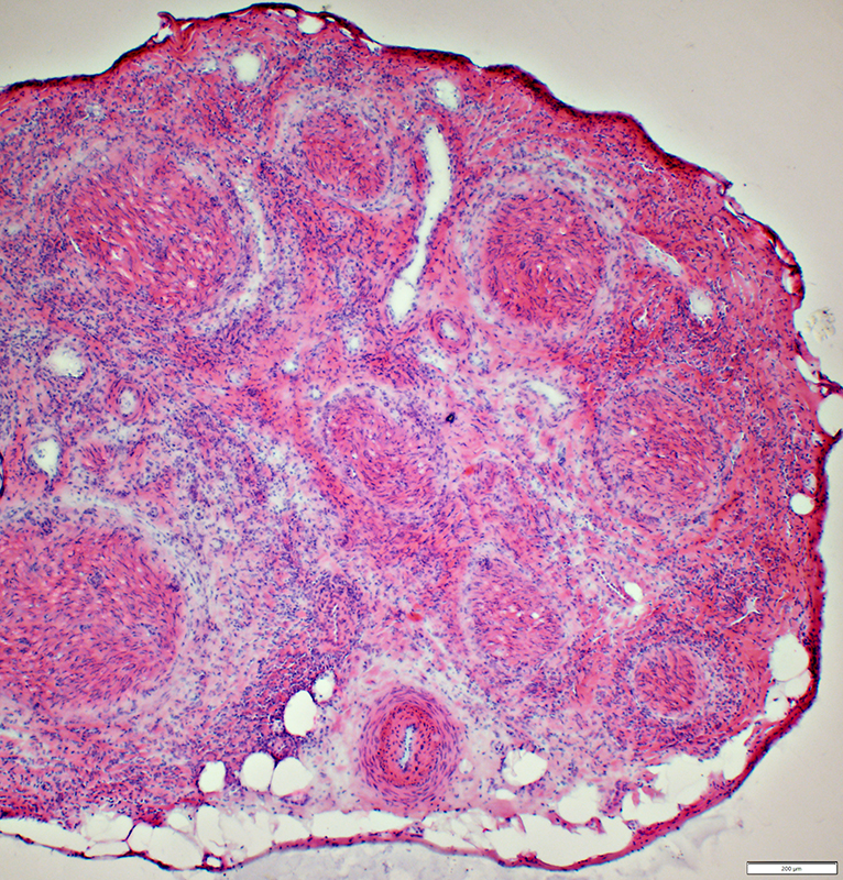
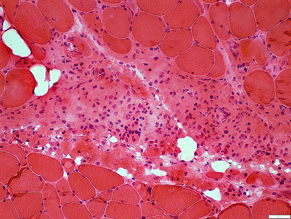
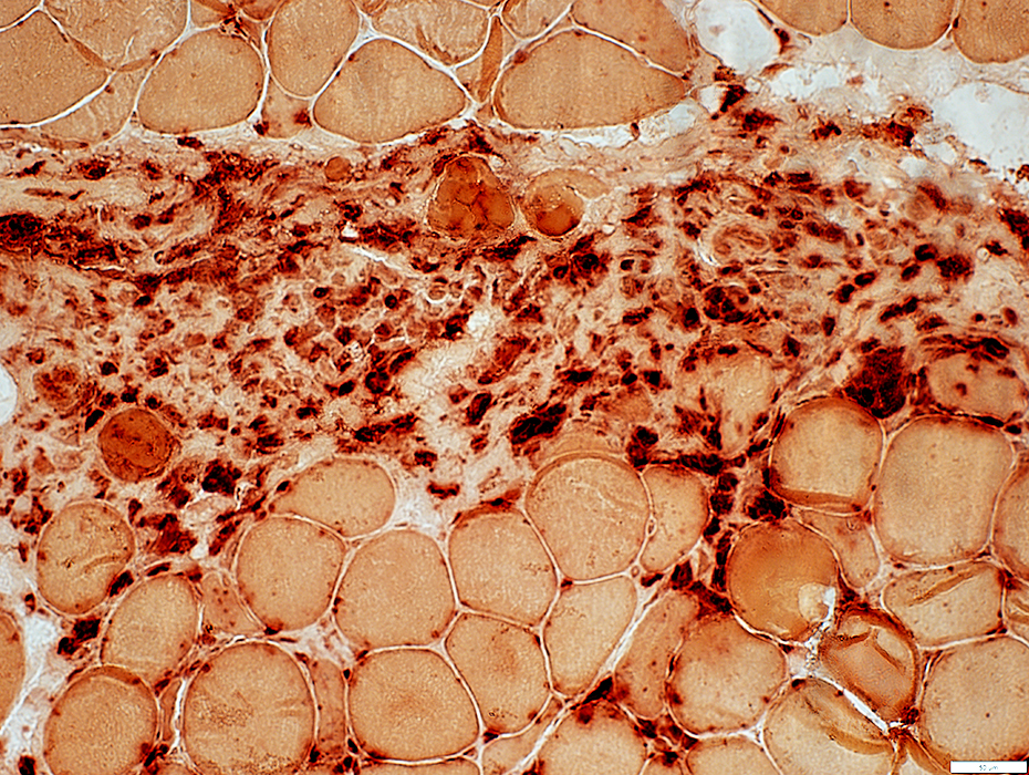
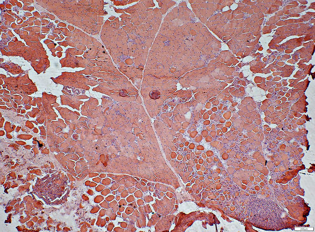
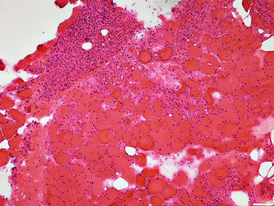
ICI Myopathy, Multifocal
| Immune ICI Myopathies, Multifocal Perimysial Cell foci, Mild fiber necrosis Cell foci, Clustered fiber necrosis Clustered fiber necrosis, Small |
ICI Myopathy, Multifocal 1
CellsHistiocyte foci, Perimysial
Muscle fibers Necrosis, Mild
MHC Class I: Increased in clusters of fibers
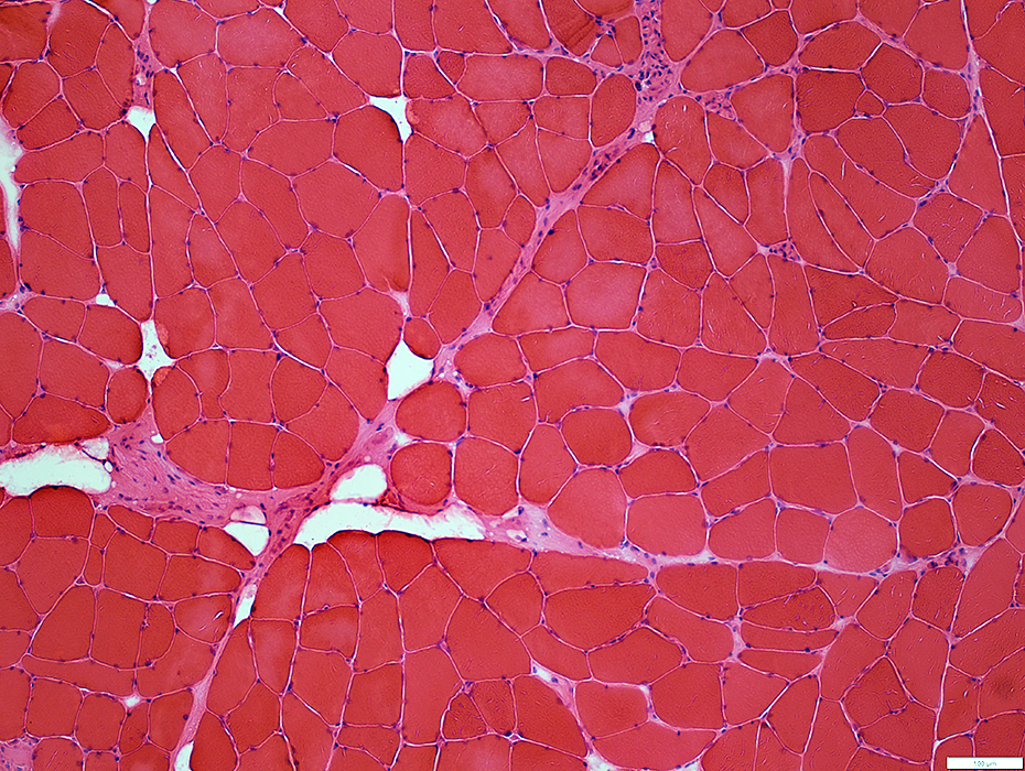
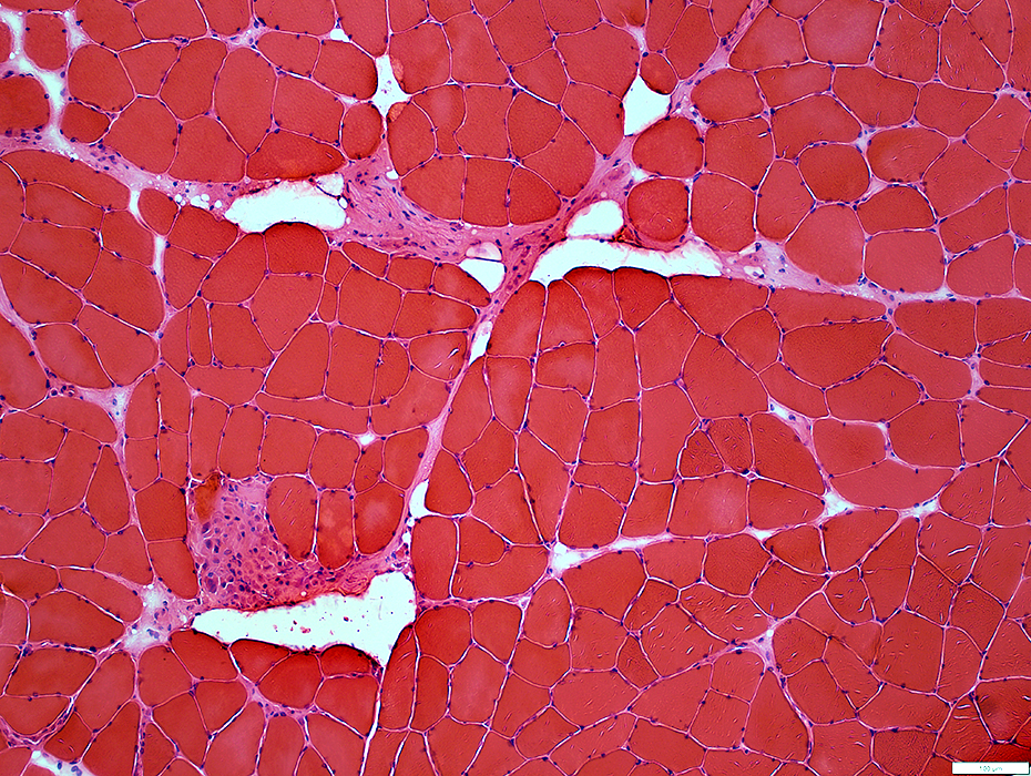
 H&E stain |
Widened: focal regions
Cellular, Histiocytes
Muscle fibers
Perifascicular pathology
Scattered, small fibers at edge of some fascicles, near perimysial damage
 H&E stain |
 Acid phosphase stain |
 Esterase stain |
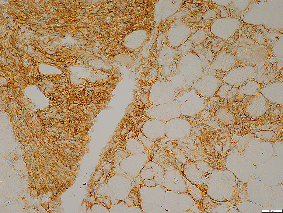
Alkaline Phosphatase stains
Perimysium
Endomysial capillaries
 Alkaline Phosphatase stain |
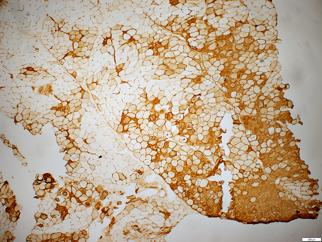
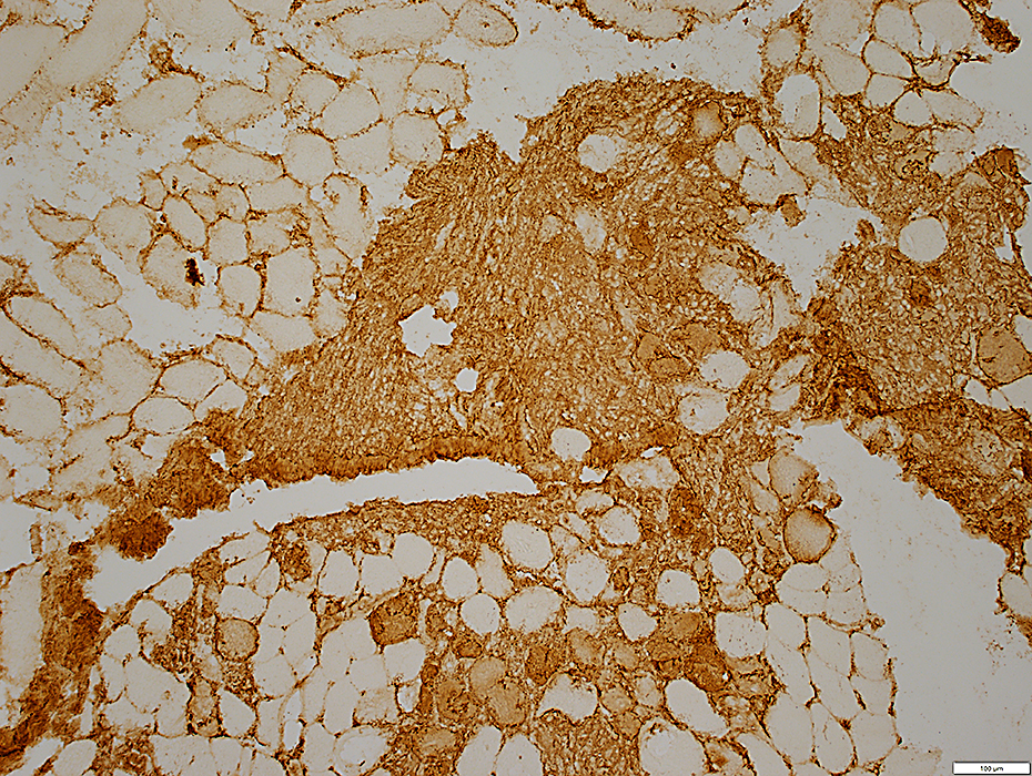
Multifocal involvement of Muscle fibers
 MHC I stain |
Necrotic muscle fibers: C5b-9 stained
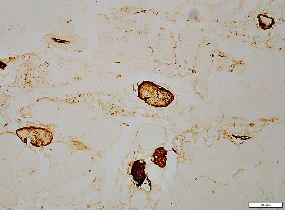 C5b-9 stain |
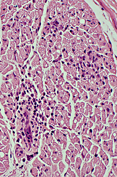
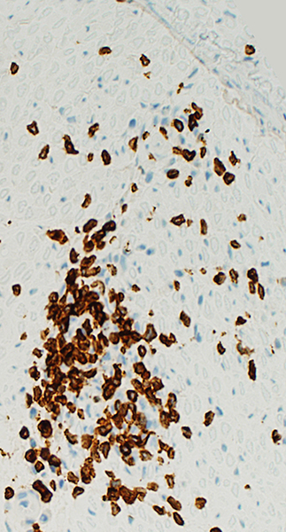
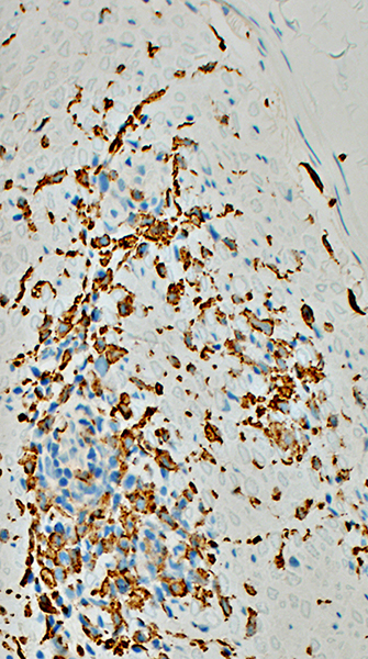
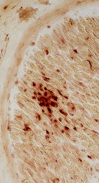
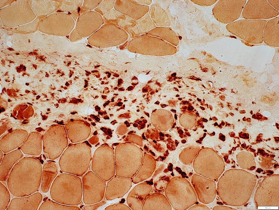
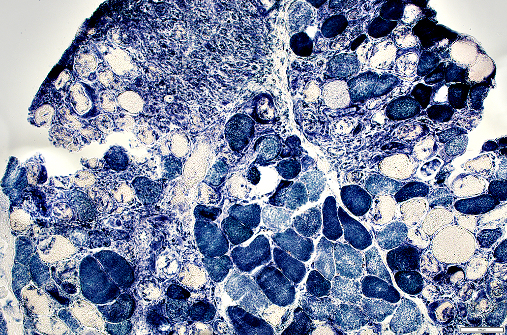
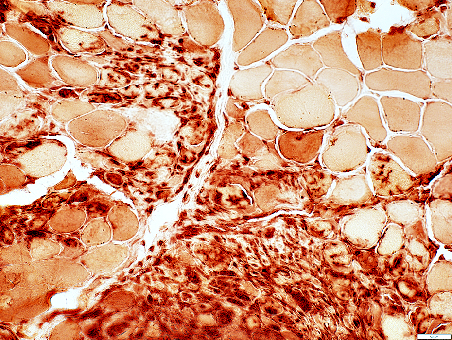
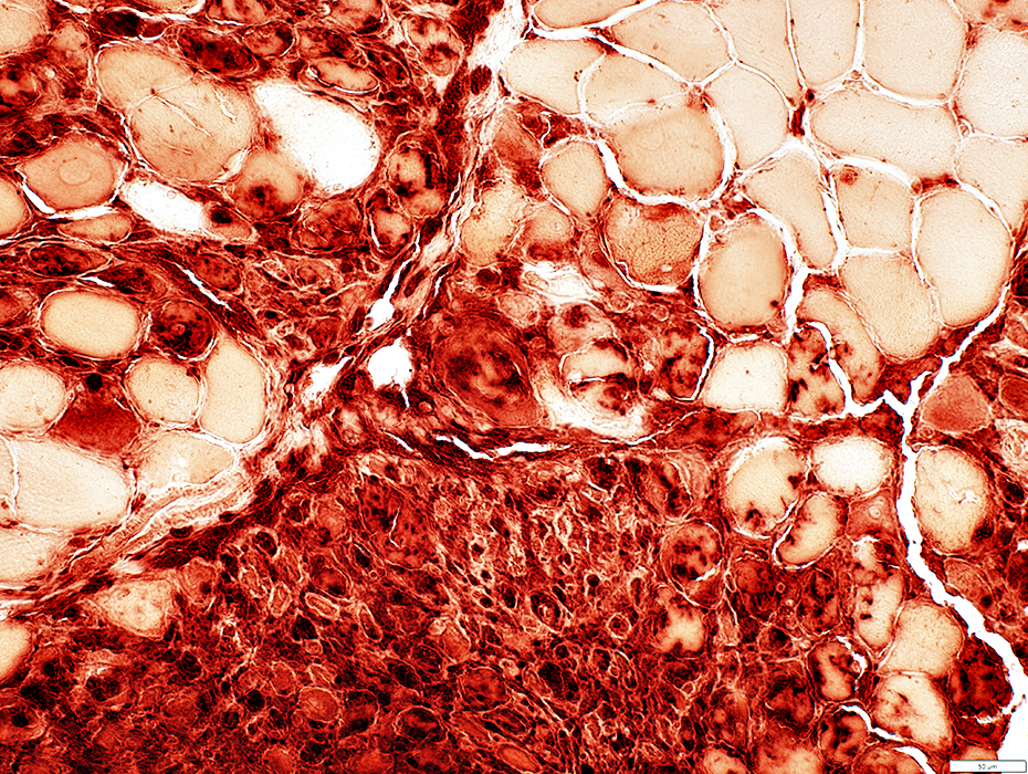
ICI Myopathy, Multifocal 2
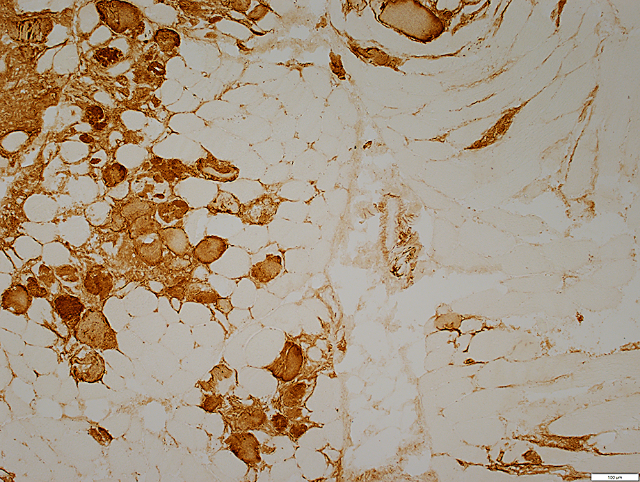
CellsHistiocyte Foci
Muscle Fibers Necrosis, Clustered
MHC Class I: Increased in clusters of fibers
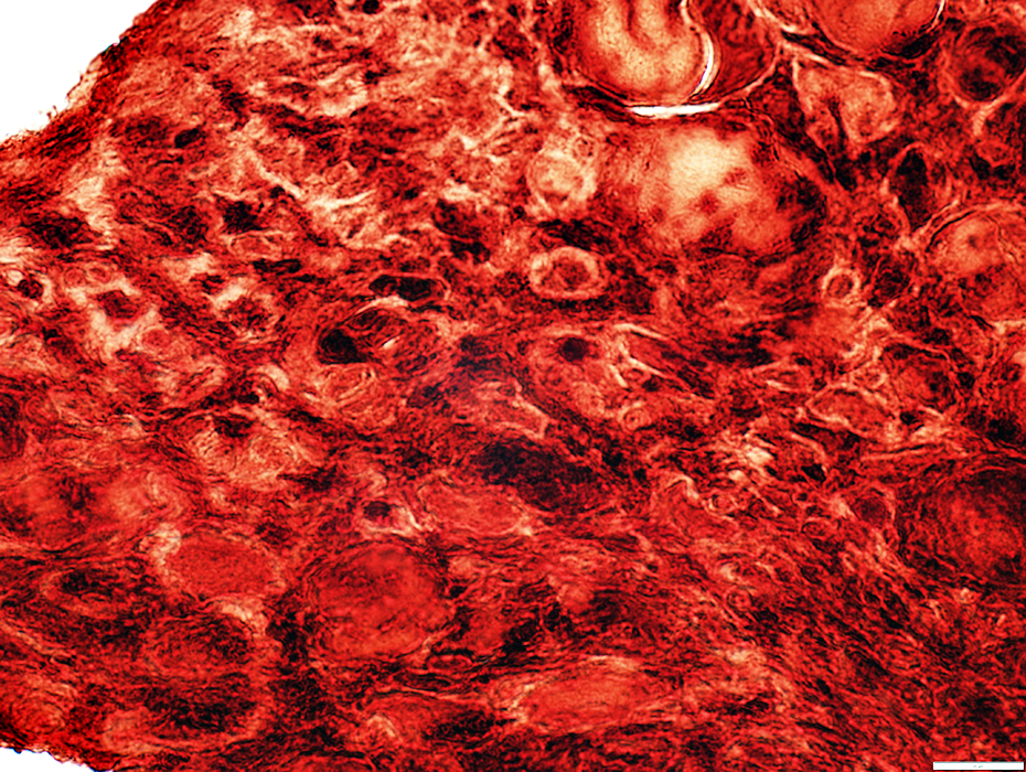
 Congo red stain |
Regions of: Necrotic fibers; Cell clusters
 NADH stain |
 C5b-9 stain |
Regions of: Necrotic or Damaged muscle fibers
 MHC Class 1 stain |
 C5b-9 stain |
 NADH stain |
 H&E stain |
 Congo Red stain |
 Alkaline phosphatase stain |
Stain for alkaline phosphatase
Contain many capillaries
 UEA I stain |
 H&E stain |
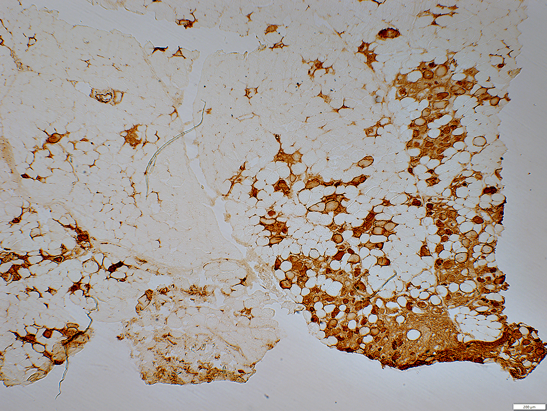
Contain mostly histiocytes
 HAM56 stain |
 CD4 stain |
Necrotic muscle fibers: Clustered
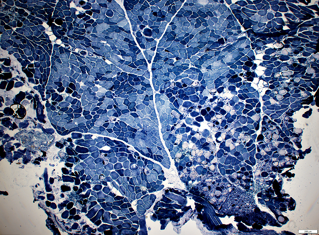
 NADH stain |
 VvG stain |
 Esterase stain |
 Acid phosphatase stain |
 Acid phosphatase stain |
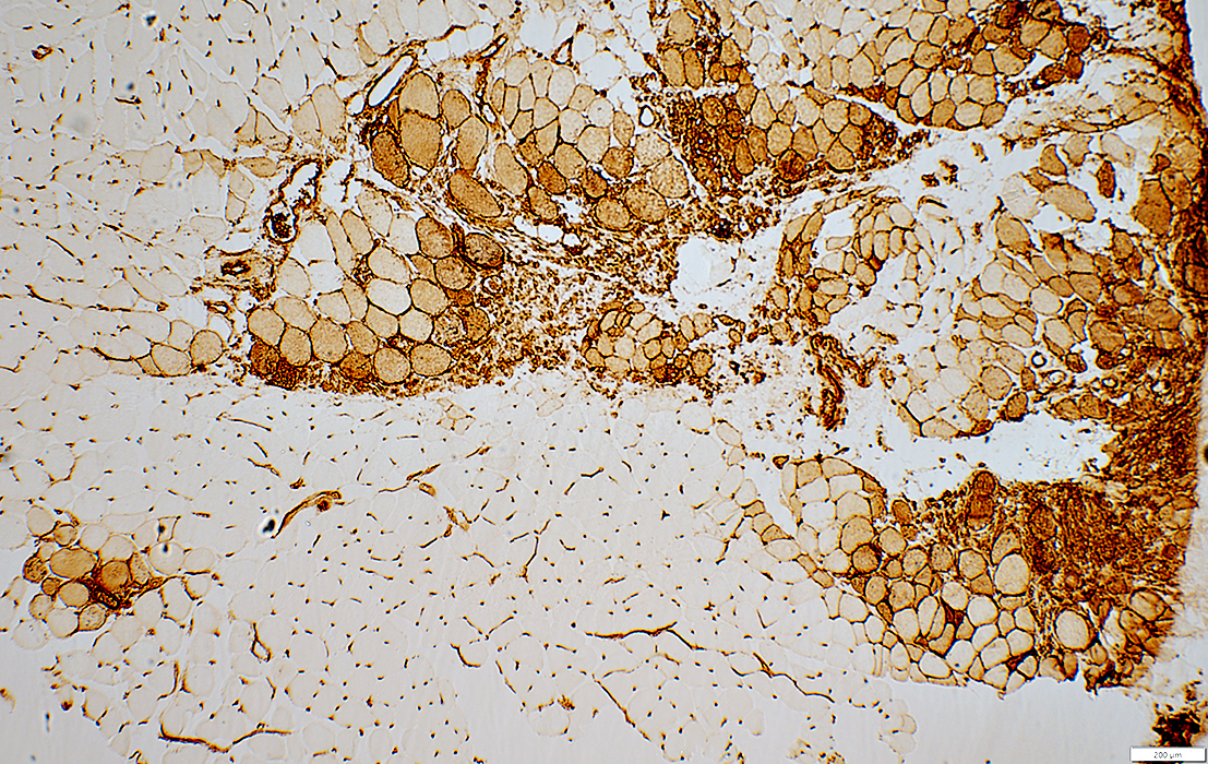
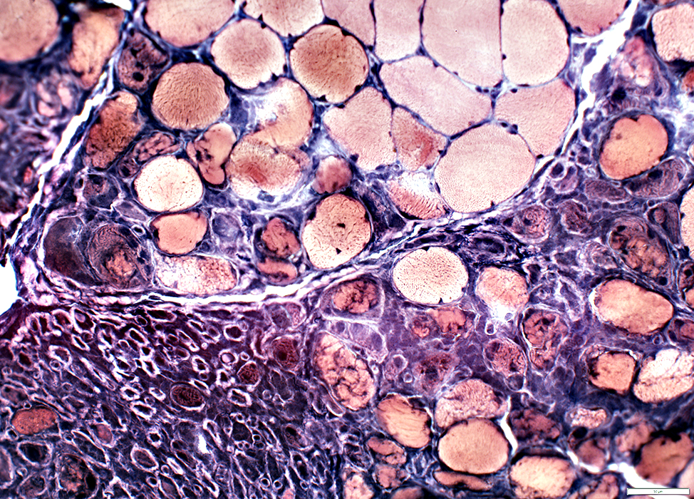
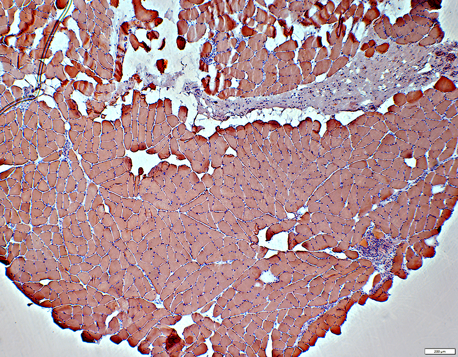
ICI Myopathy, Multifocal 3
Muscle Fiber MorphologyNecrosis, Clustered, Small Region
Most fibers: Normal morphology
MHC Class I: Increased in clusters of fibers
 Congo red stain |
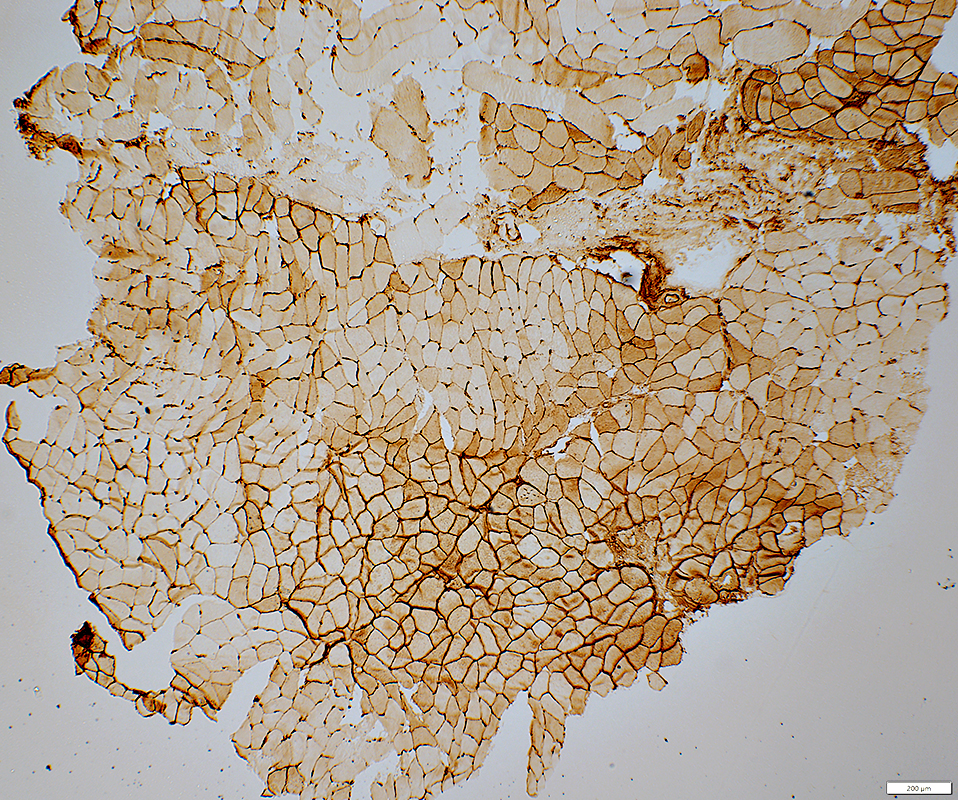 MHC Class I stain |
 H&E stain |
 H&E stain |
 stain |
Return to Ipilimumab/ICI
Return to Neuromuscular Home Page
Return to Pathology index
9/26/2025