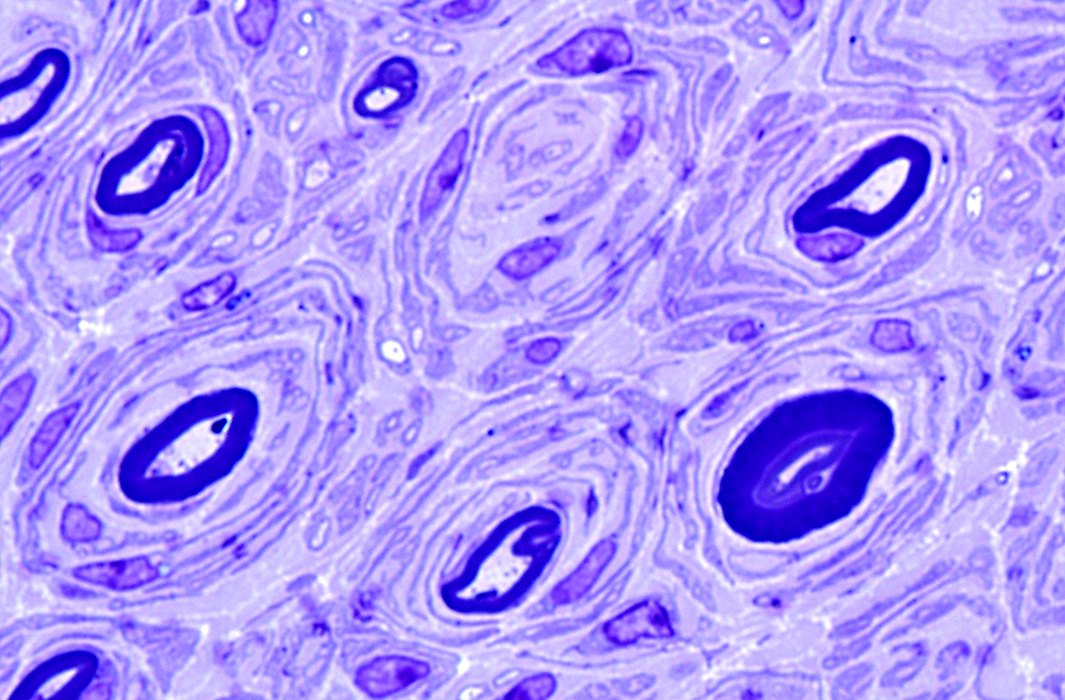
CMT 1A
|
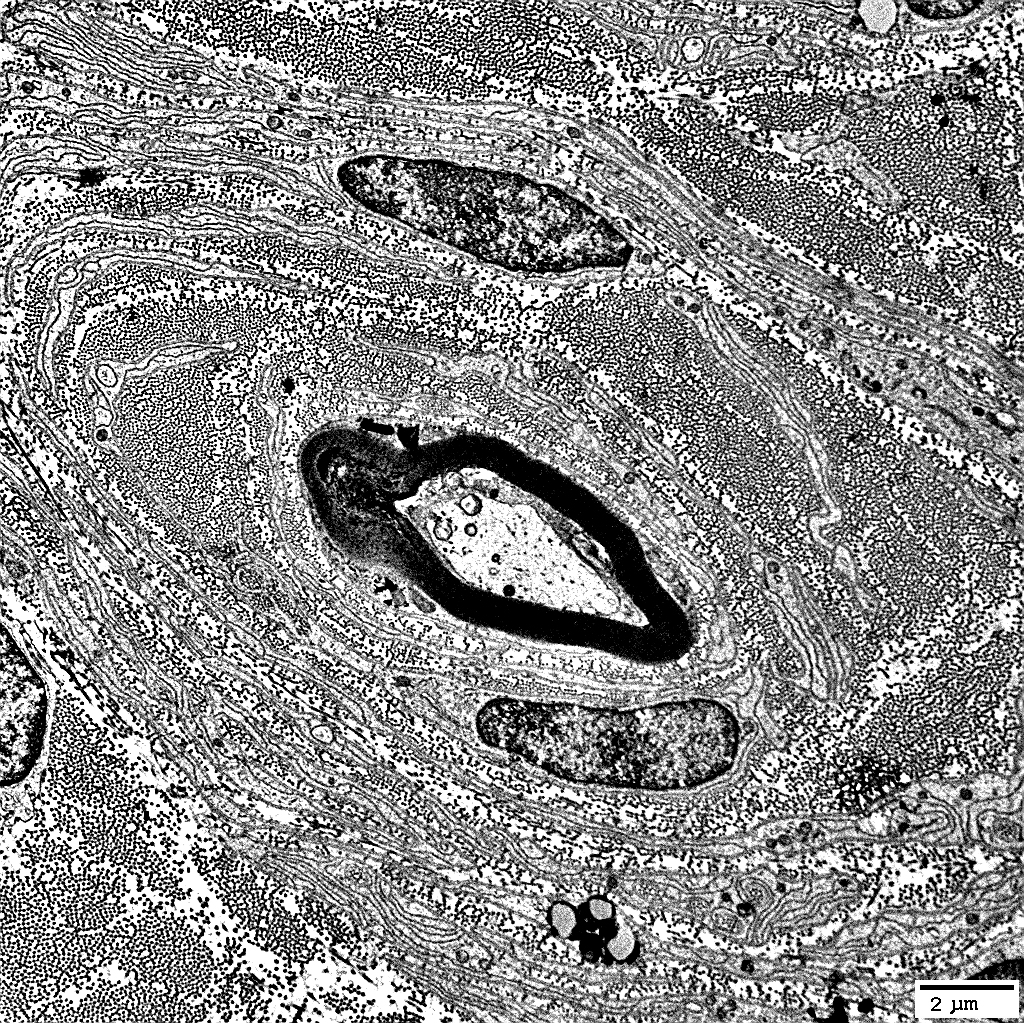
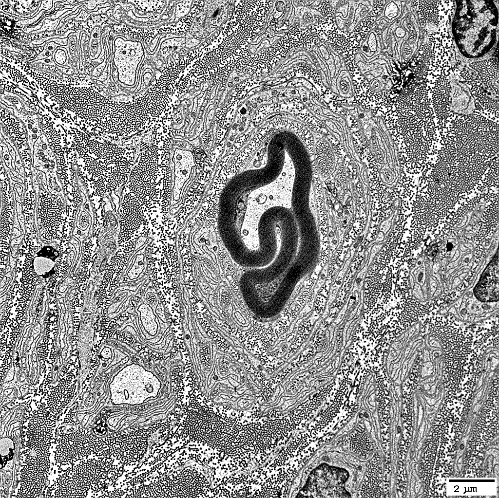
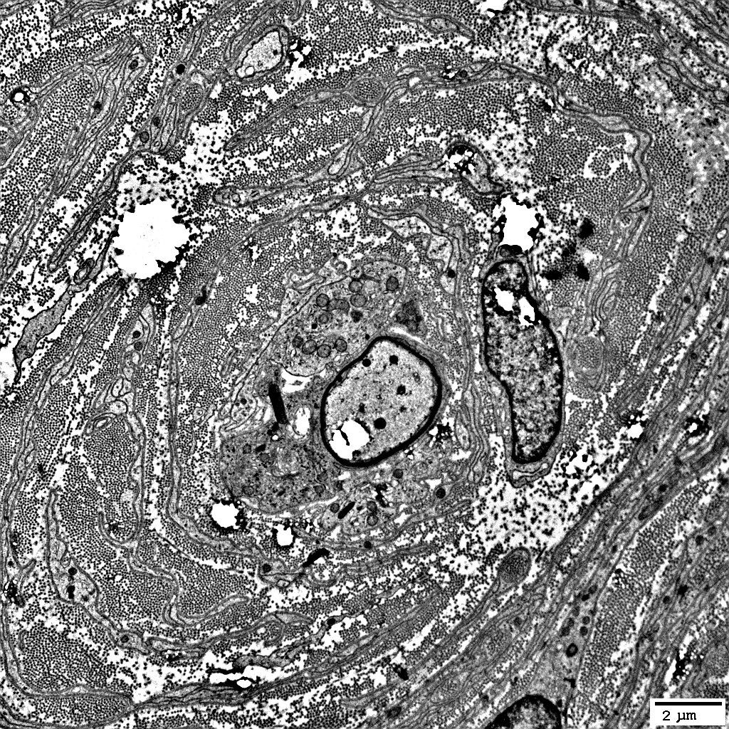
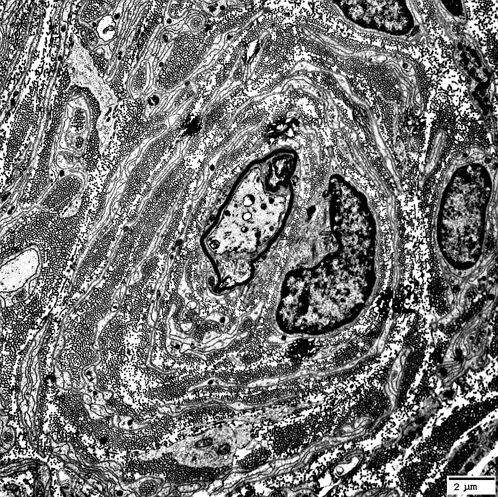
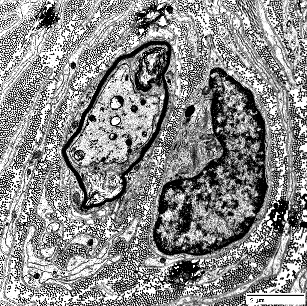
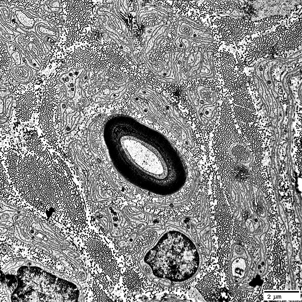
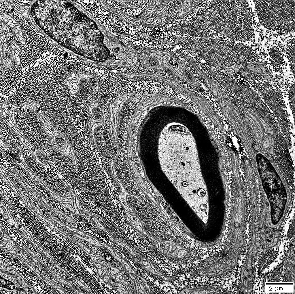
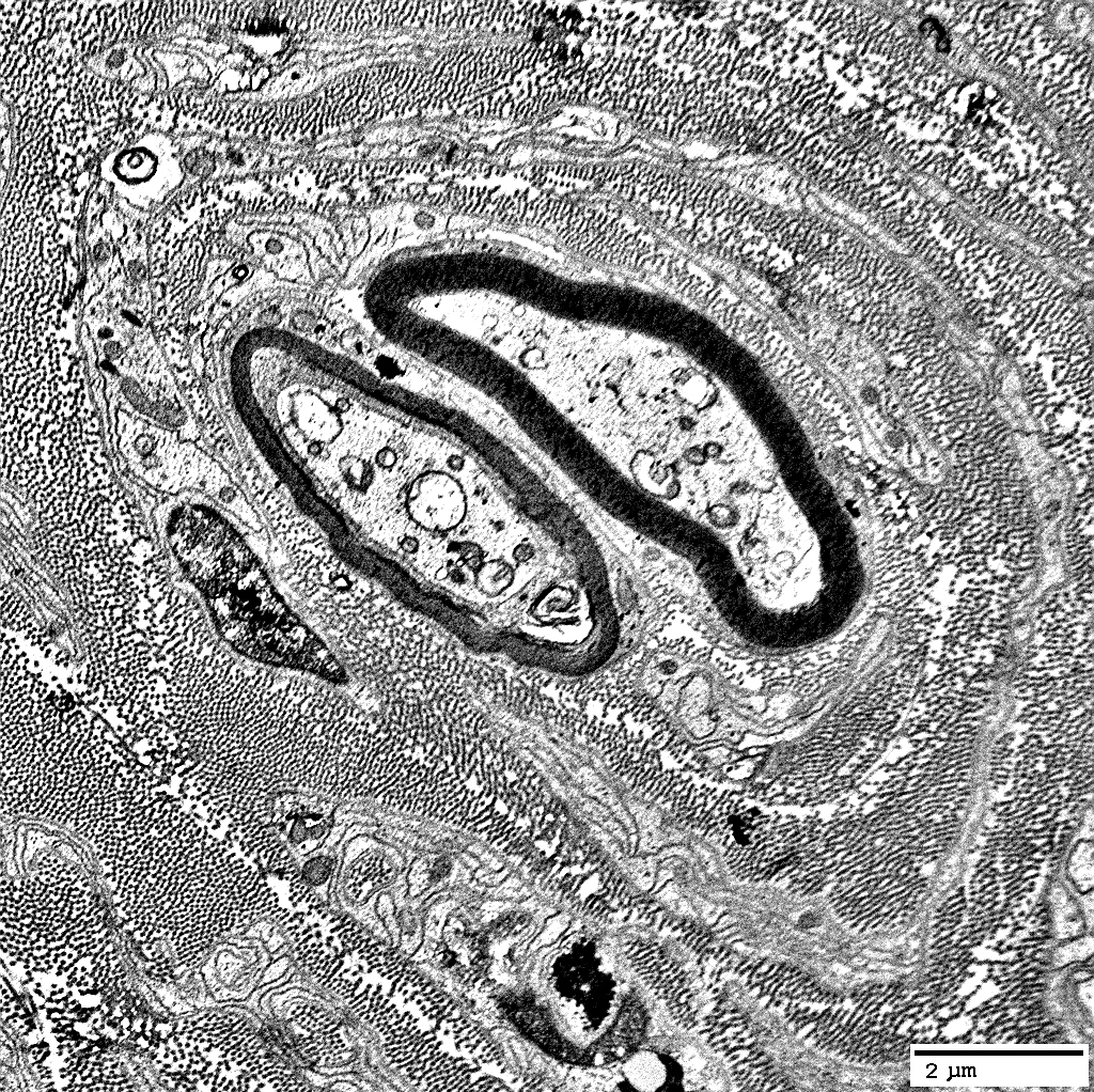
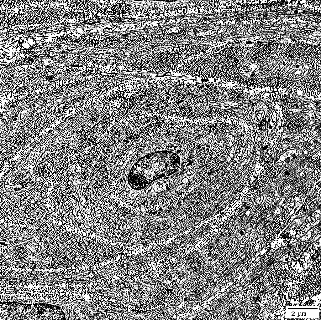
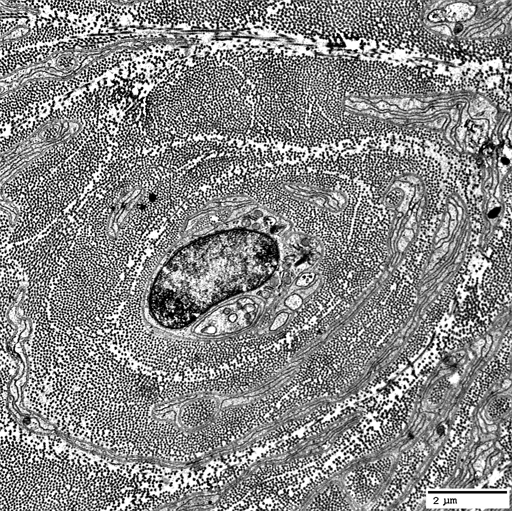
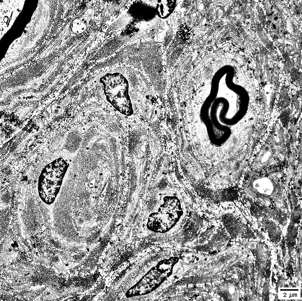
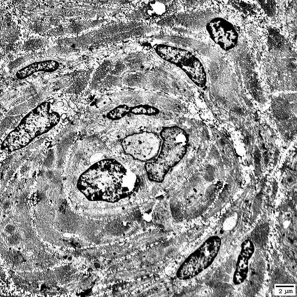
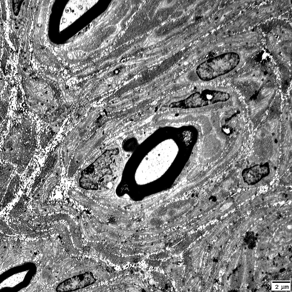
Muscle Nerve Schwann cells Ultrastructure: Onion bulbs |
Nerve
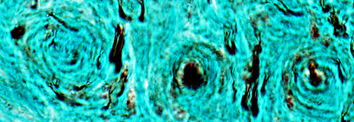
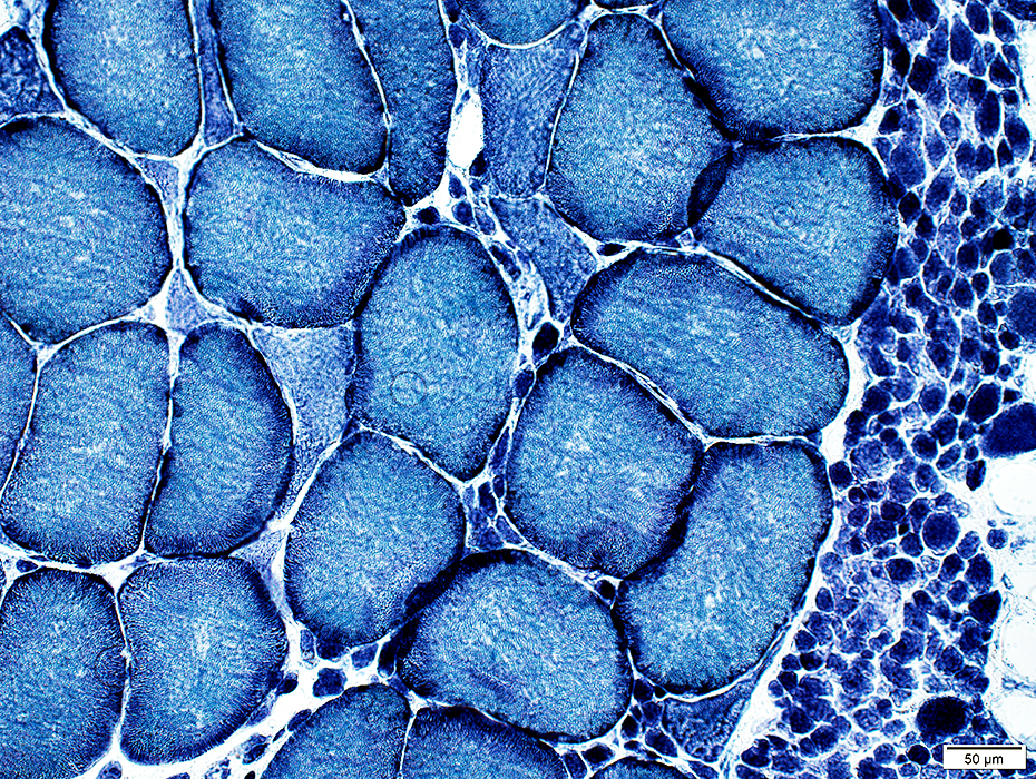
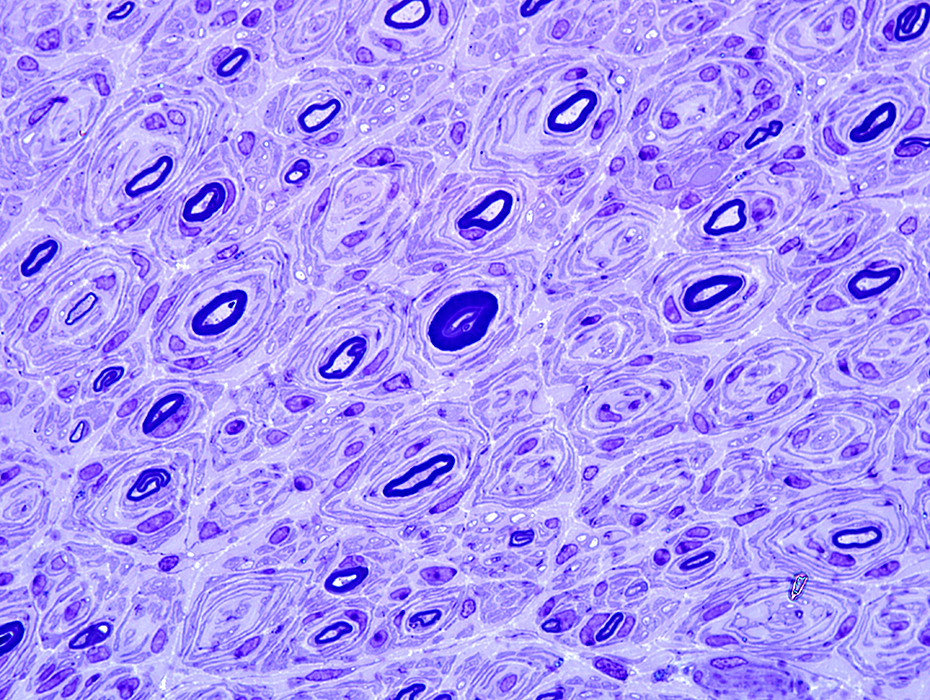 Toluidine blue |
Some contain myelinated axons; Others are empty ("Obsolete")
Myelin sheaths: Thick; Normal or Thin
Contain abundant non-myelinating Schwann cells
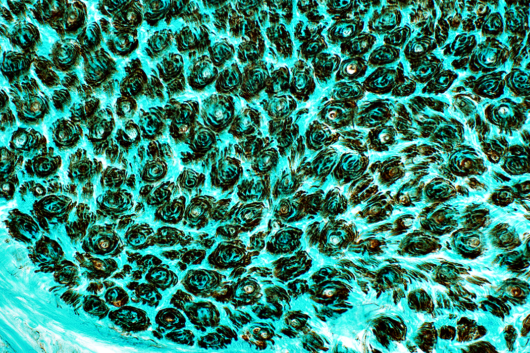
 Toluidine blue |
Axons
Reduced numbers: Large & Small
Some onion bulbs have no axons
Some small axons are present in layers within & around onion bulbs
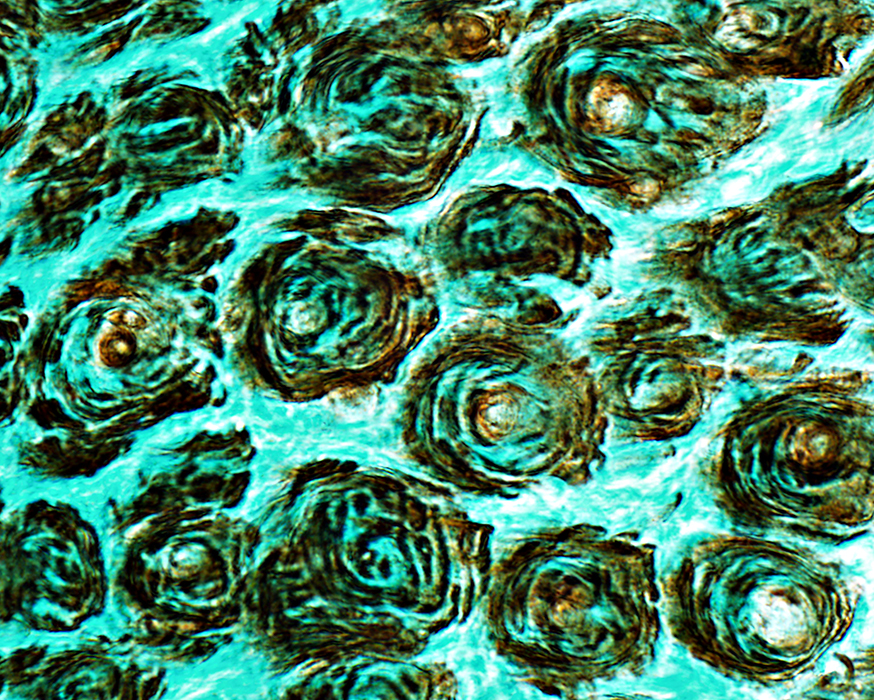
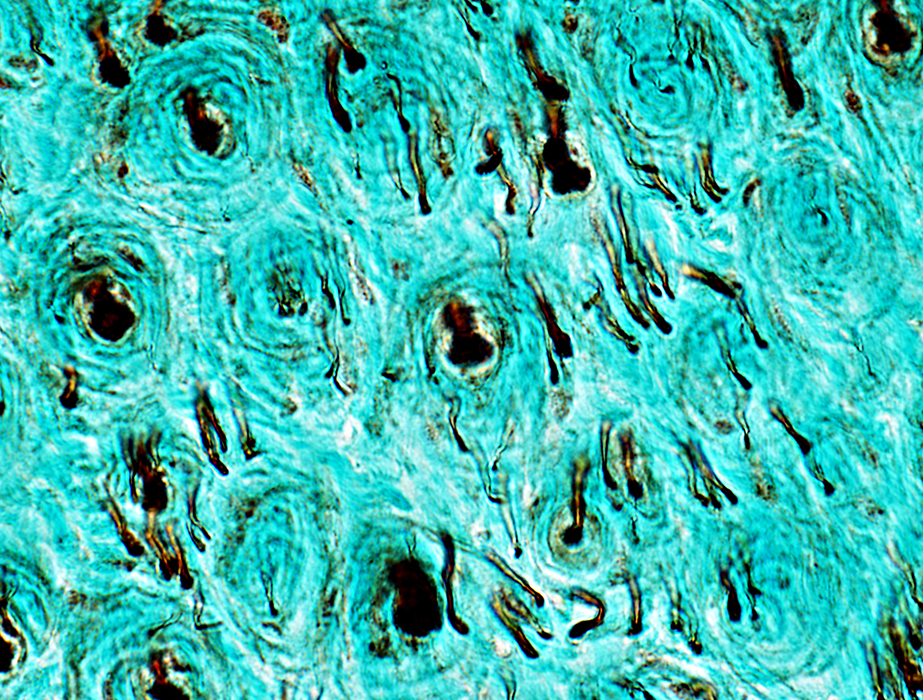 SMI31: Neurofilament |
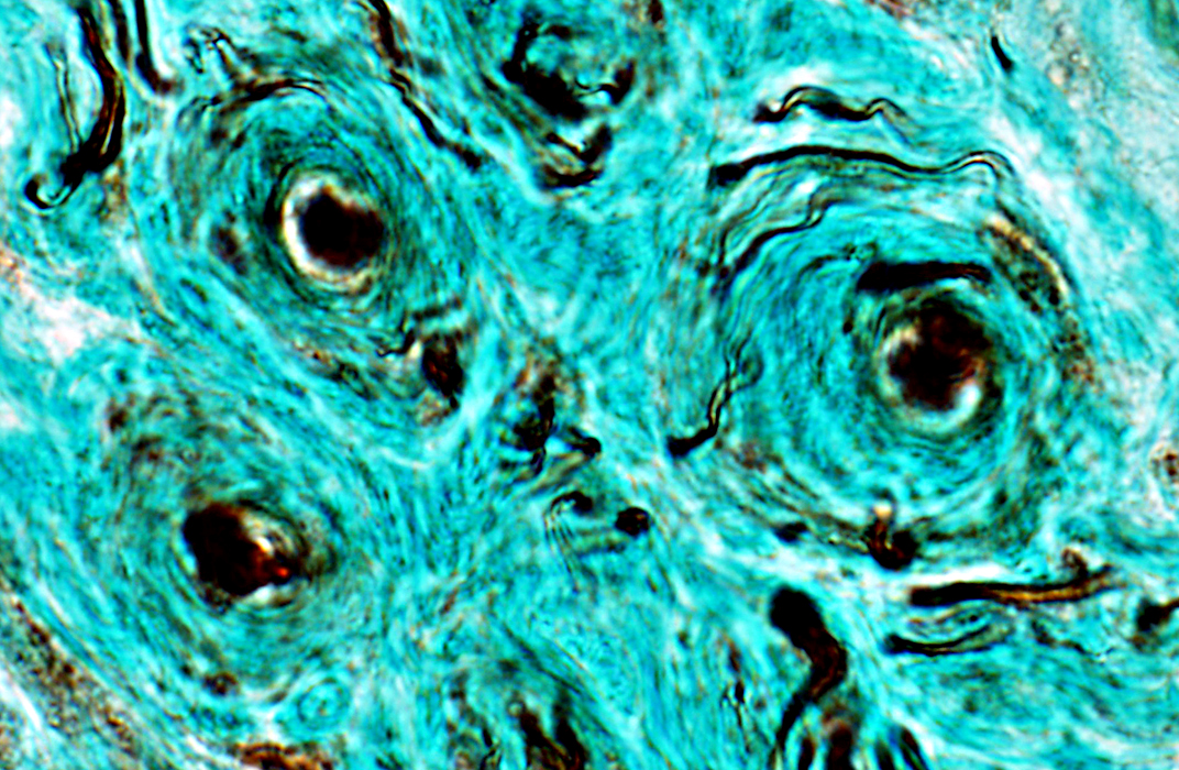 SMI31: Neurofilament |
 SMI31: Neurofilament |
Schwann cells in Onion bulbs
Contain: NCAM & P0 protein
 NCAM |
Myelin around central axons: Abnormal; Stains for NCAM
 NCAM |
Schwann cells in Onion bulbs also contain P0 protein
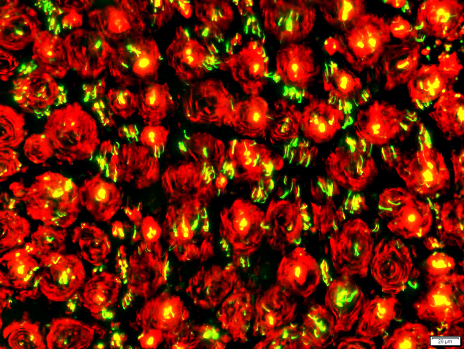 Neurofilament (Green) & P0 (Red) stains |
 |
 |
 |
 |
 |
 |
 |
 |
 |
 |
 |
 |
 |
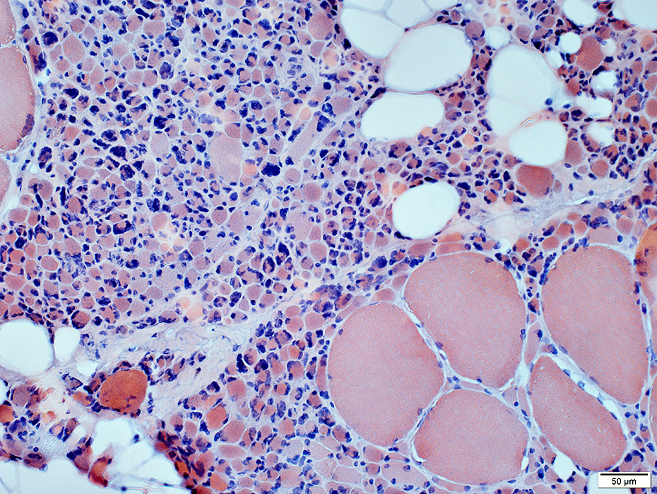
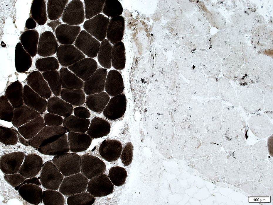
CMT 1A: Muscle
Fiber sizeLarge fibers: Hypertrophied
Small fibers: Very small; Grouped; Some pyknotic nuclear clumps
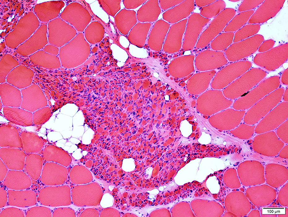 H&E |
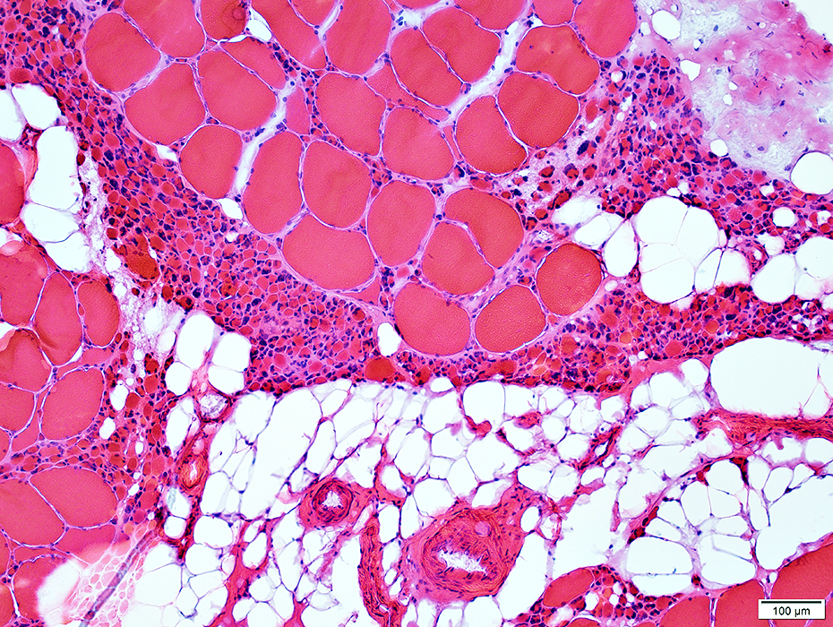 H&E |
Perimysium: Replaced by fat
 Congo red |
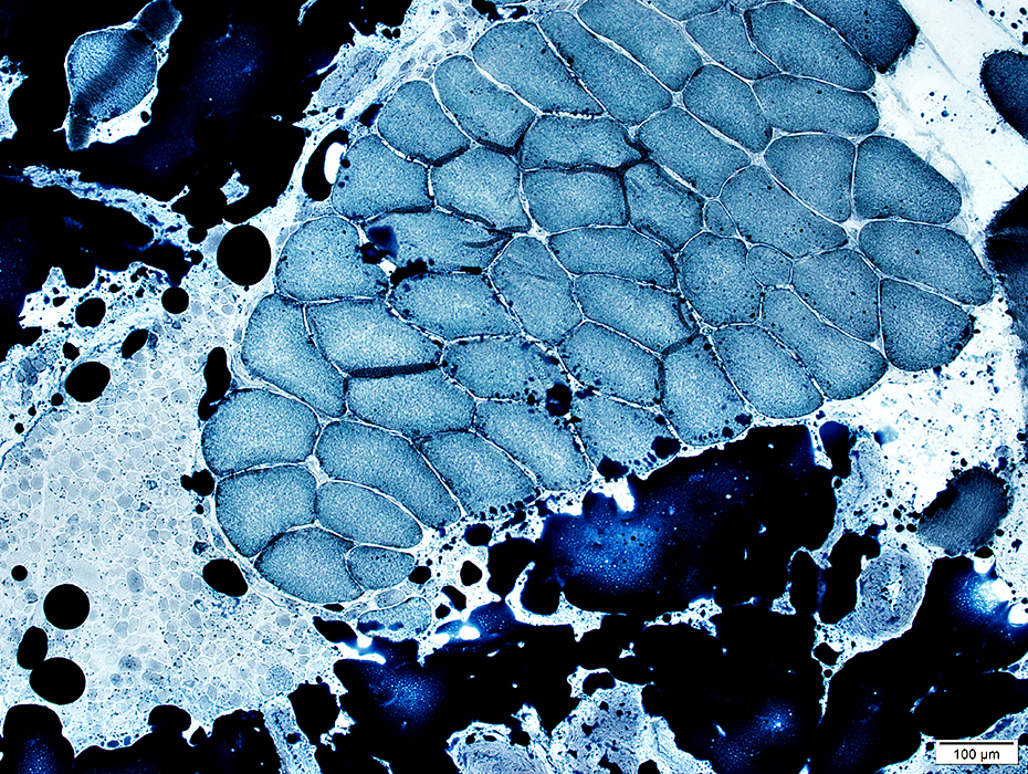
Small muscle fibers: Dark on NADH stain
 NADH |
Fiber types
Large fibers: Type grouping
Small fibers: Grouped; Type 2
 ATPase pH 4.3 |
Chronic changes: Perimysium is replaced by fat
 Sudan black |
Return to Normal nerve biosies
Return to Biopsy illustrations
Return to CMT 1A
Return to Neuromuscular Home Page
Return to Nerve biopsy
Return to Demyelinating neuropathies
Return to Chronic demyelination
1/18/2017