Dermatomyopathies with TIF1-γ (TRIM33) Antibodies
- Pathology patterns: General
- Anatomic distribution: Regionally varied pathology
- Vessel pathology
- Inflammation & damage of vessel bundles in perimysium
- Capillaries: Near atrophic muscle fibers
- Muscle fibers: Perifascicular myopathy & atrophy near avascular perimysium
- Perimysium: Inflammation
- Lymphocytes: Most in vascular perimysium
- Histiocytes: May occur in avascular perimysium near atrophic muscle fibers
- Vessel pathology
- Muscle fiber pathology: Regional
- Vacuoles
- Aggregates: LC3
- Internal architecture: Irregular
- COX stain: Reduced in regions of fiber atrophy (DM-VP syndromes)
- Necrosis: Varied
- Little or None: DM-VP syndromes
- Regional: RIIM syndromes
- Myonuclei: Enlarged
- Vessels
- Capillaries (Endomysial)
- C5b-9 stain: In regions of muscle fiber pathology
- Ulex stain: Reduced numbers
- Esterase stain: Increased
- Alkaline phosphatase stain: Increased
- Tubuloreticular profiles in endothelial cells
- Perivascular (Perimysial) inflammation: Some patients
- Lymphocytes: Some foci may contain CD20 cells
- Capillaries (Endomysial)
- Perimysium
- Inflammation: Mosly in vascular perimysium
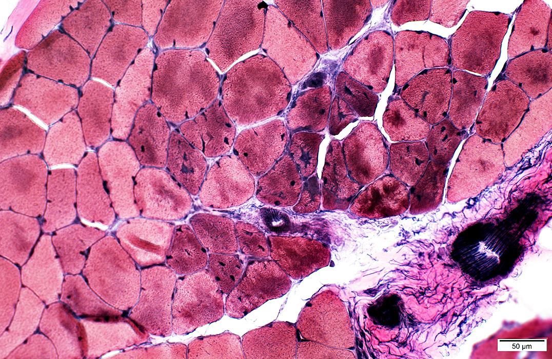
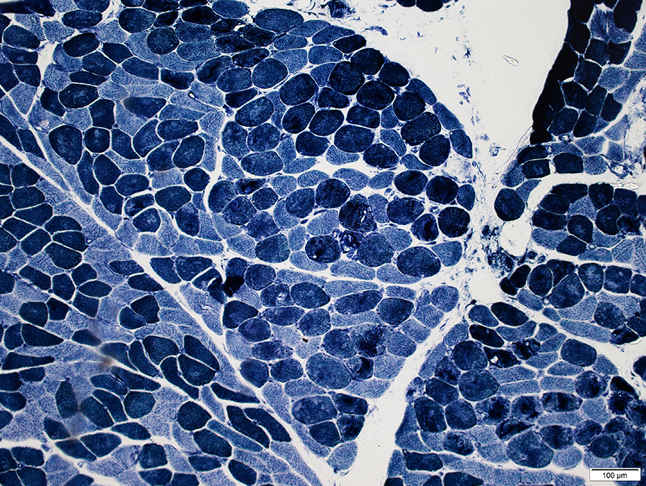
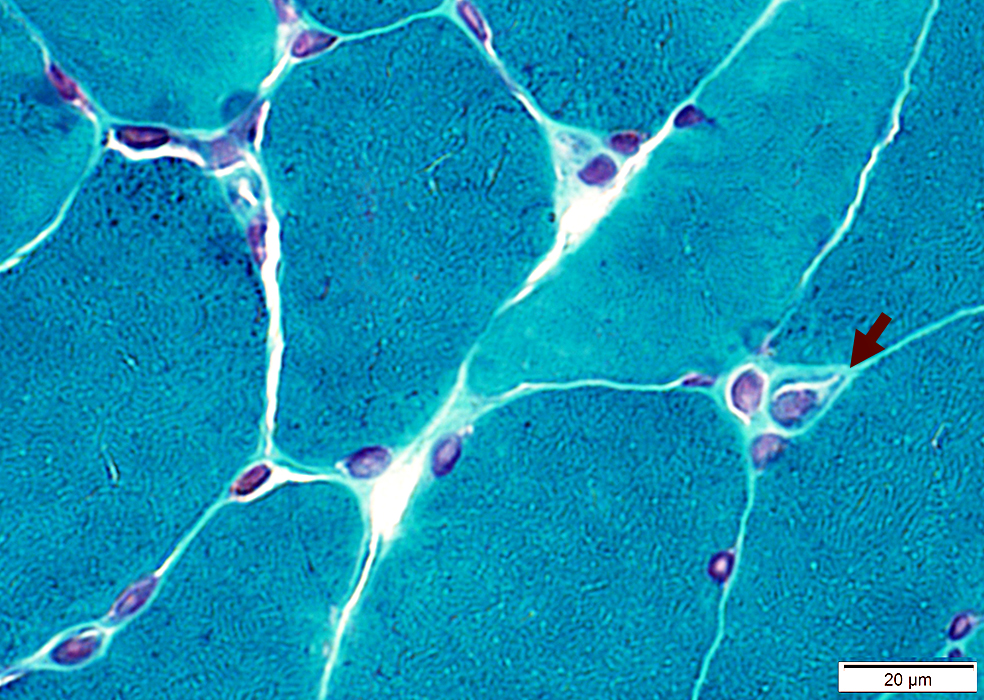
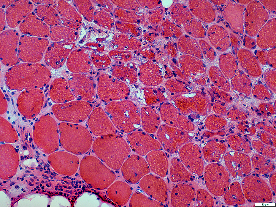
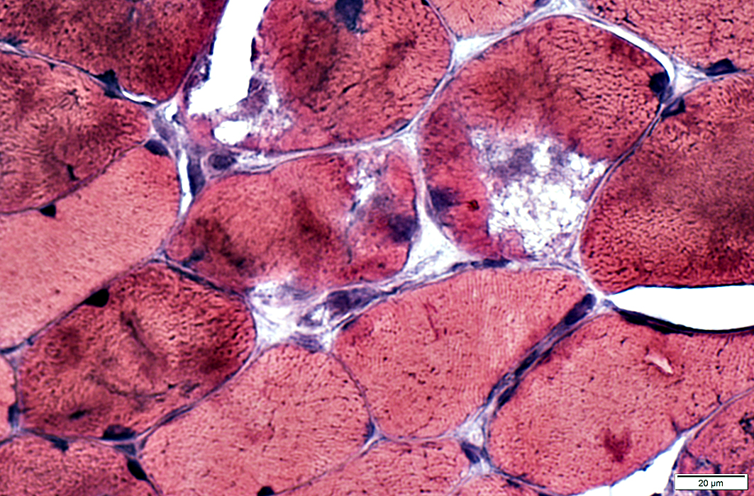
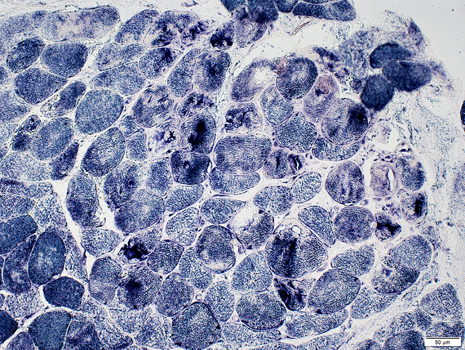
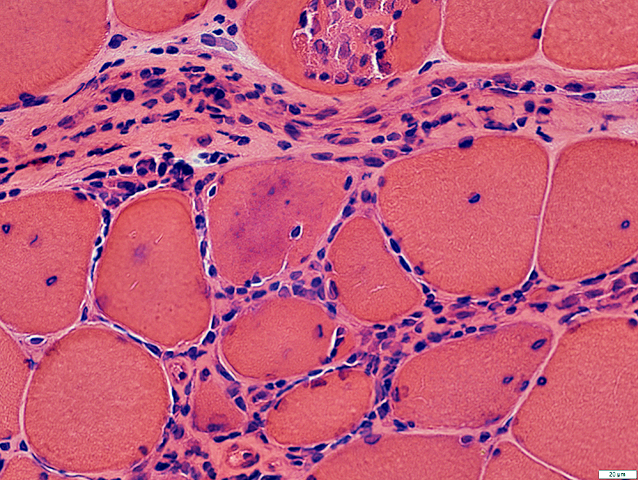
Muscle Fiber Pathology Distribution: Perifascicular Atrophy near Avascular Perimysium
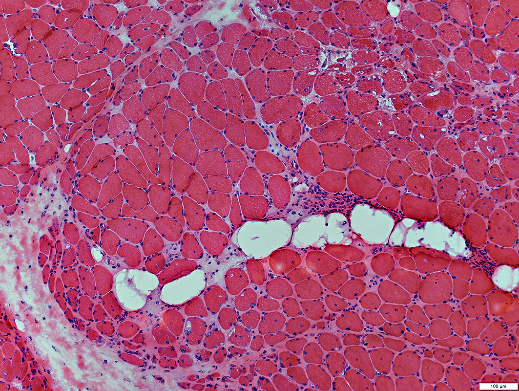 H&E stain |
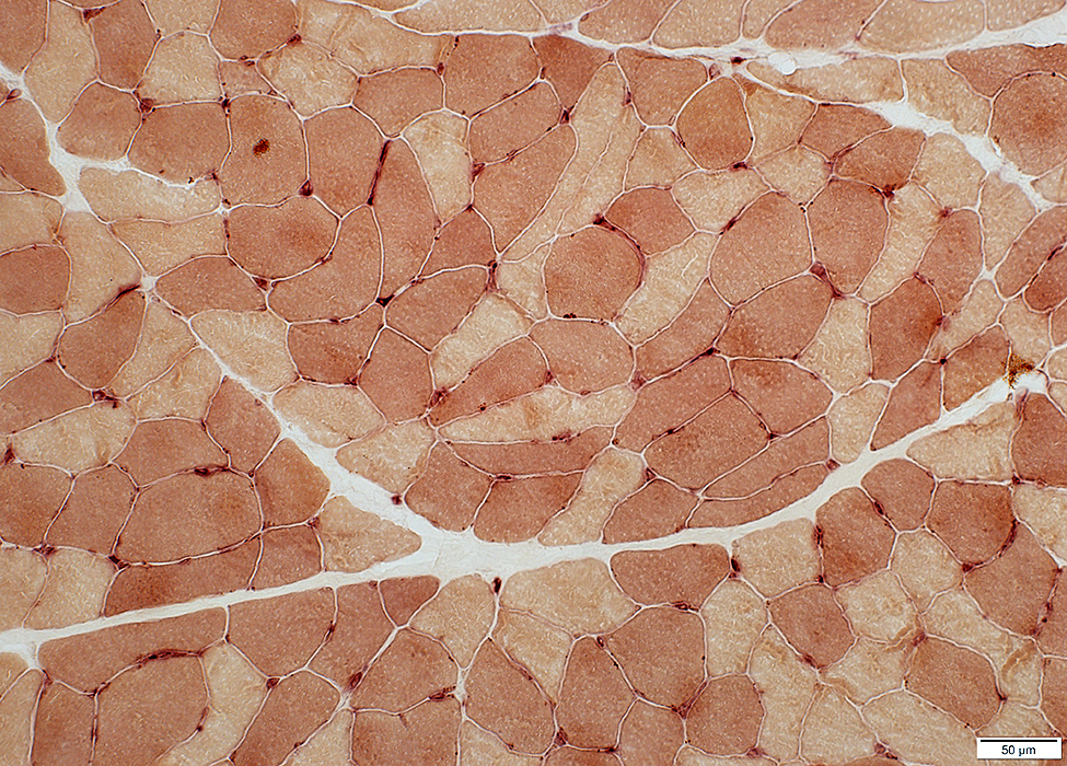
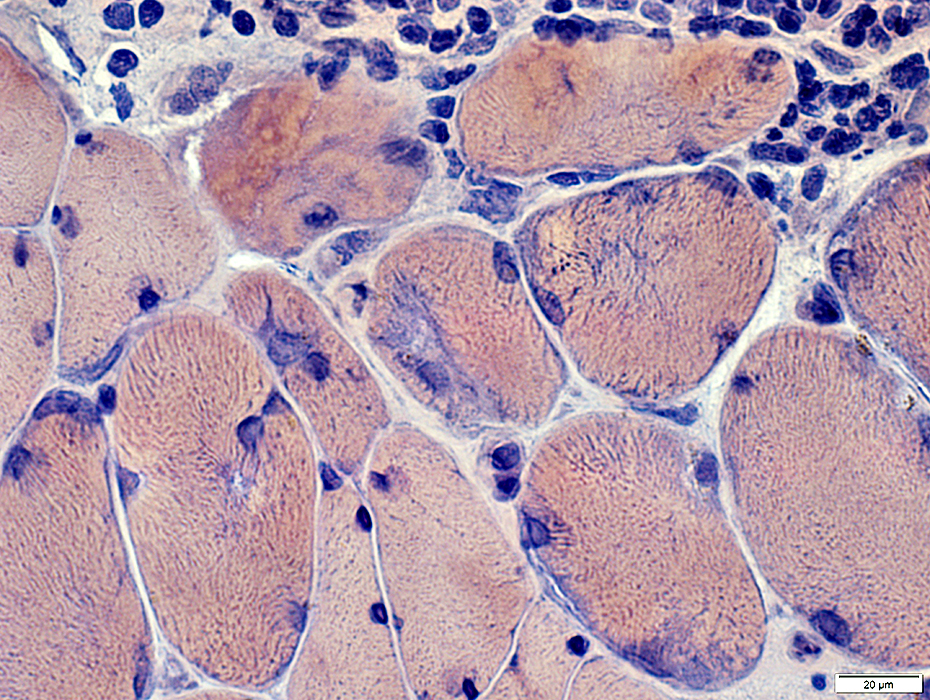
Muscle Fiber Pathology Distribution: Near or Around Perimysial vessels
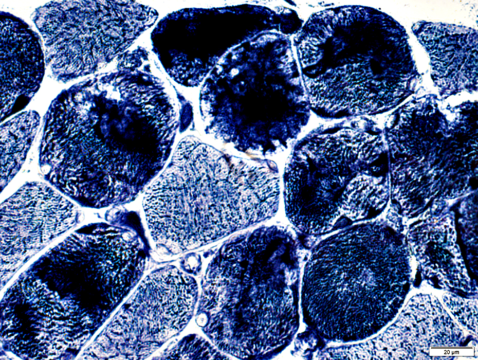
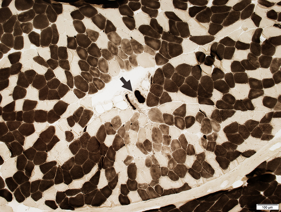 ATPase pH 4.3 stain |
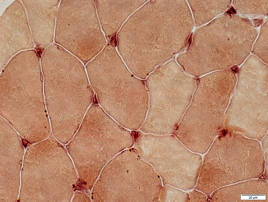
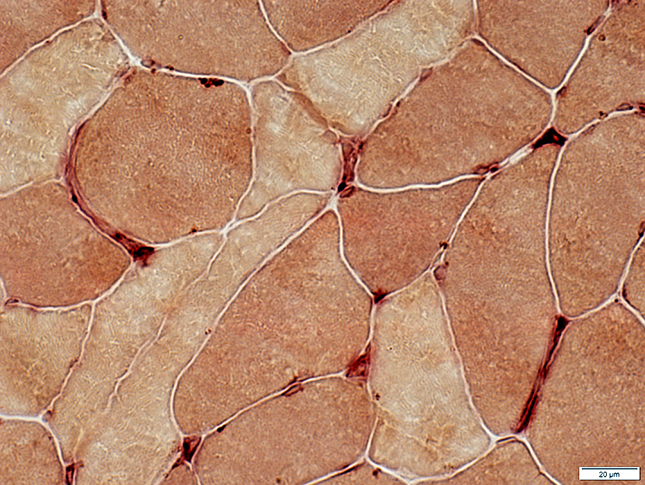
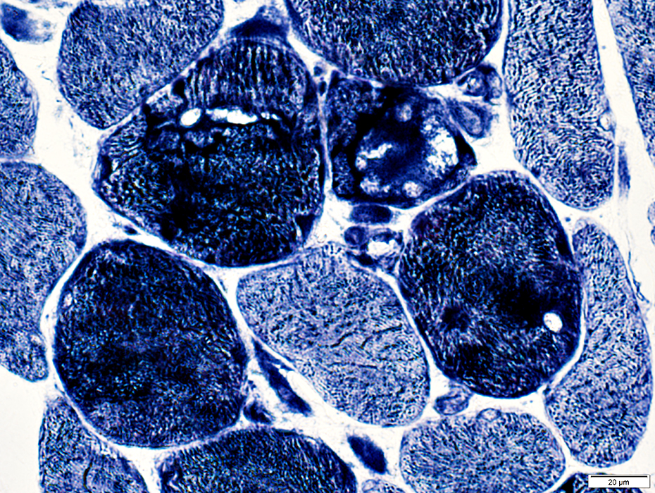
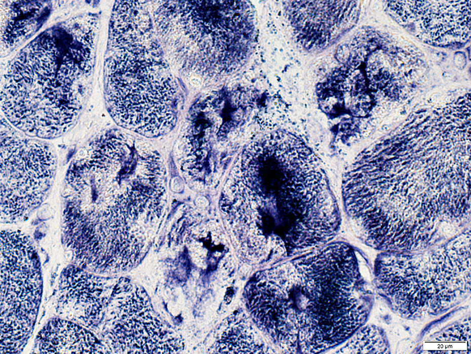
Muscle fibers with abnormal internal architecture near Perimysial vessels
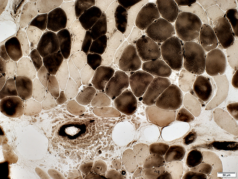
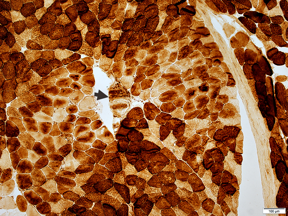 Cytochrome Oxidase stain |
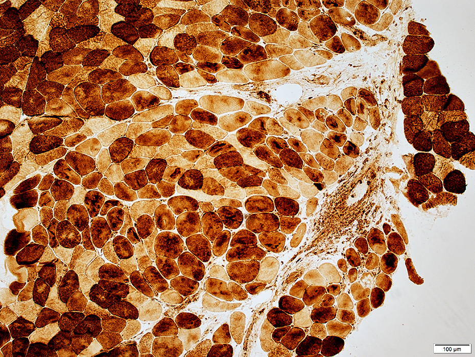 Cytochrome Oxidase stain |
 ATPase pH 4.3 stain |
 Congo red stain |
 VvG stain |
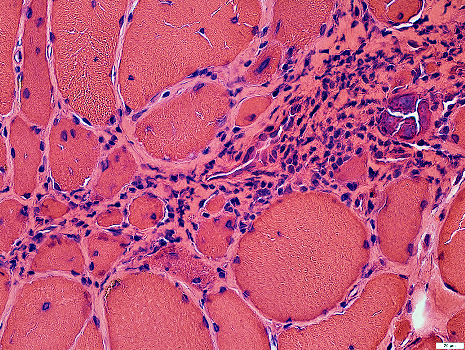
Perivascular Pathology
Abnormal (small) muscle fibers & capillary loss in region neighboring intermediate sized vessels
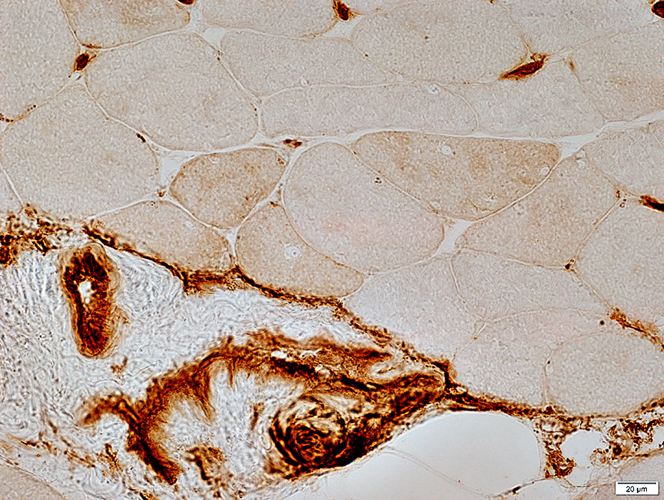 UEAI stain |
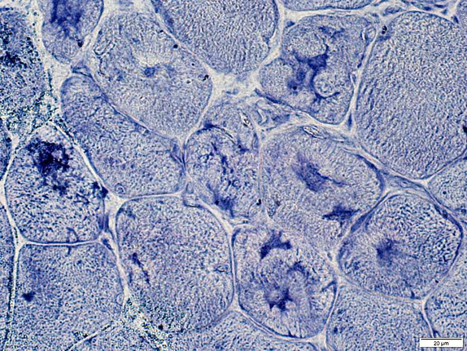
Perifascicular Atrophy, Mild
 NADH stain |
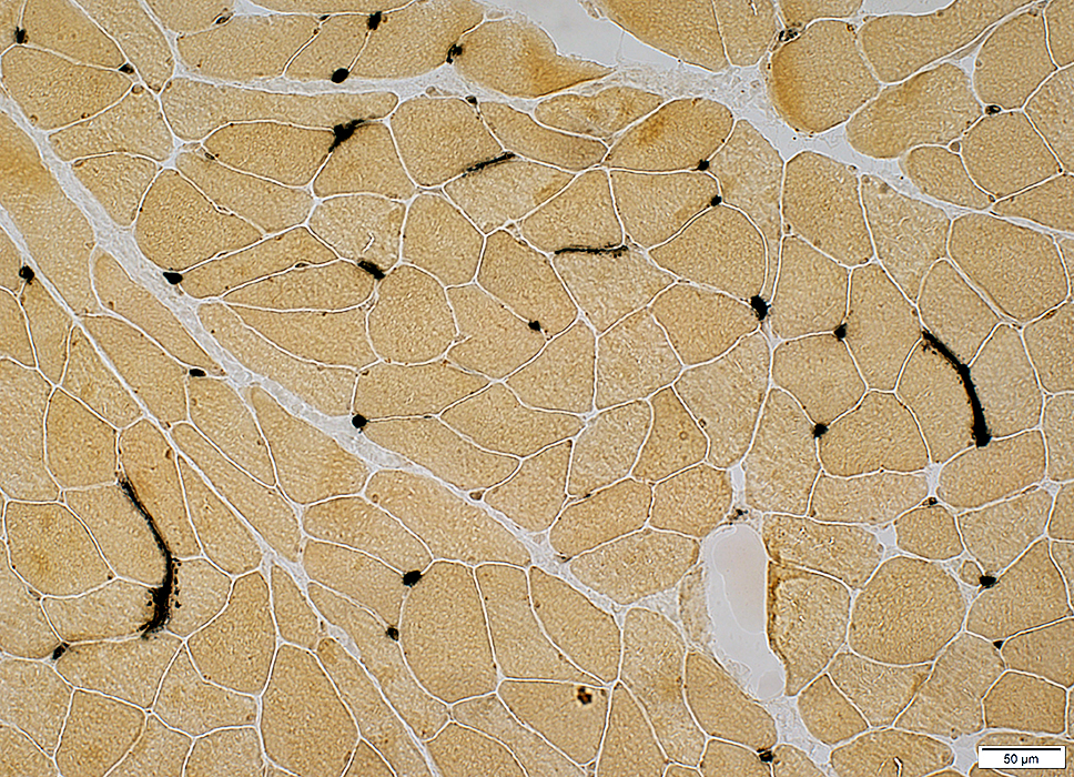
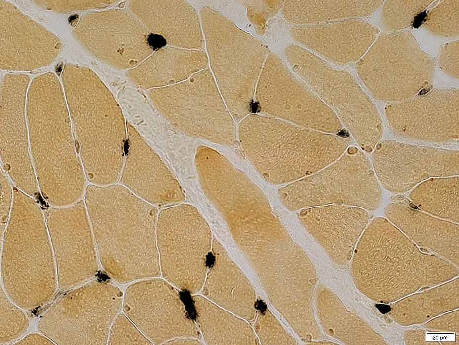
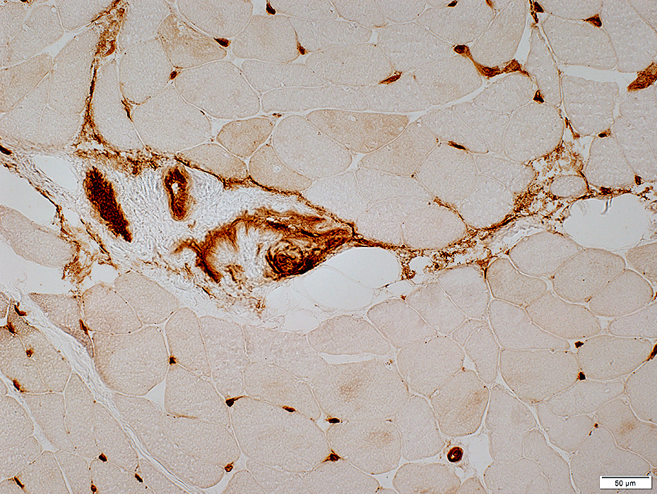
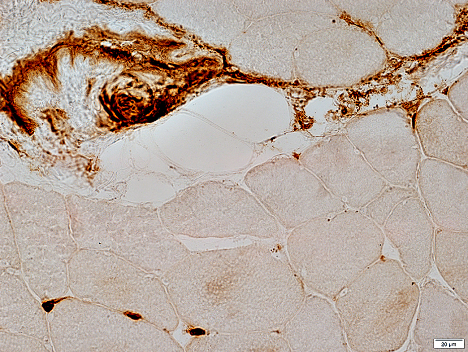
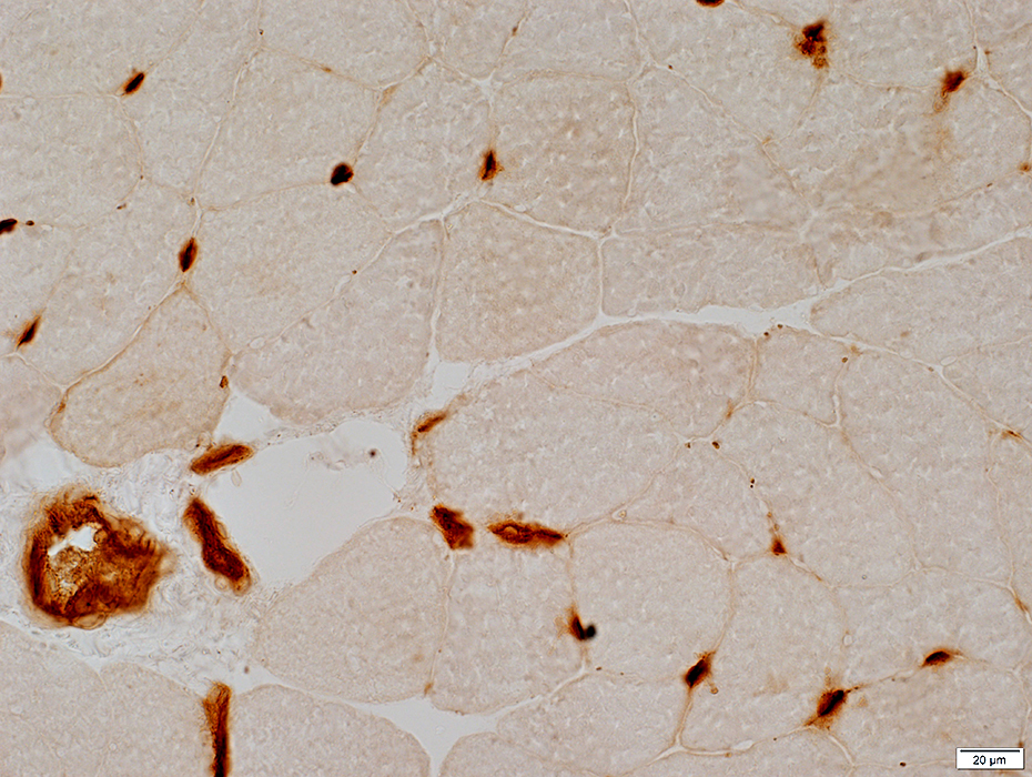
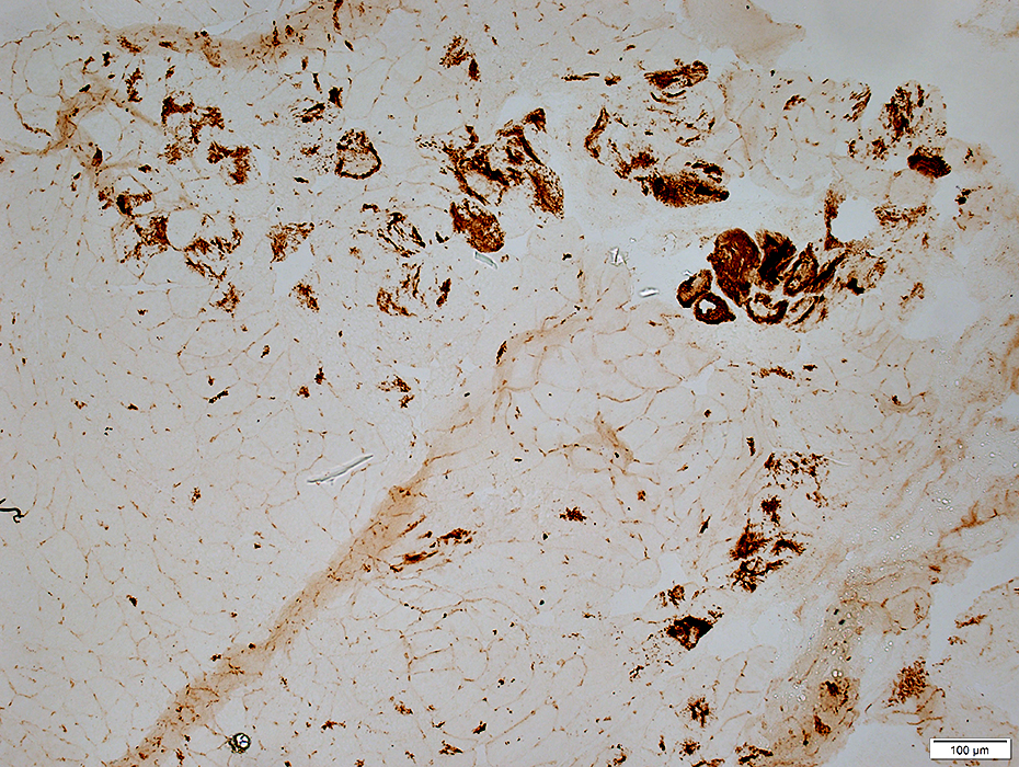
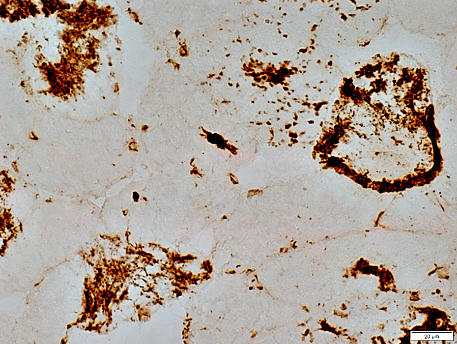
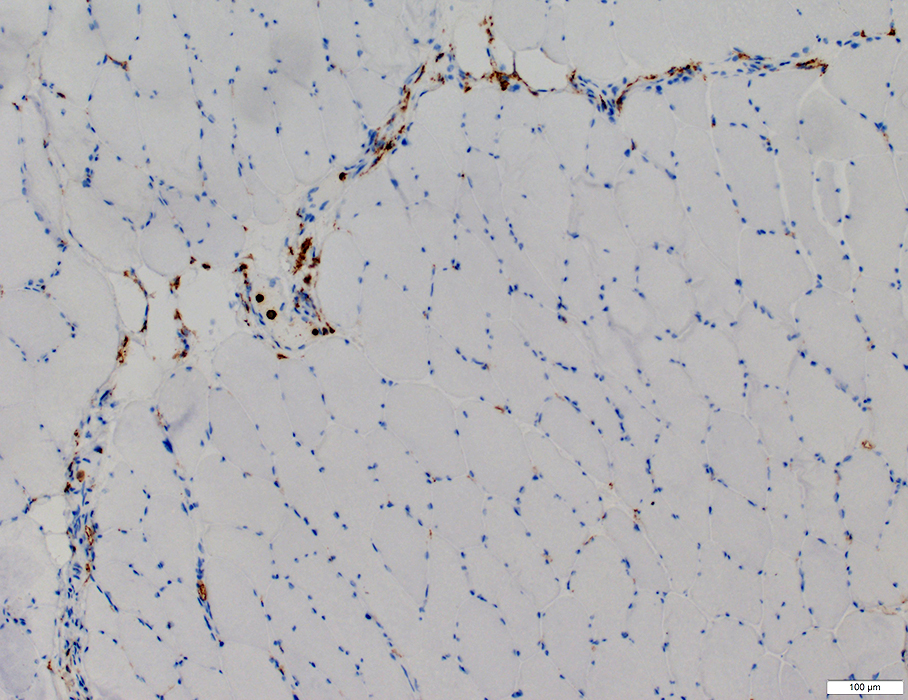
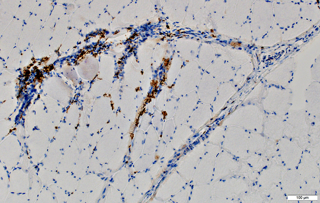
Capillary Pathology
 Esterase stain |
Stain abnormally for esterase
 Esterase stain |
 Esterase stain |
Endomysial capillaries
Mildly large
 Gomori trichrome stain stain |
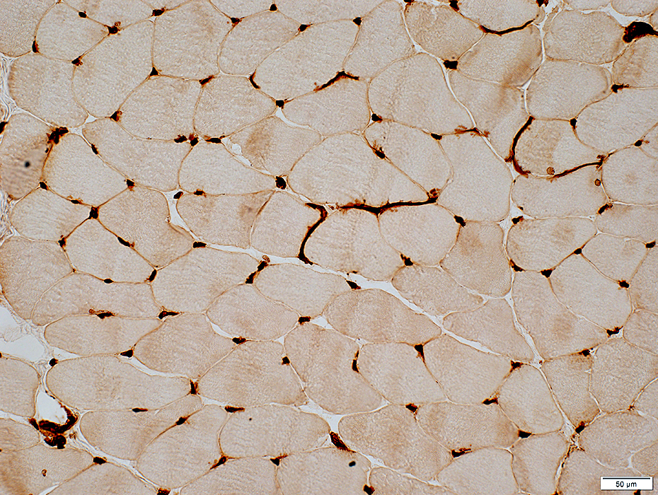
 Alkaline phosphatase stain |
Increased numbers stain with alkaline phosphatase
 Alkaline phosphatase stain |
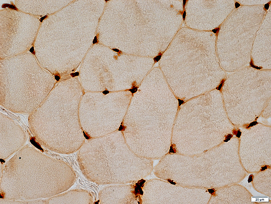
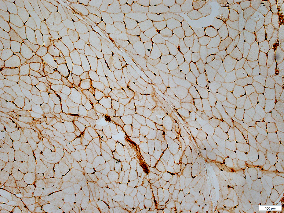
Endomysial capillaries
Staining for C5b-9 in regions with muscle fiber pathology
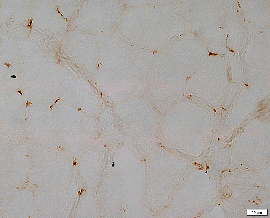 C5b-9 stain |
 UEA1 stain |
Reduced regional staining in TIF1-γ DM patient (Above)
Normal adult: Every muscle fiber has at least one adjacent capillary (Below)
 UEA1 stain |
 UEA1 stain |
Reduced numbers of stained endomysial capillaries in region near perimysial vessel
 UEA1 stain |
 UEA1 stain |
Muscle Fiber Pathology: Vacuoles & Aggregates
 H&E stain |
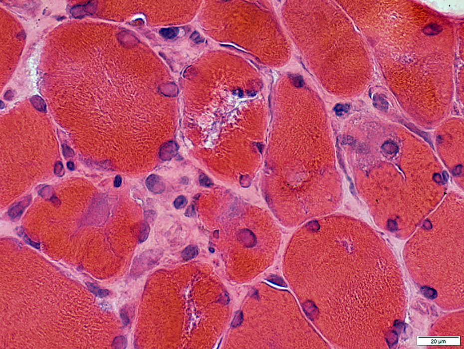 H&E stain |
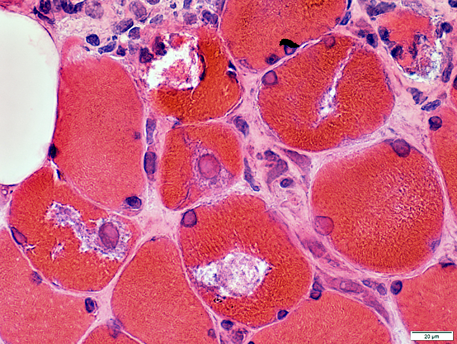 H&E stain |
 VvG stain |
 Congo red stain |
 Congo red stain |
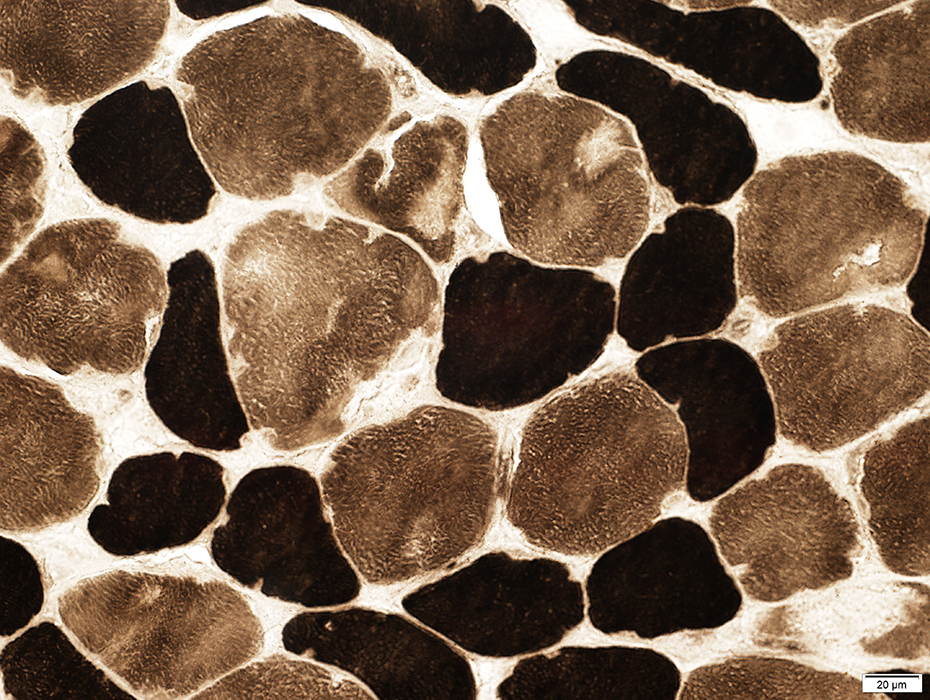 ATPase pH 9.4 stain |
More abnormal in type 1 muscle fibers
 NADH stain |
 NADH stain |
 AMPDA trichrome stain |
One muscle fiber has cytoplassmic bodies (Below)
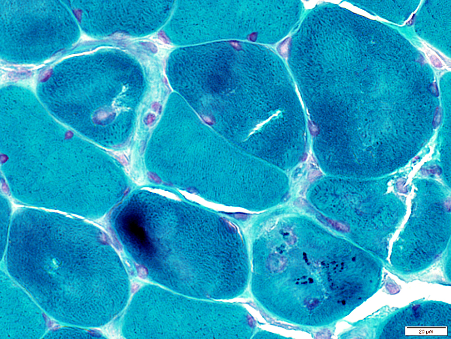 Gomori trichrome stain |
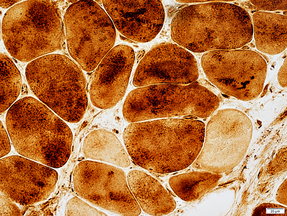
 SDH stain |
 SDH stain |
SDH: Irregular internal architecture; Normal levels of staining
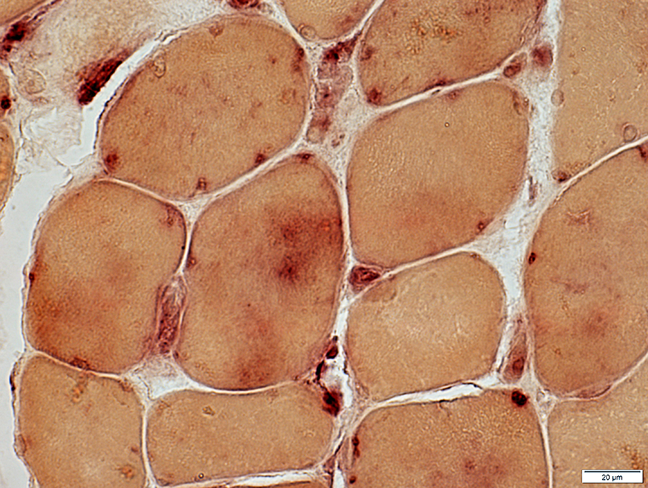
COX: Irregular internal architecture; Variably reduced levels of staining
 COX stain |
 Acid Phosphatase stain |
 LC3 stain |
Similar patterns may occur in DM-VP
 LC3 stain |
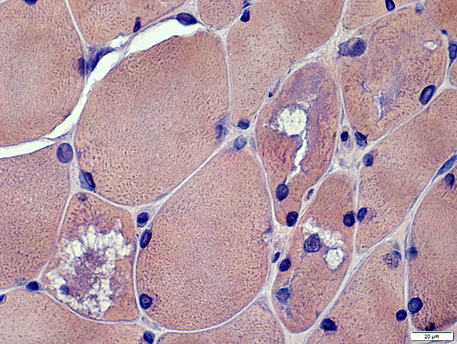
MHC Class I: Upregulation by all muscle fibers
 MHC Class I stain |
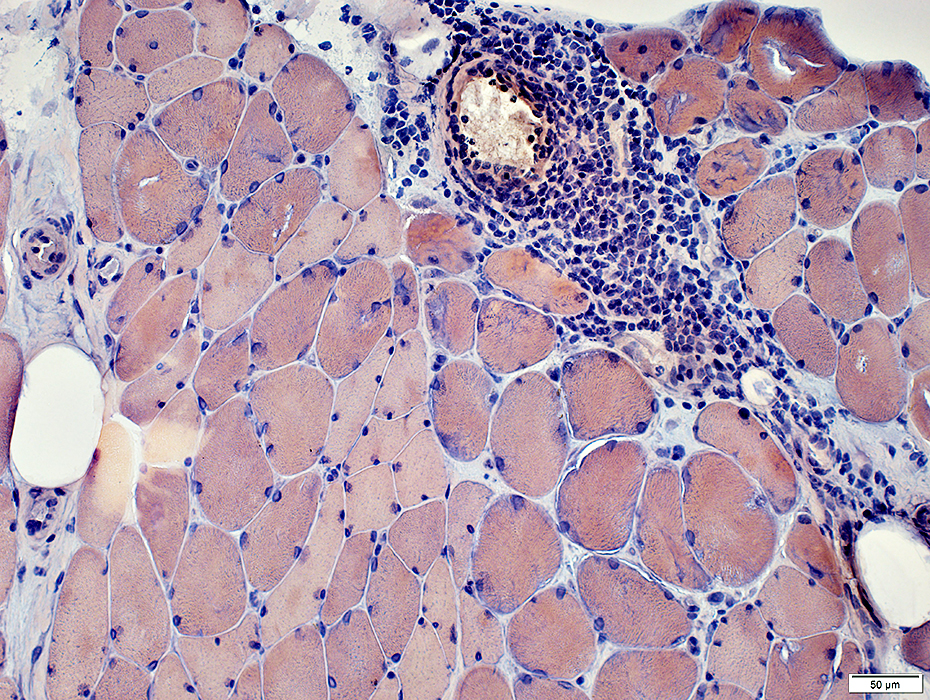
Perimysial Pathology
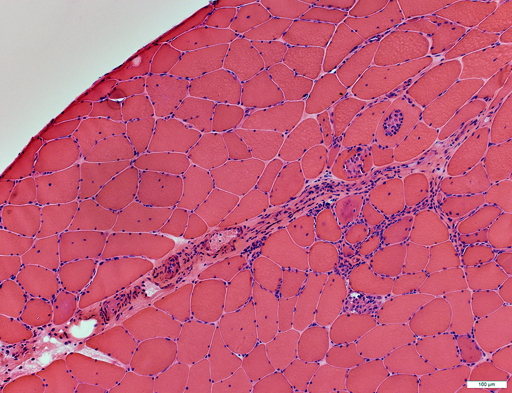 H&E stain |
 H&E stain |
 H&E stain |
 CD45 stain |
 CD8 stain |
Return to Dermatomyositis with TIF1-γ (TRIM33) Antibodies
Return to Neuromuscular Home Page
Return to Pathology index
11/1/2025