Immune Myopathies with Perimysial Pathology (IMPP): PL-12 antibody associated
|
Also see IMPP Dermatomyositis: Adult, IMPP type Jo-1 muscle pathology |
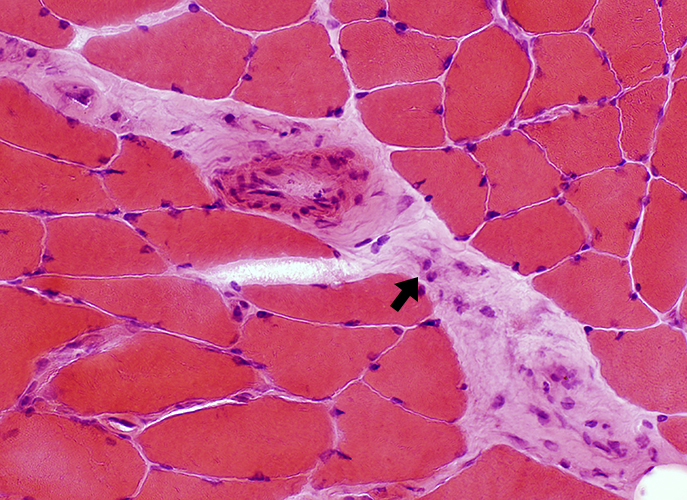 H&E stain Perimysial cellularity
 |
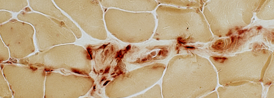 Acid phosphatase stain Perimysial cells: Acid phosphatase positive 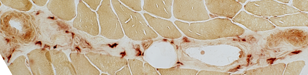 Acid phosphatase |
|
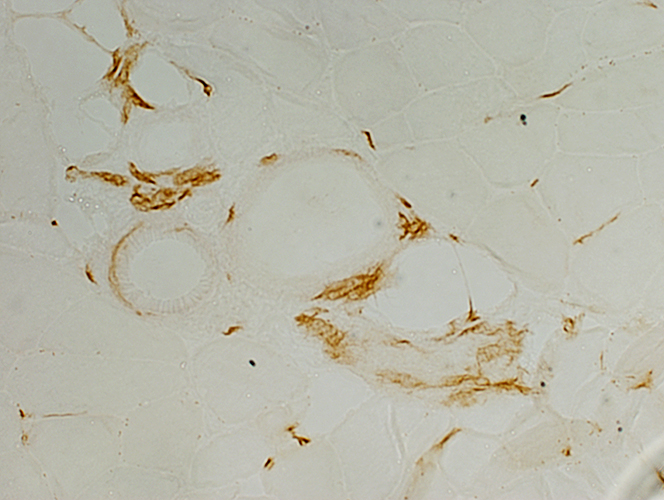 CD4 stain CD4+ cells: Perimysial, & Scattered endomysial | |
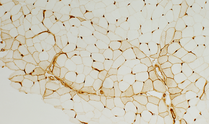 MHC Class I Perifascicular muscle fibers: Express MHC Class I 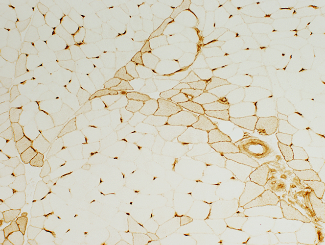 MHC Class I |
C5b-9 deposition on Perimysium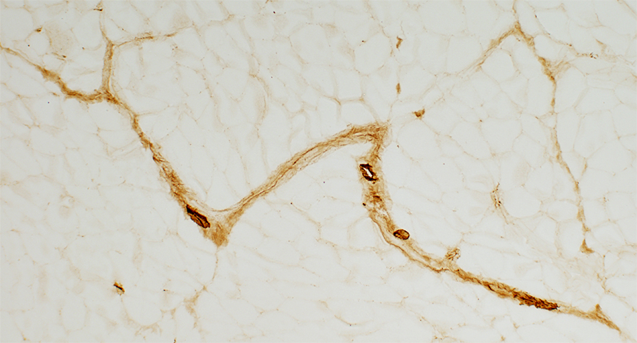 C5b-9 stain |
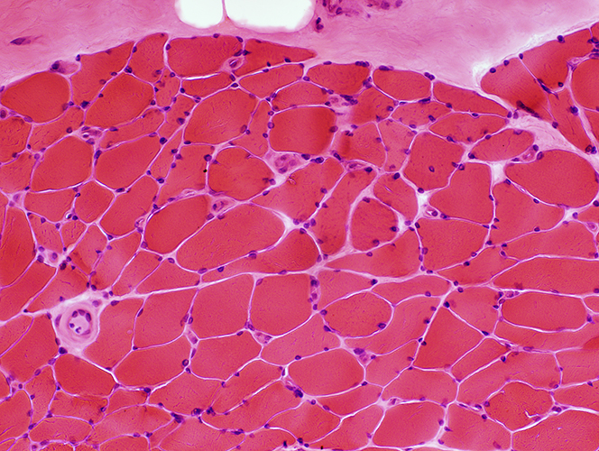
Varied fiber size: Some small fibers are type 2C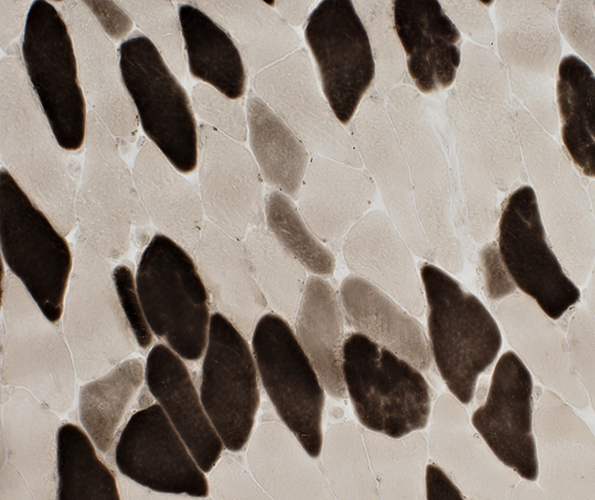 ATPase pH 4.3 stain |
Return to Inflammatory myopathies
Return to IMPP
References
1. Curr Opin Rheumatol 2011;23:595-604
2/24/2013