NXP-2 Antibody: Myopathy Patterns
|
NXP-2 Myopathology Patterns: 3 types Regional Necrosis Regional Ischemic Immune Myopathy (RIIM) Other RIIM pathology Perifascicular Myopathy Dermatomyositis + Vascular Pathology (DM-VP) Minimal Myopathy with Capillary Pathology (MMCP) |
Regional Ischemic Immune Myopathy (RIIM): NXP-2 Antobody-associated
Clusters of Necrotic & Regenerating Muscle Fibers
Necrosis: Fibers in each cluster are in similar stages of necrosis or regeneration
Anatomy: Clusters of damaged muscle fibers may not be at the very edge of fascicles
Perimysial Connective Tissue
Wide
Fragmented
No immune cells
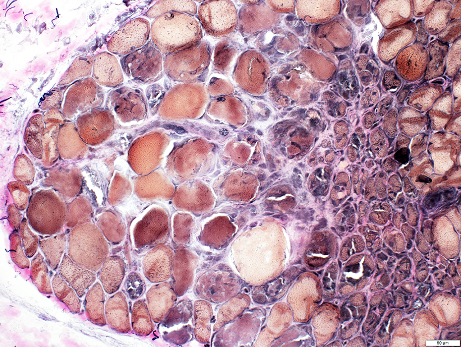
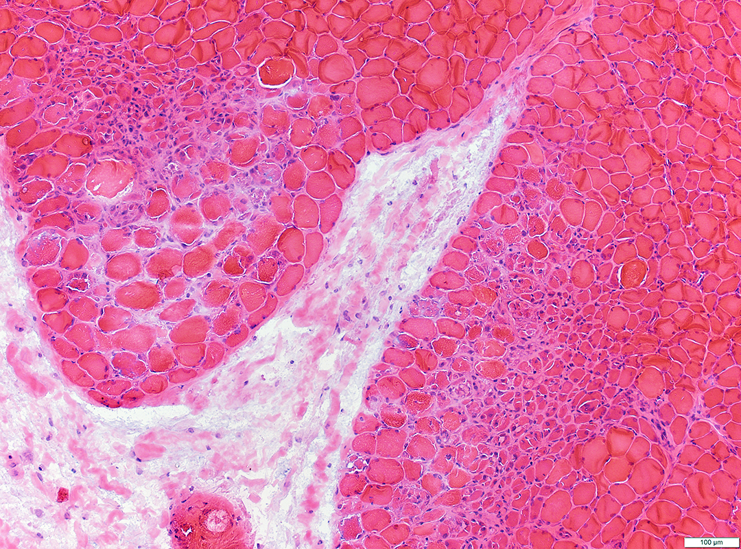 H & E stain |
 Congo red stain |
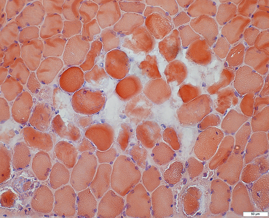
Necrotic fibers often have moderately dark, featureless cytoplasm with no visible myonuclei
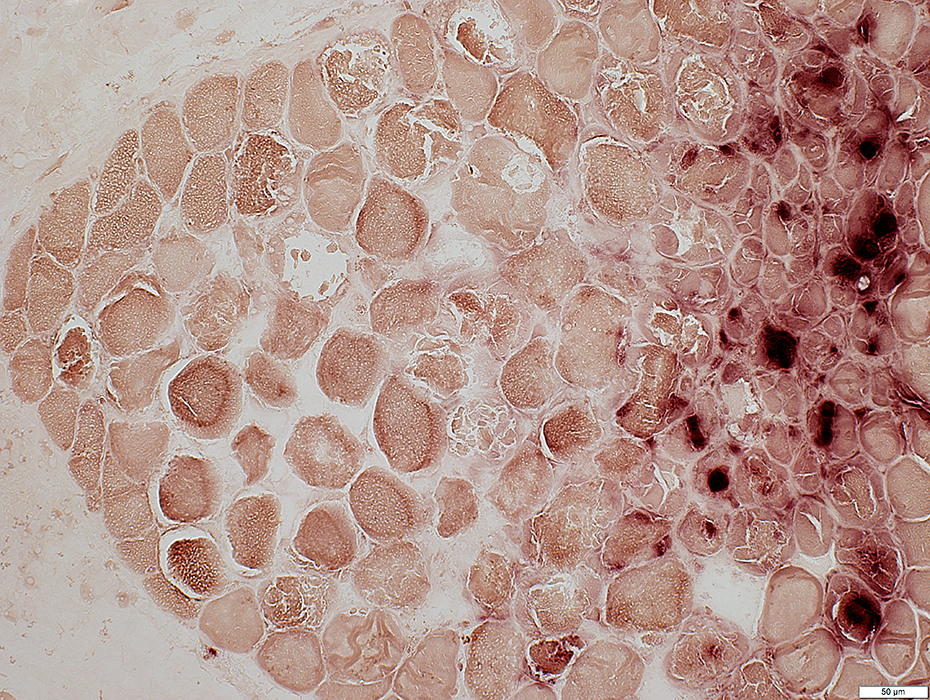
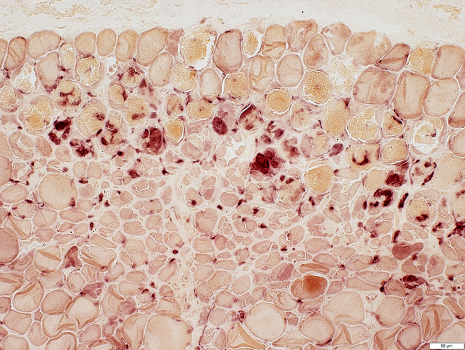
 C5b-9 complement stain |
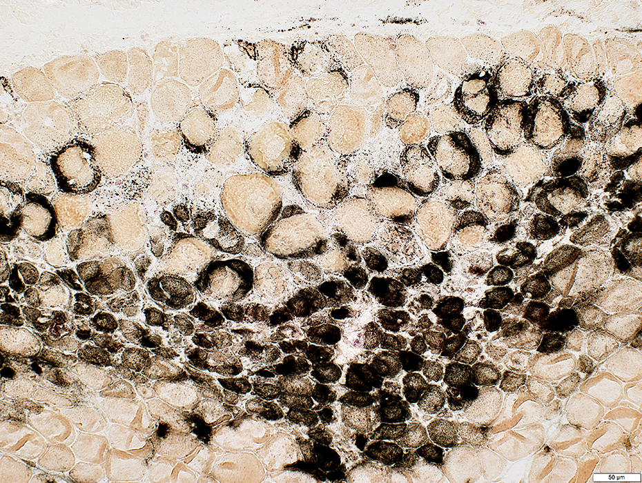
Muscle fiber features
C5b-9 staining of muscle fiber cytoplasm: diffuse
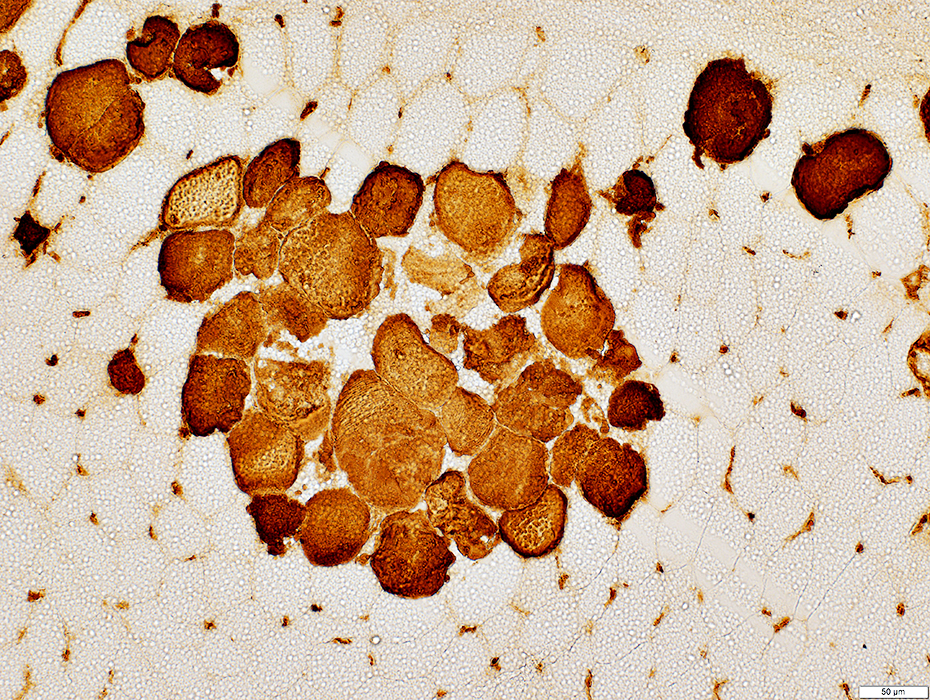
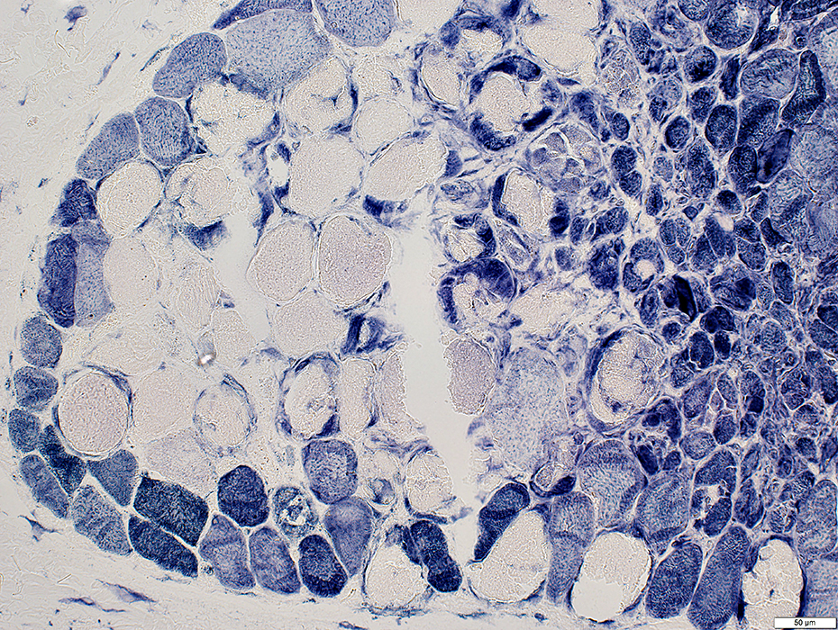
NADH staining of cytoplasm is absent
Anatomy
Necrotic fibers are in clusters
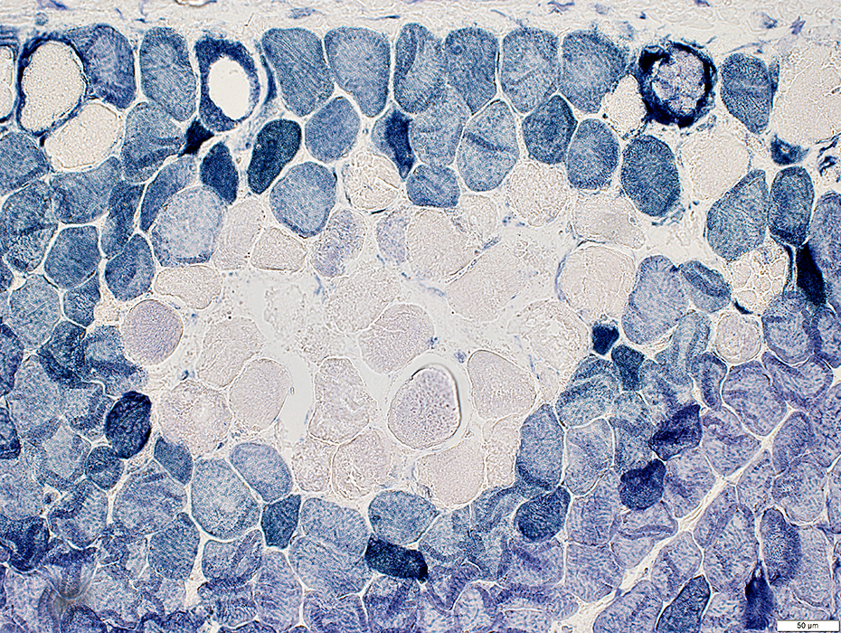 NADH stain |
 NADH stain |
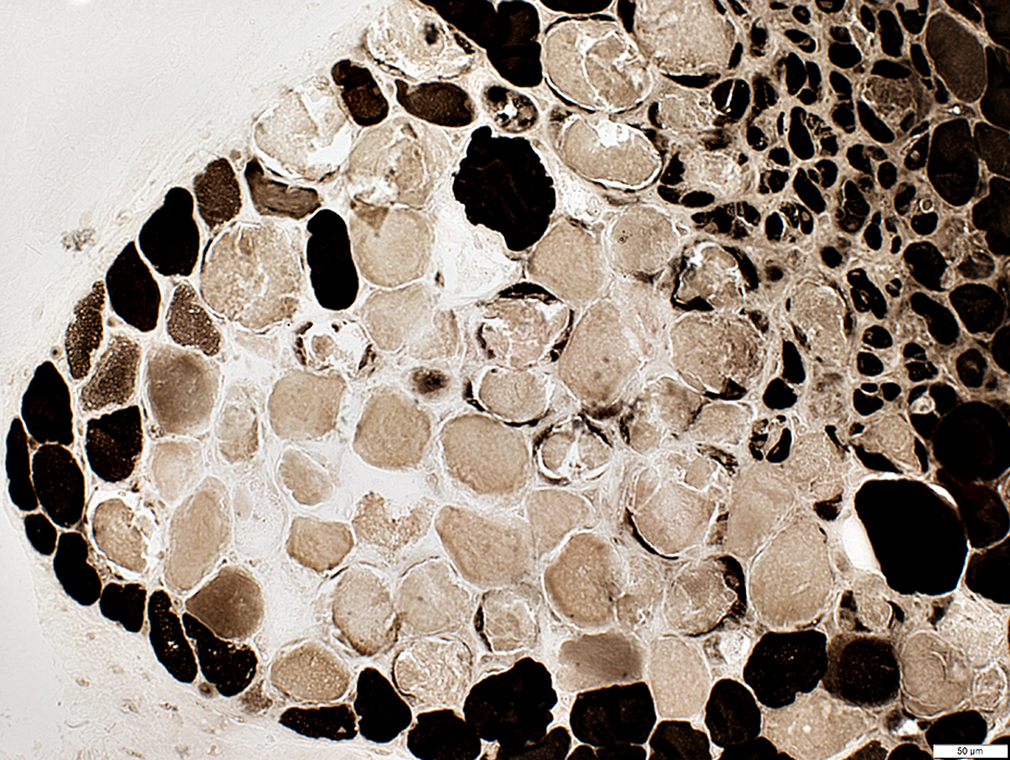
 Esterase stain |
 VvG stain |
 ATPase pH 9.4 stain |
 Alkaline phospohatase stain |
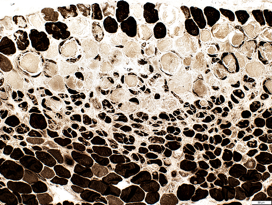 ATPase pH 9.4 stain |
 Acid phosphatase stain |
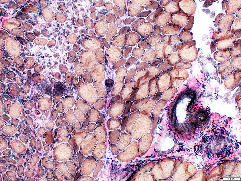 VvG stain |
Vessels
Artery, Large (Left): Normal structure
Artery, Small (Center): Abnormal wall structure
Fibril (Elastin) layer: Incomplete (White arrow)
Connective tissue inside fibril layer
Vein (Right; White arrow) Pale wall inside fibril layer
No visible lumen
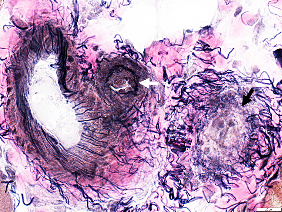 VvG stain |
Minimal Myopathy with Capillary Pathology: NXP-2 Antibody-associated
Features
Muscle
Fiber morphology: Lipid droplets present in some fibers
Fiber sizes: Varied
MHC Class I stain: Increased on muscle fiber surfaces
Capillaries
C5b-9 staining
Sizes: Mildly large
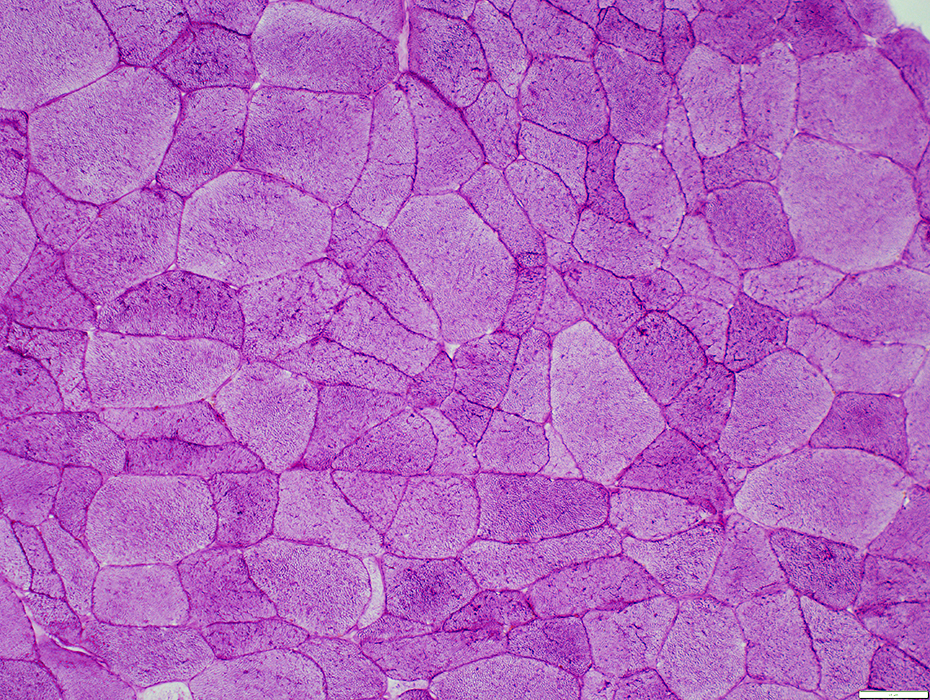
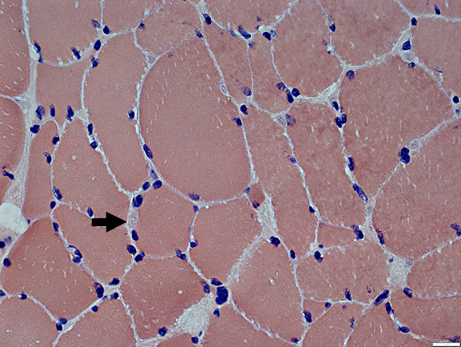
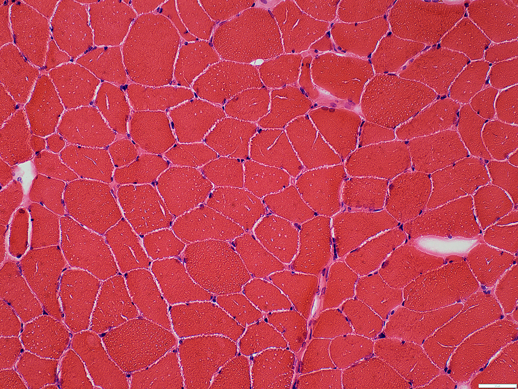 H&E stain |
Muscle fibers
Sizes: Moderate variation
No necrosis or immaturity
Endomysial connective tissue: Normal
 H&E stain |
 Gomori trichrome stain |
Muscle fibers
Sizes: Moderate variation
Smaller fibers are dark on PAS stain
No necrosis or immaturity
Endomysial connective tissue: Normal
 PAS stain |
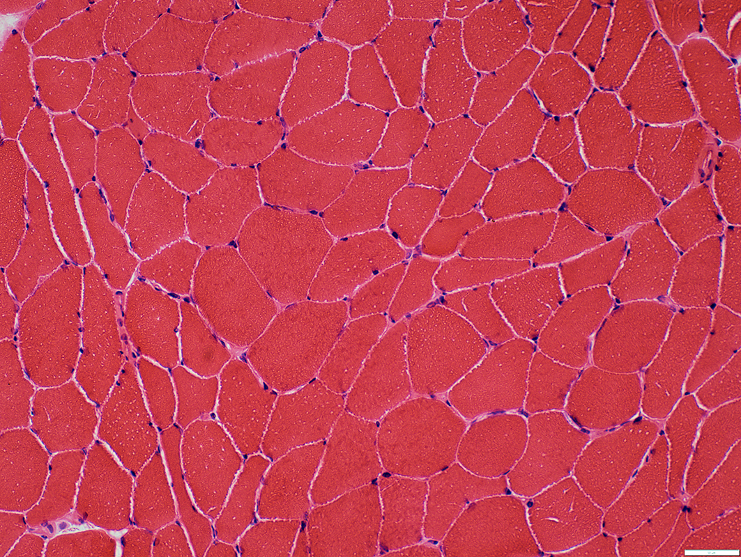
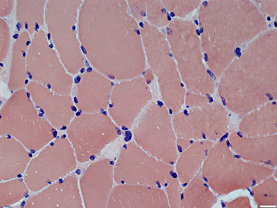
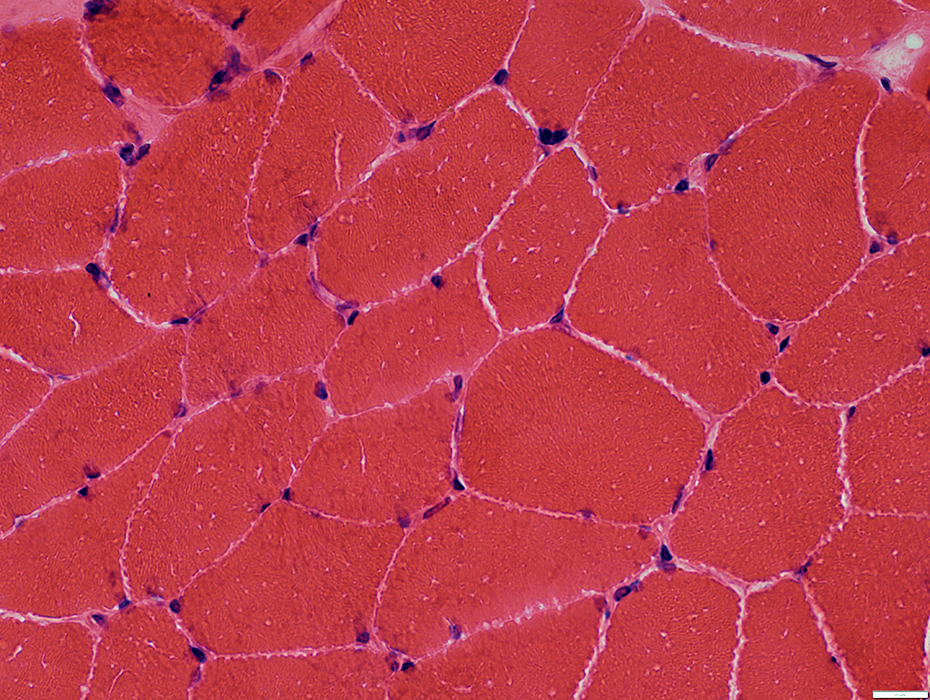 H&E stain |
Muscle fibers
Sizes: Moderate variation
No necrosis or immaturity
Endomysial connective tissue: Normal
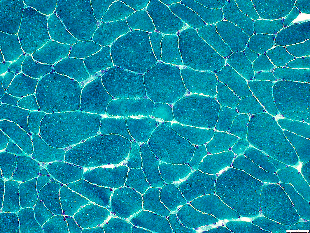
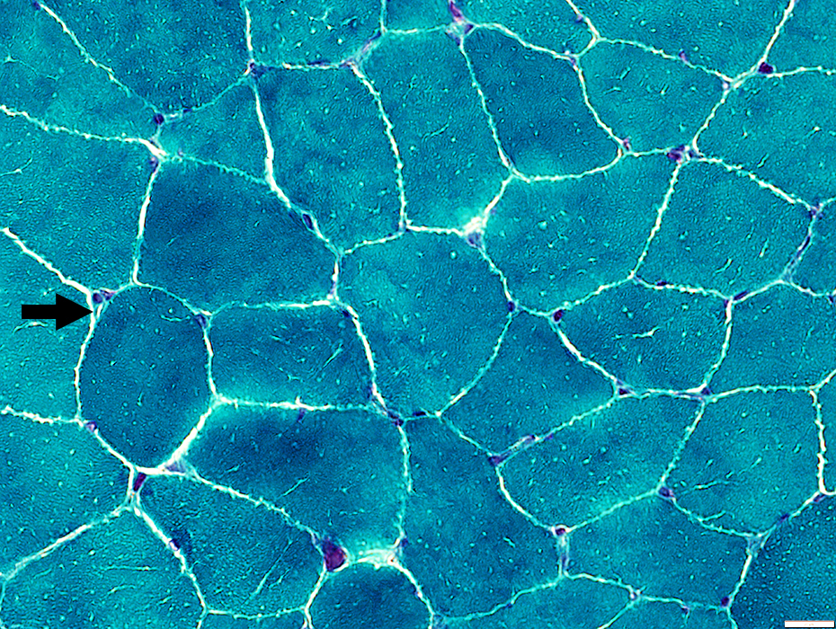 Gomori trichrome stain |
Muscle fibers
Sizes: Moderate variation
No necrosis or immaturity
Endomysial connective tissue: Normal
Endomysial capillaries: Normal to Slightly large (Arrow)
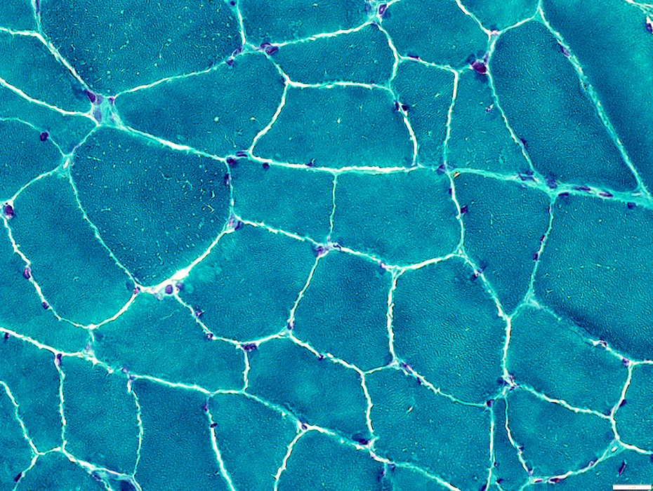 Gomori trichrome stain |
 Congo red stain |
Muscle fibers
Sizes: Moderate variation
No necrosis or immaturity
Endomysial connective tissue: Normal
Endomysial capillaries: Normal to Slightly large (Arrow)
 Congo red stain |
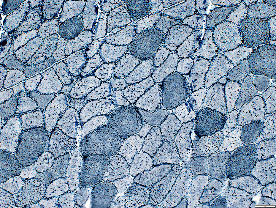
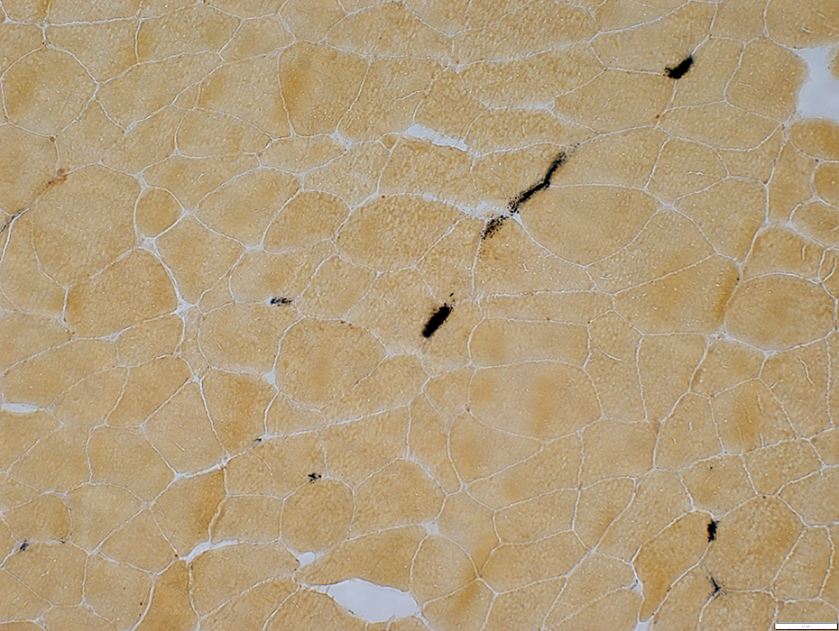
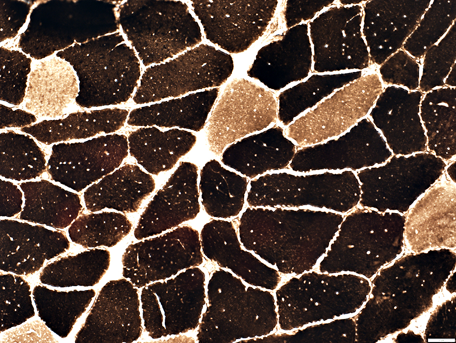 ATPase pH 9.4 stain |
Muscle fibers
Lipid droplets in type 2 (Dark) fibers
Type 2 fiber predominance
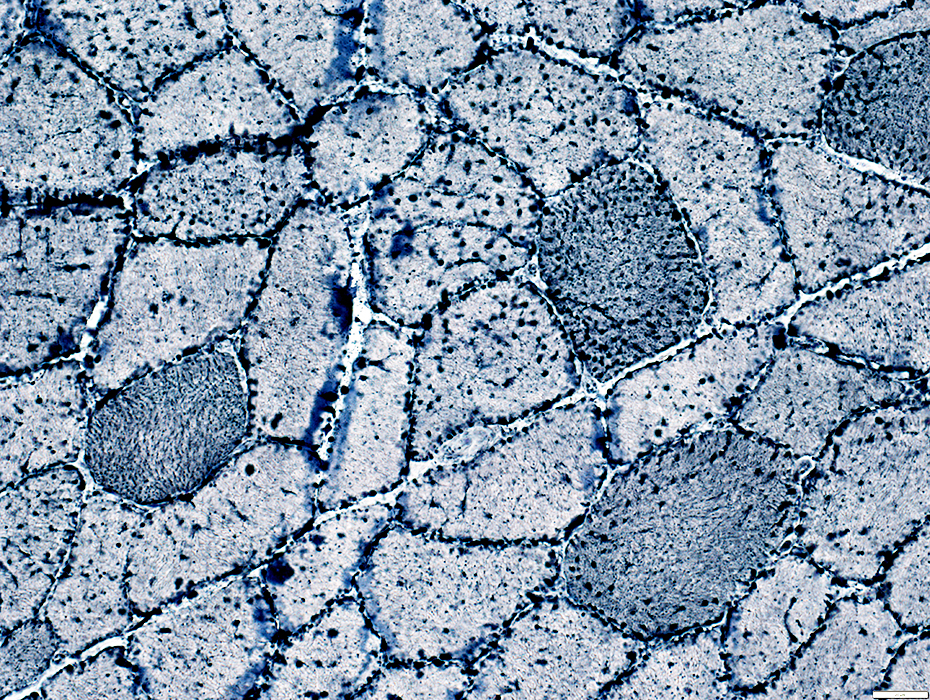
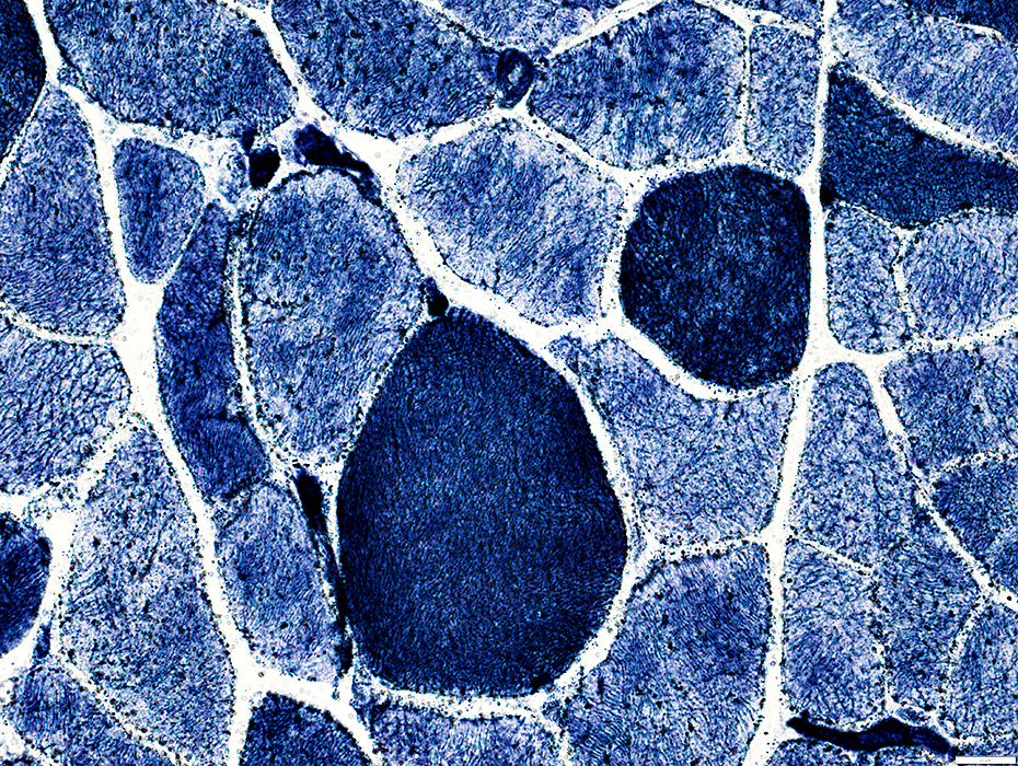
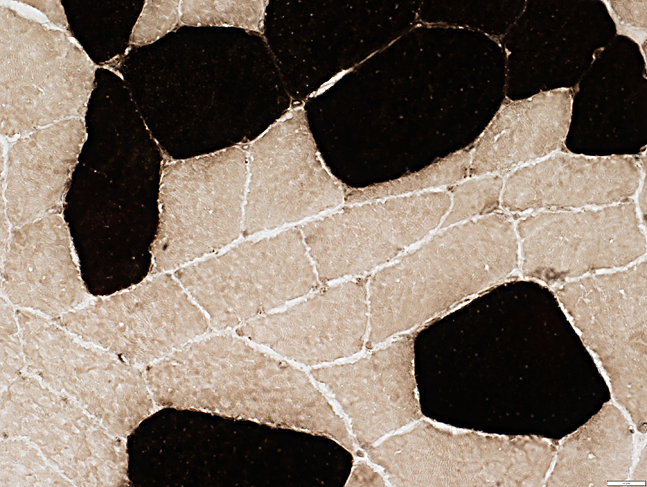 ATPase pH 4.3 stain |
Muscle fibers
Type 2 fiber predominance
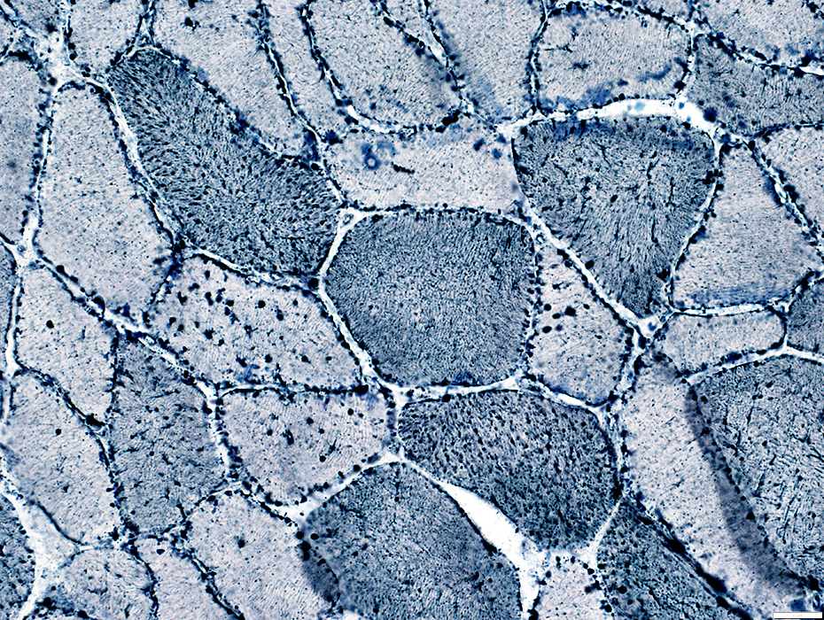
 Sudan black stain |
Muscle fibers
Lipid droplets more prominent in type 2 (Pale) fibers
 Sudan black stain |
 Sudan black stain |
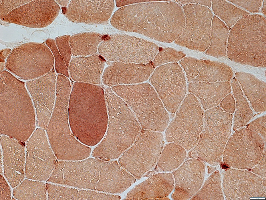
 MHC Class I stain |
Muscle fibers: MHC I abnormally expressed on the surface of most fibers
 MHC Class I stain |
Muscle fibers: MHC I abnormally expressed on the surface of most fibers
 MHC Class I stain |
 Esterase stain |
Esterase stains some endomysial capillaries
 Acid phosphatase stain |
No prominent staining of endomysial capillaries with Acid or Alkaline phosphatase
 Alkaline phosphatase stain |
 NADH stain |
NADH stains some endomysial capillaries
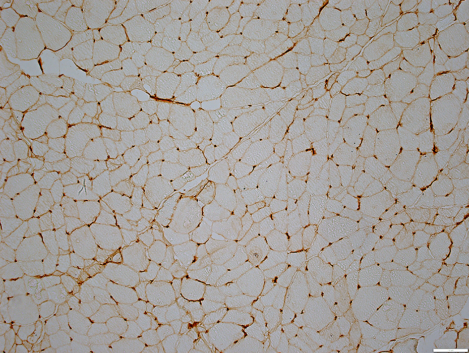
 UEA I stain |
UEA I normally stains endomysial capillaries
Some small muscle fibers have no adjacent capillary (Arrows)
 UEA I stain |
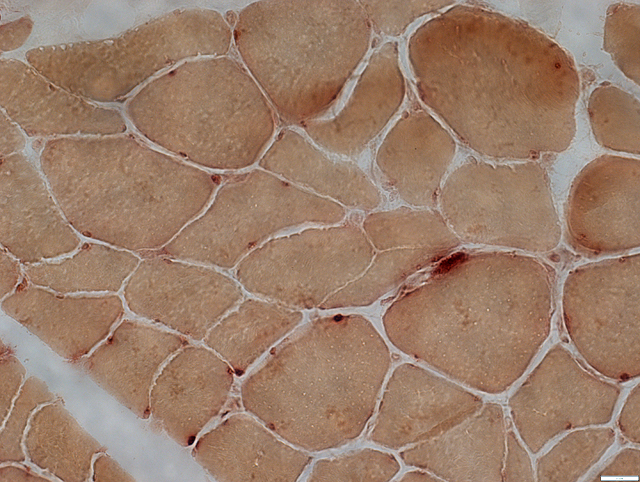
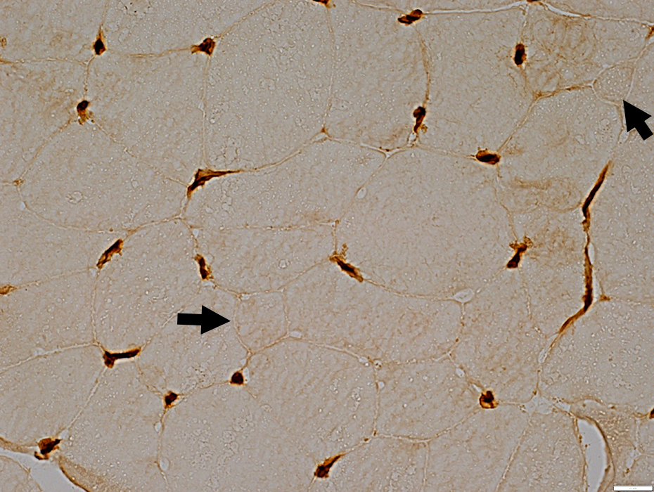
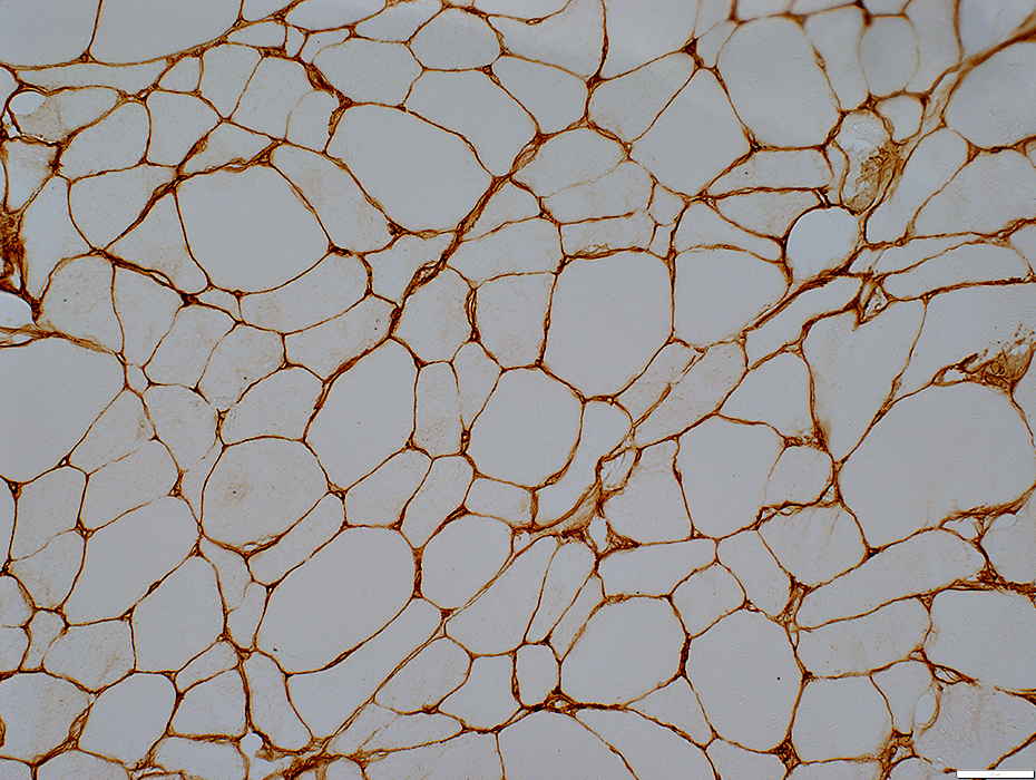 Decorin stain |
Endomysial capillaries are midly large
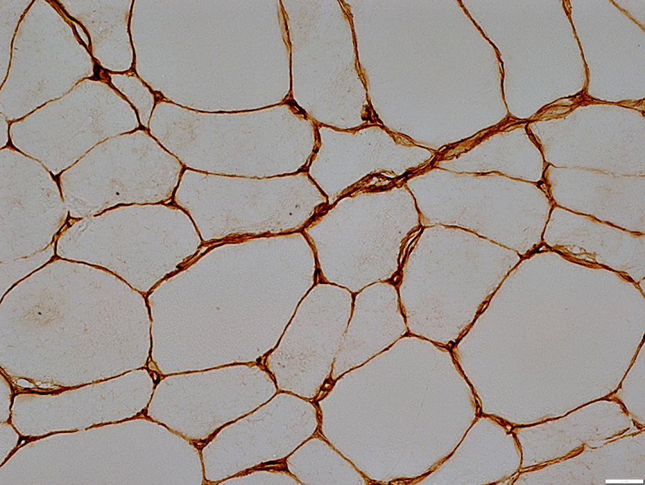 Decorin stain |
Endomysial capillaries are midly large (Arrow)
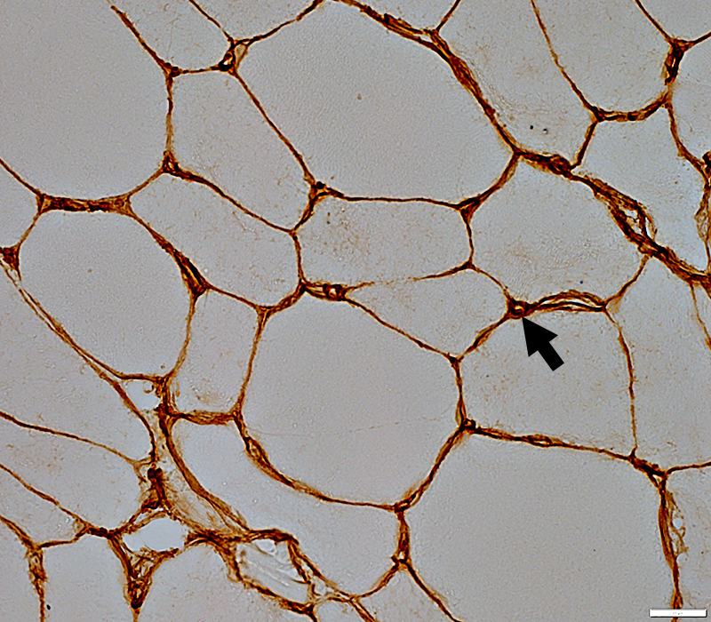 Decorin stain |
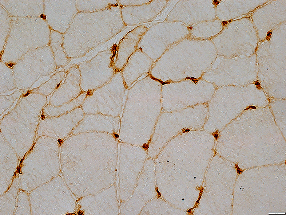
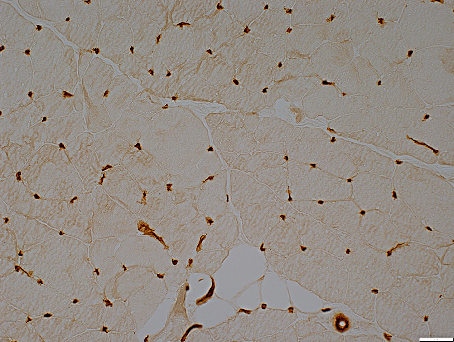
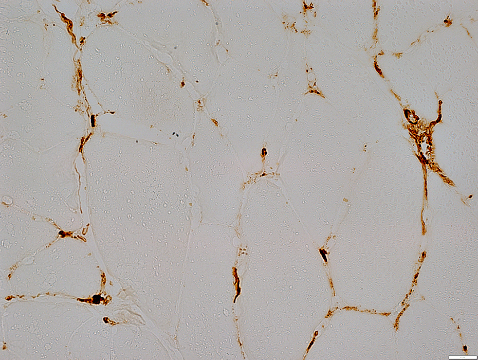
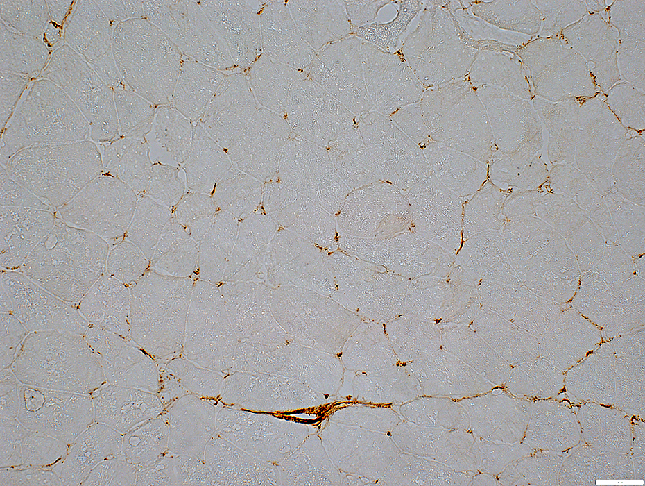 C5b-9 stain |
C5b-9 is stained on many endomysial capillaries
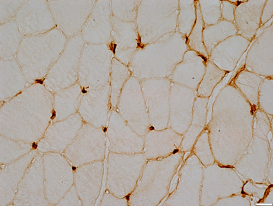
 C5b-9 stain |
 C5b-9 stain |
C5b-9 is stained on many endomysial capillaries
Return to NXP-2
Return to Neuromuscular Home Page
11/28/2022