Chronic Neuropathy with IgG vs Neurofascin 140, 155 & 186
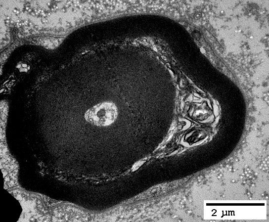
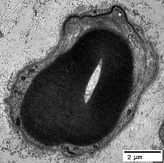
| Ultrastructure |
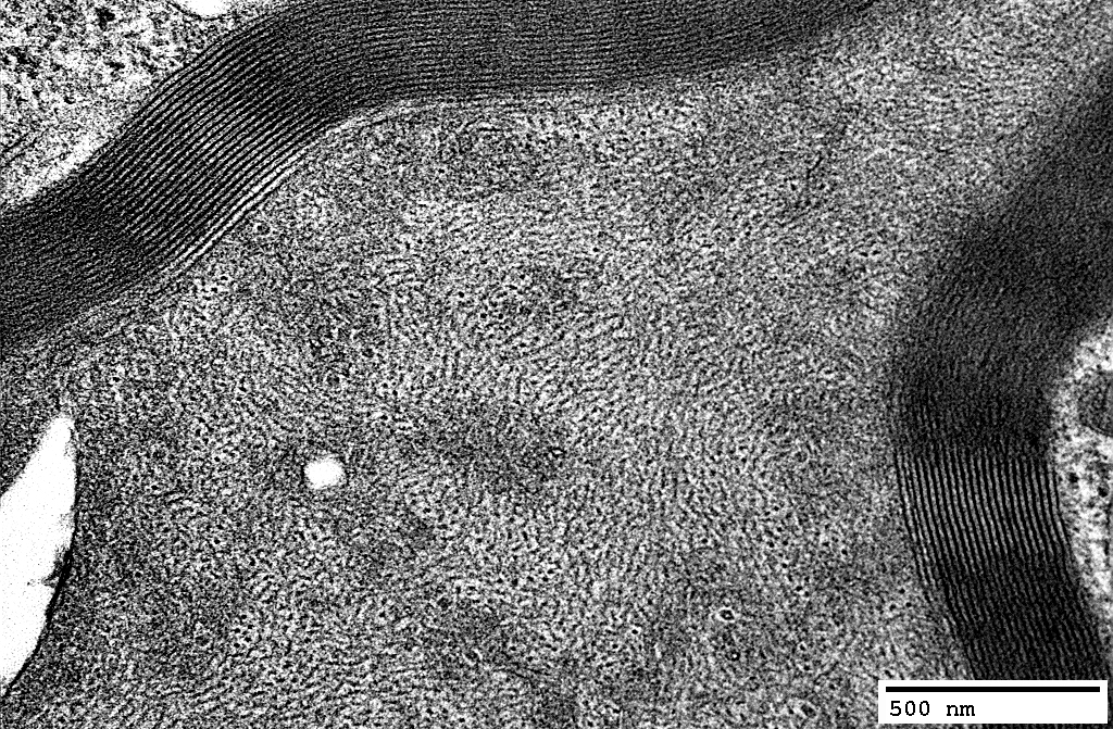
Myelin Pathology
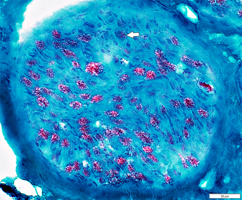 Gomori trichrome stain |
Irregular structure (Arrow; Below)
 VvG stain |
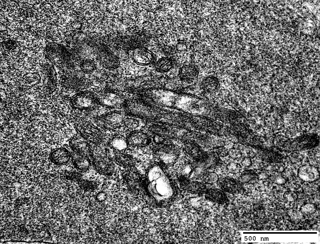
Autophagic Myelin
Myelin sheaths stain for acid phosphatase
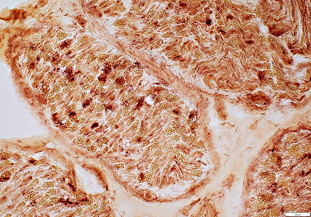 Acid phosphatase stain |
Irregular Myelin Structure
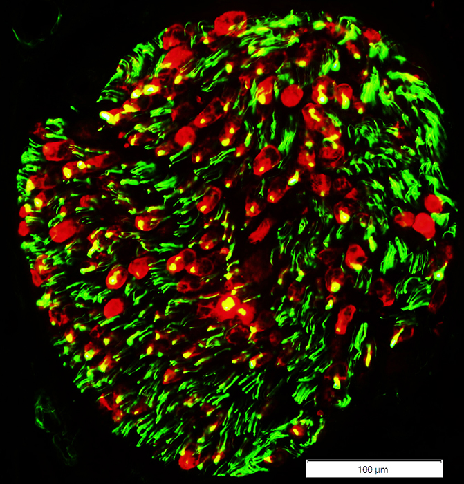
Irregular co-localization of P0 & MBP (Red areas within myelin sheaths)
See: Control nerve
MBP(r)nfascpan.jpg) P0 (Green) + MBP (Red) stain |
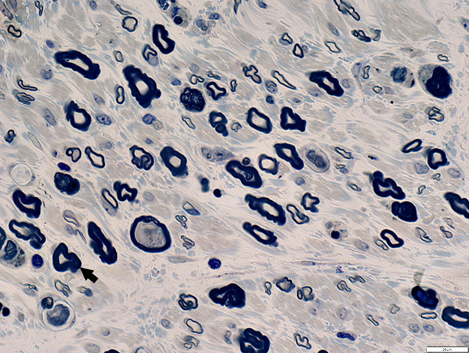
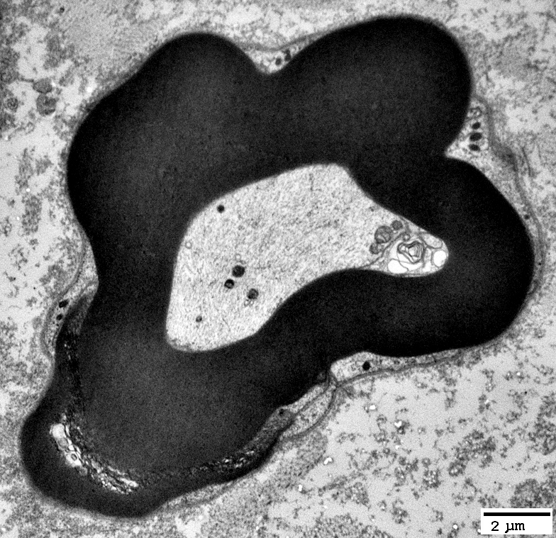
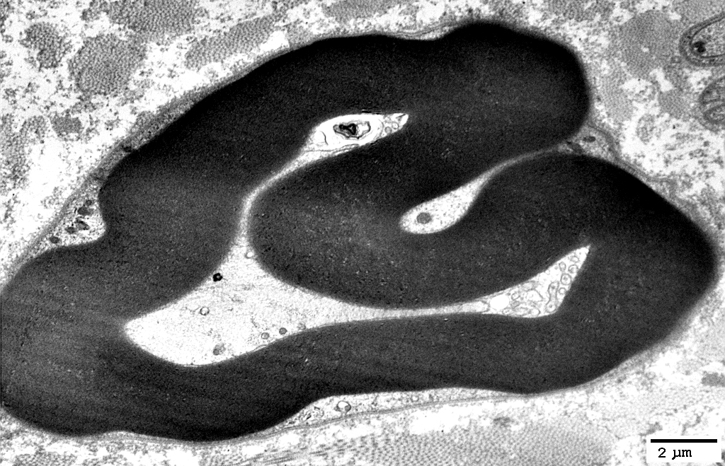
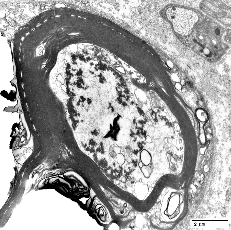
Myelin Sheaths: Structure & Shape Irregular
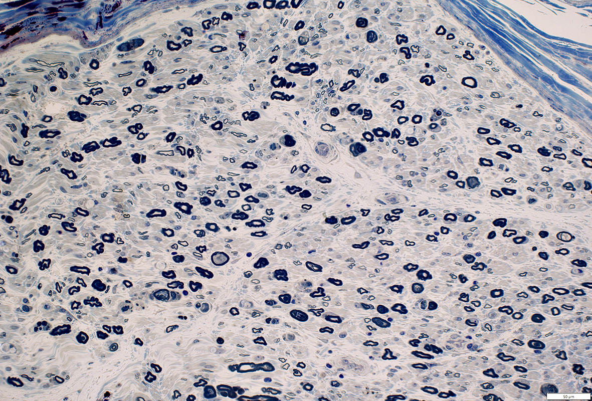 Toluidine blue stain |
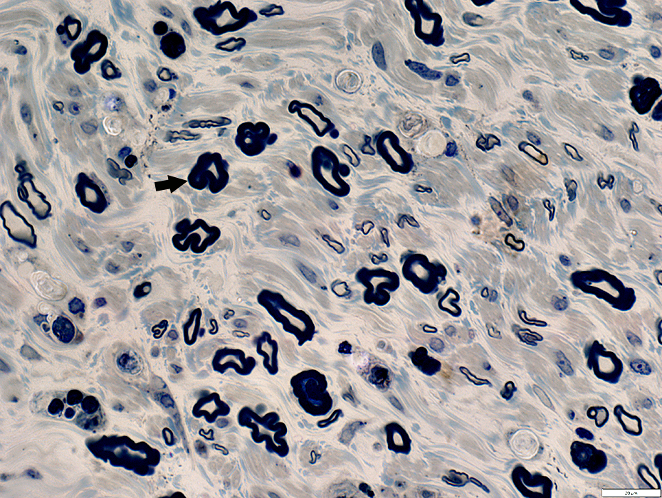 Toluidine blue stain |
 Toluidine blue stain |
 Neurofilament (Green) + Myelin Basic Protein (Red) |
P0(r)nfascpan.jpg) Neurofilament (Green) + P0 Protein (Red) |
Myelin Sheaths vs Axons
 Myelin Sheath Morphology Thick for axon size Shapes: Irregular |
|

|
Thick for axon size
Shapes: Irregular
|

|
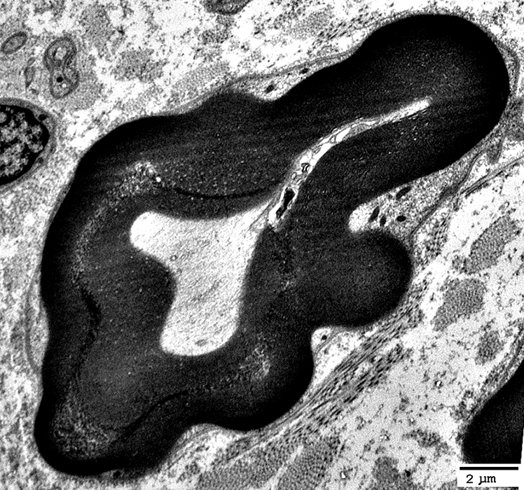
Thick for axon size
Shapes: Irregular

|
|
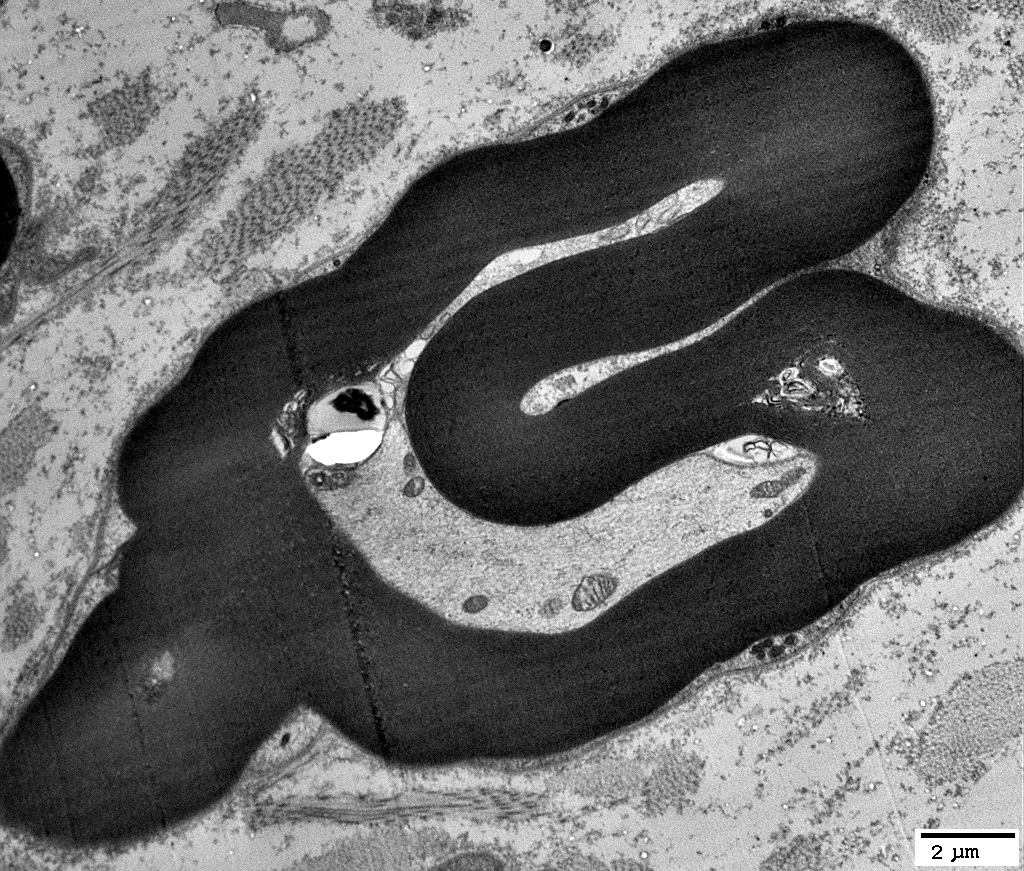
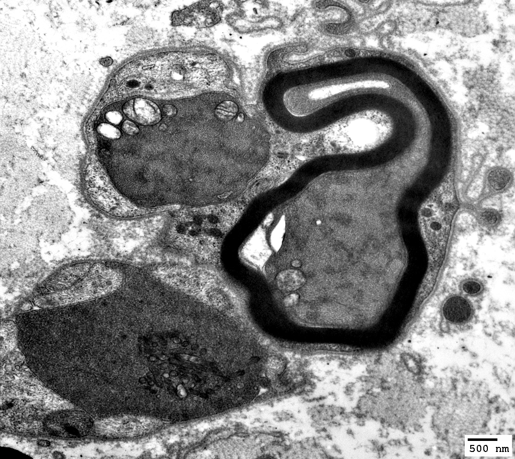
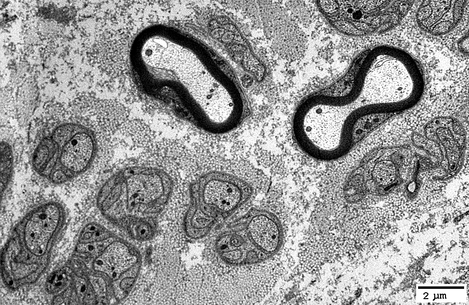
Myelin Sheath Morphology
Thick for axon size
Shapes: Irregular, Folded
|
|
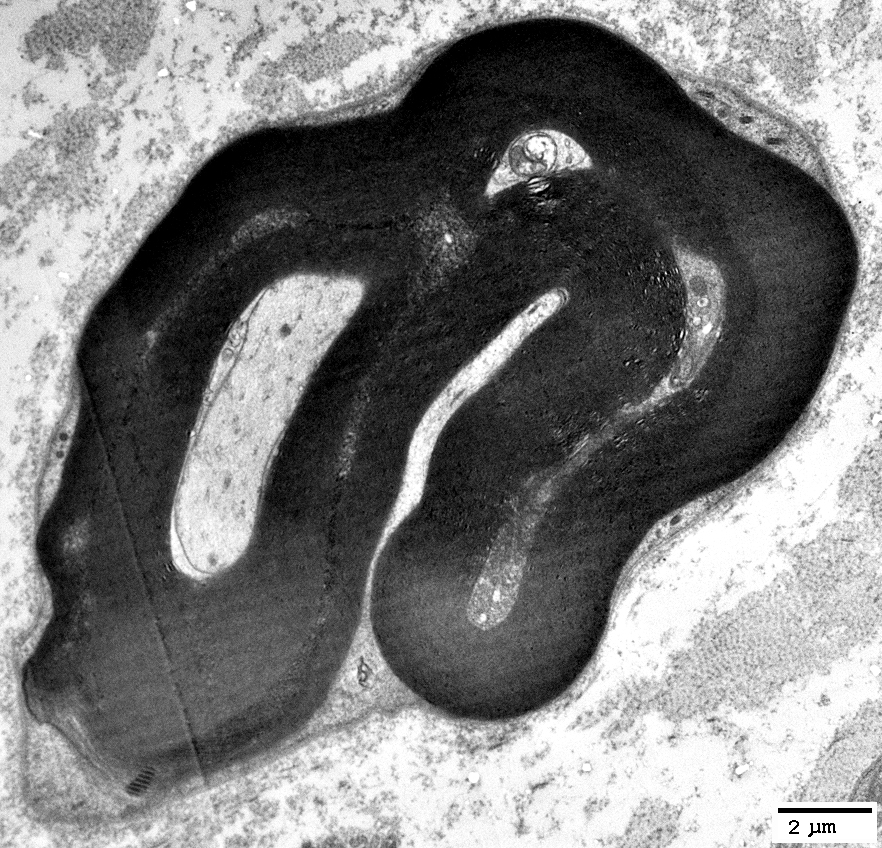
Thick for axon size
Shapes: Irregular
|
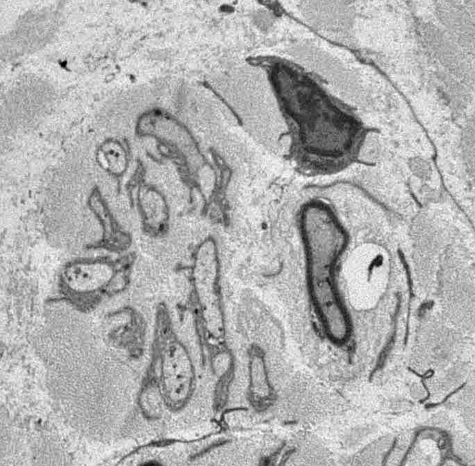
Axons
Scattered axons: Other morphologyDystrophic
Dark axoplasm
Thinly myelinated
Small, unmyelinated axons may be
Reduced in number
Singletons: Only one small axon within non-myelinating Schwann cell
|
Dark cytoplasm with tubuloreticular profiles
Unmyelinated or thinly myelinated
Present in a cluster
|
|
Axon & Myelin sheath: Abnormal
Axon: Abnormal axoplasm
Myelin Sheath: Partial degradation
|
|
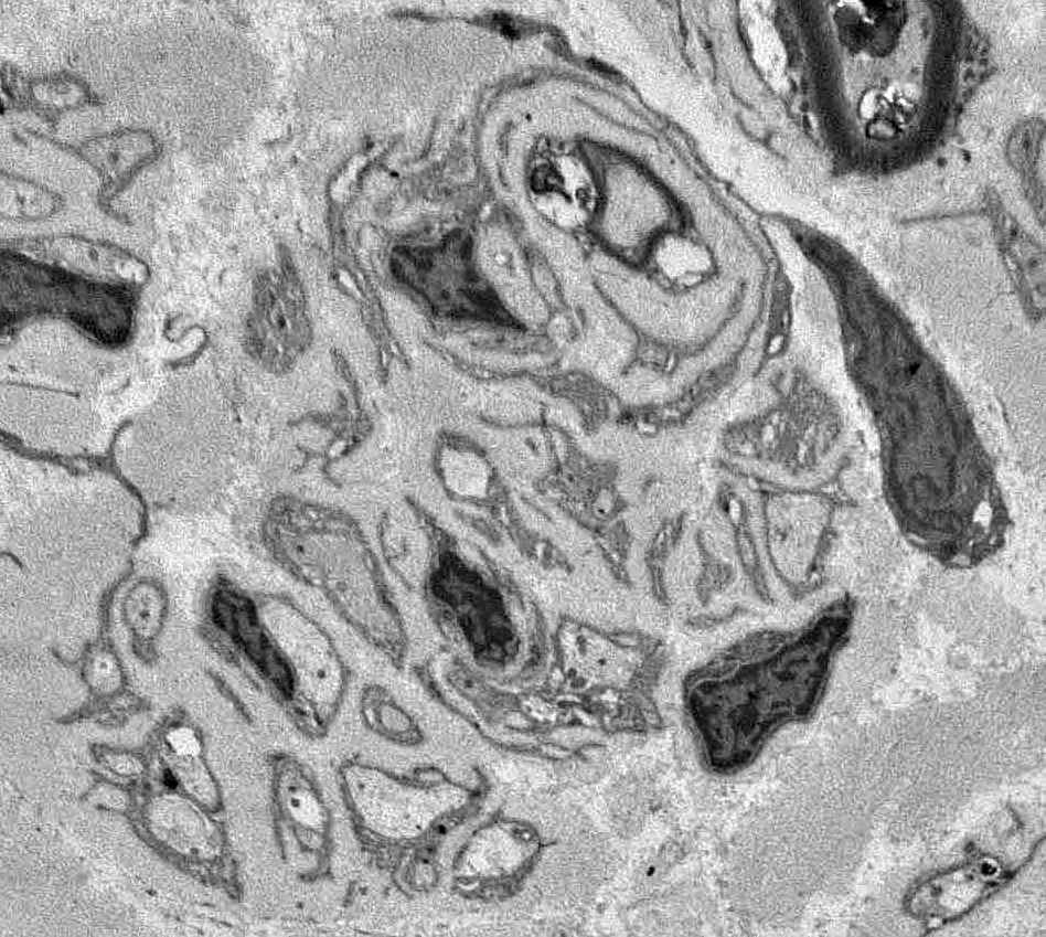
Unmyelinated axons
Reduced numbers per Schwann cell: Singletons
Axon loss
Schwann cells with: No axons; Collagen pockets
|
|
Unmyelinated axons
Reduced numbers per Schwann cell: Singletons
Axon loss
Schwann cells with: No axons; Collagen pockets

|
Loss of Small axons: Moderate
Scattered non-myelinating Schwann cells with no axon (Red)
NCAM(r)nfascpan.jpg)
|
Büngner band (Denervated) Schwann Cells: Scattered
Scattered non-myelinating Schwann cells costain for NCAM & P0
P0(r)nfascpan.jpg) |
Return to: Neurofascin Antibodies
Return to: Neuromuscular Home Page
9/14/2025