DYSMYELINATION
Metachromatic Leukodystrophy (MLD) (& Krabbe)
|
Histochemistry Myelin sheath thin: TB; EM Onion bulbs Macrophages Prismatic inclusions Schwann cells Debris & Inclusions Molecular features Tuffstone bodies |
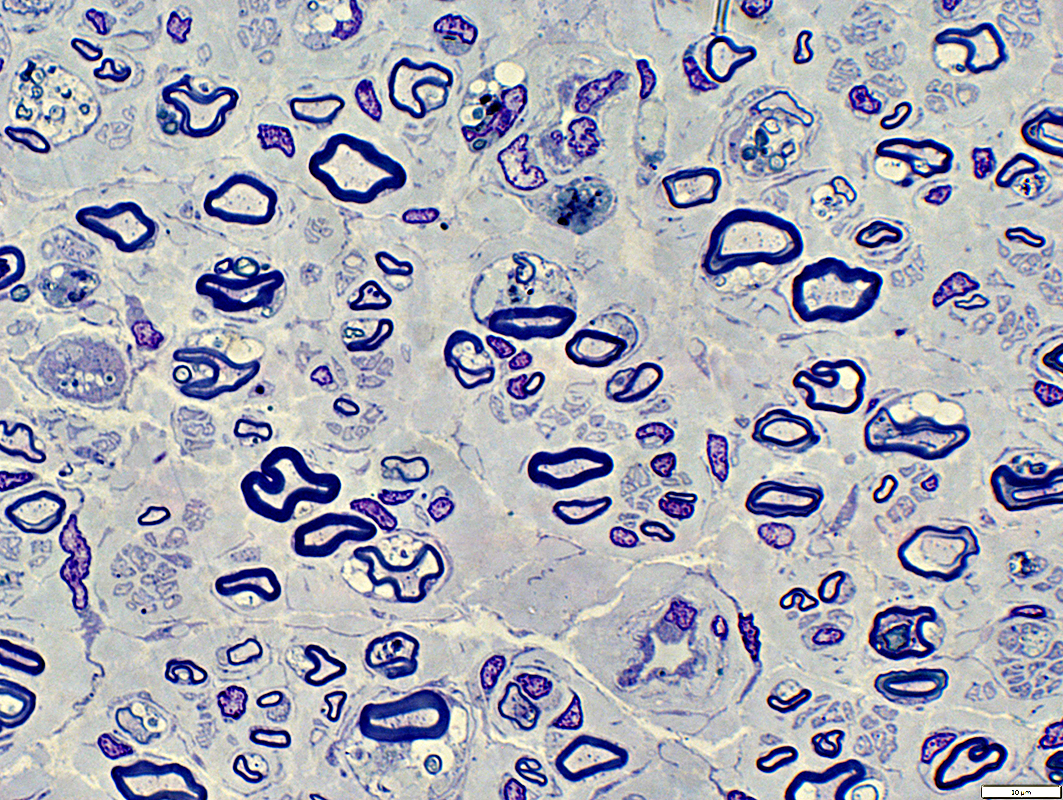
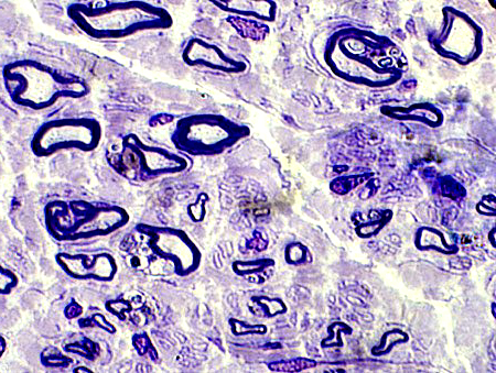
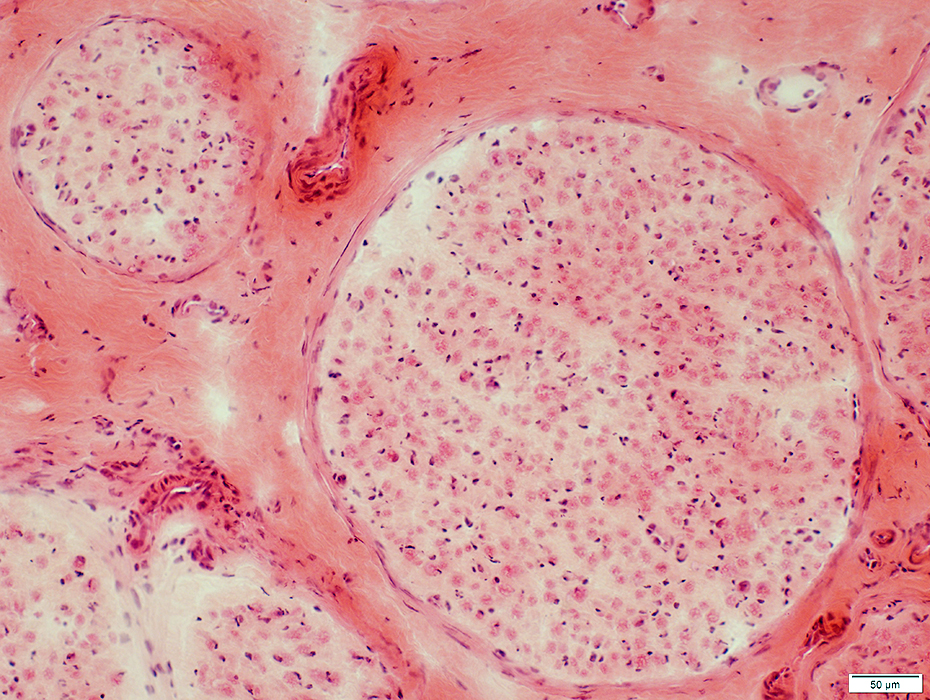
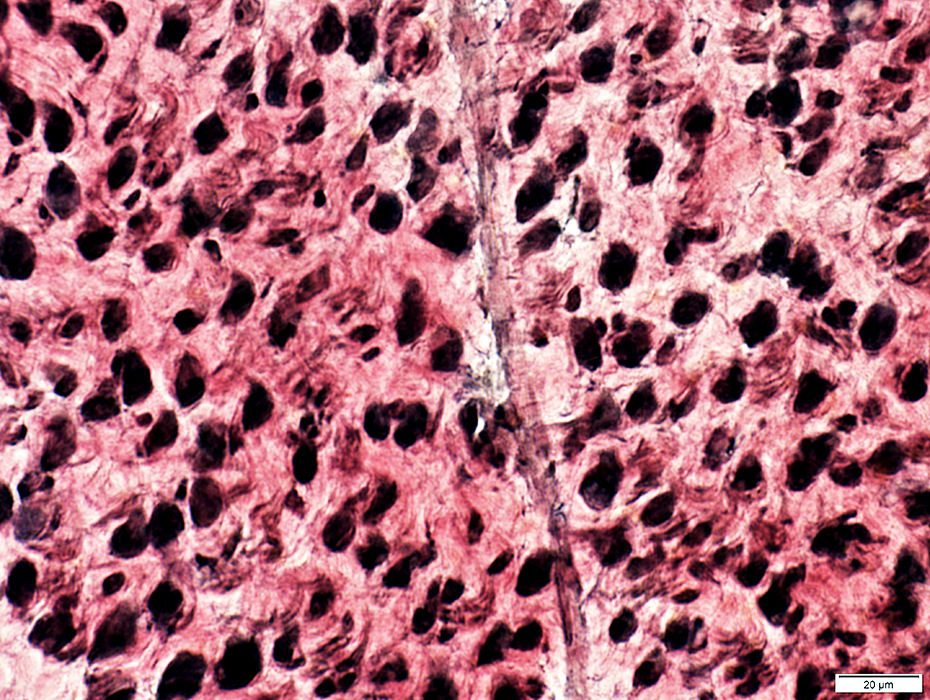
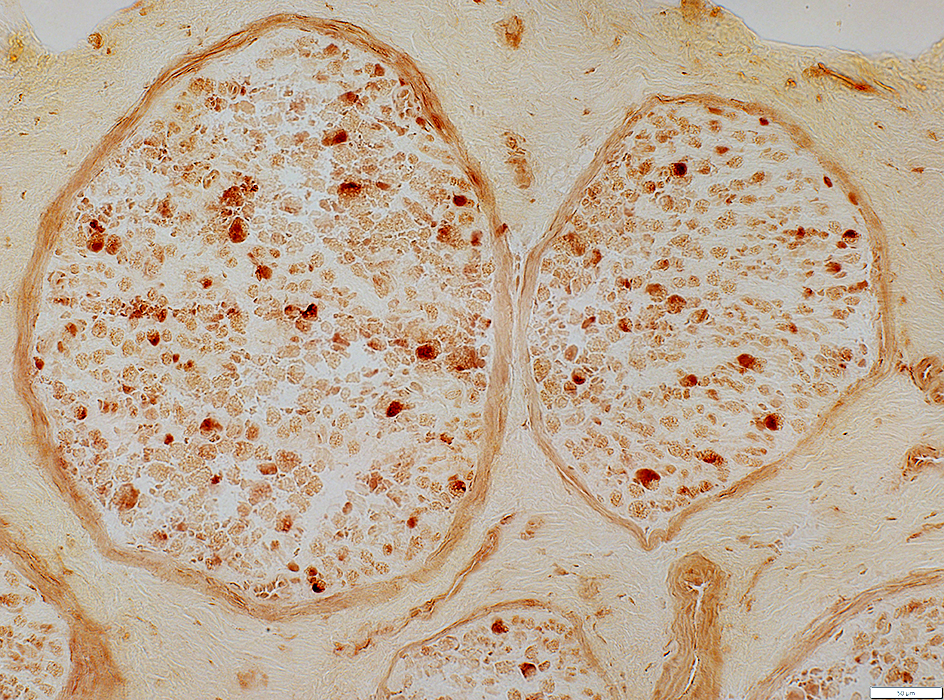
Metachromatic Leukodystrophy (MLD)
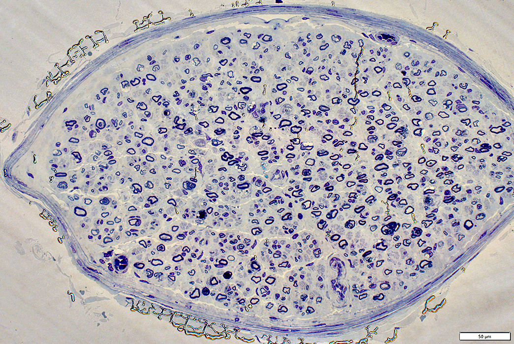 Toluidine blue stain |
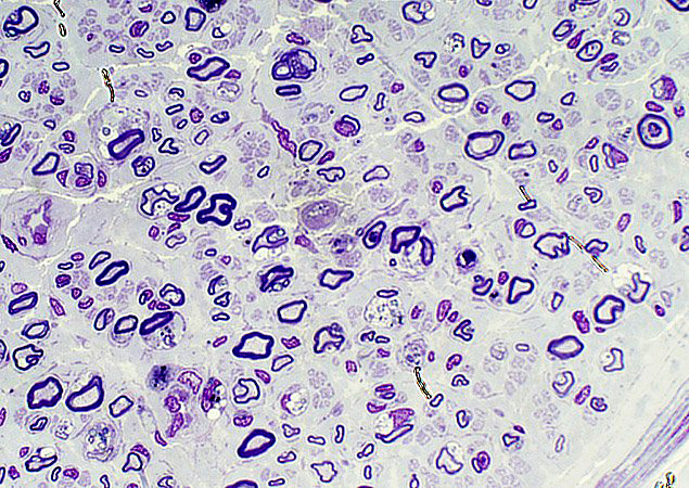 |
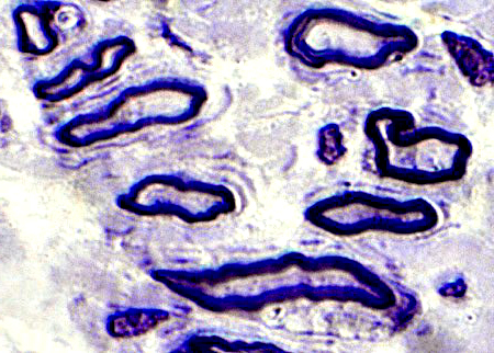
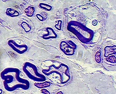
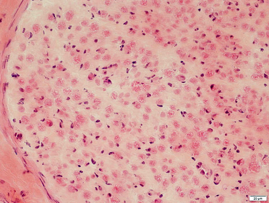
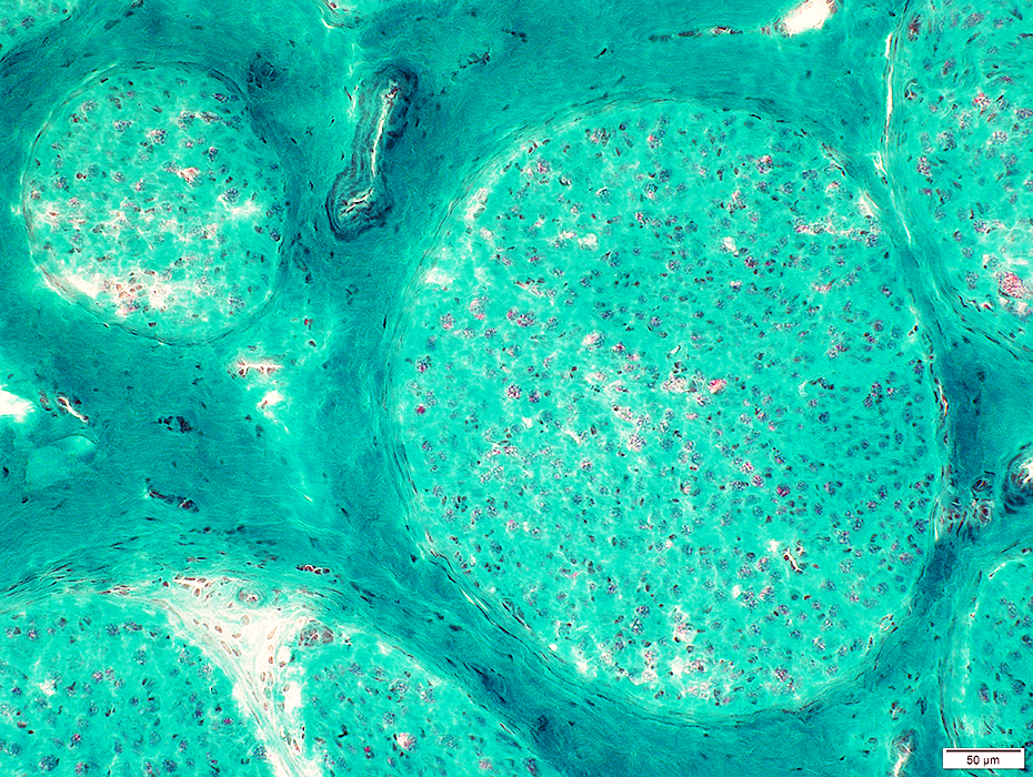
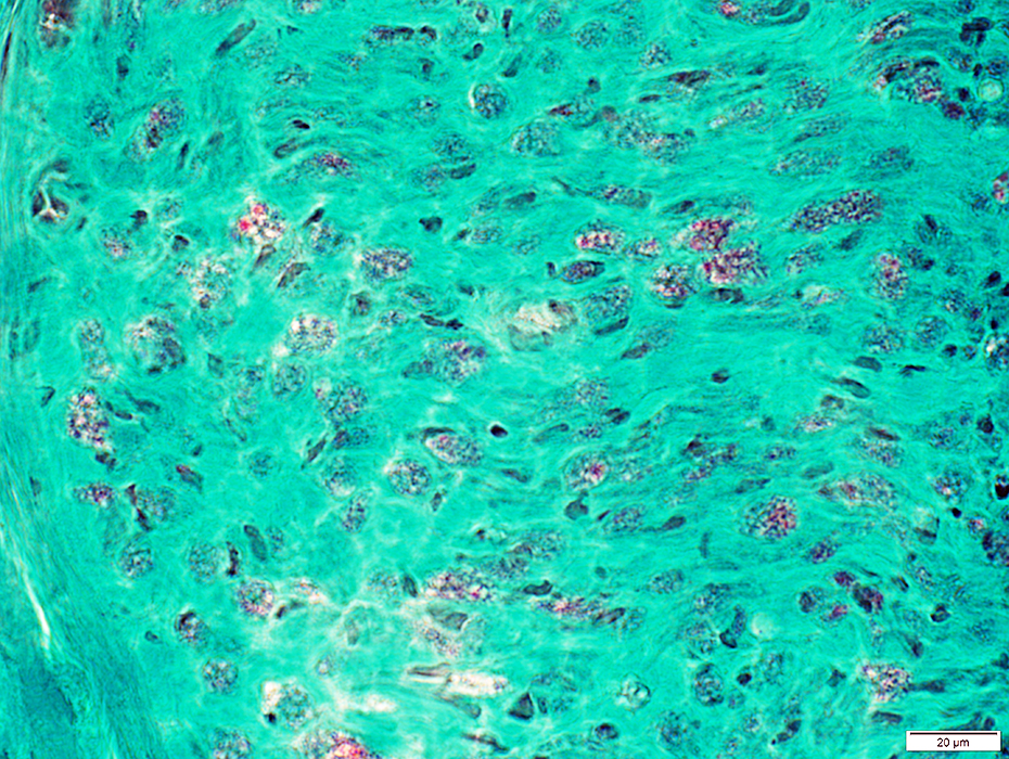
MLD Histochemistry
Pale Increased space between axons
Larger axons Increased space between axons
Smaller axons: Reduced numbers in some endoneurial areas
Lysosomal activity in some endoneurial cells
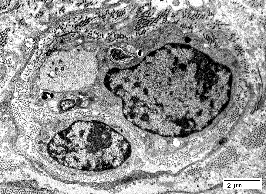
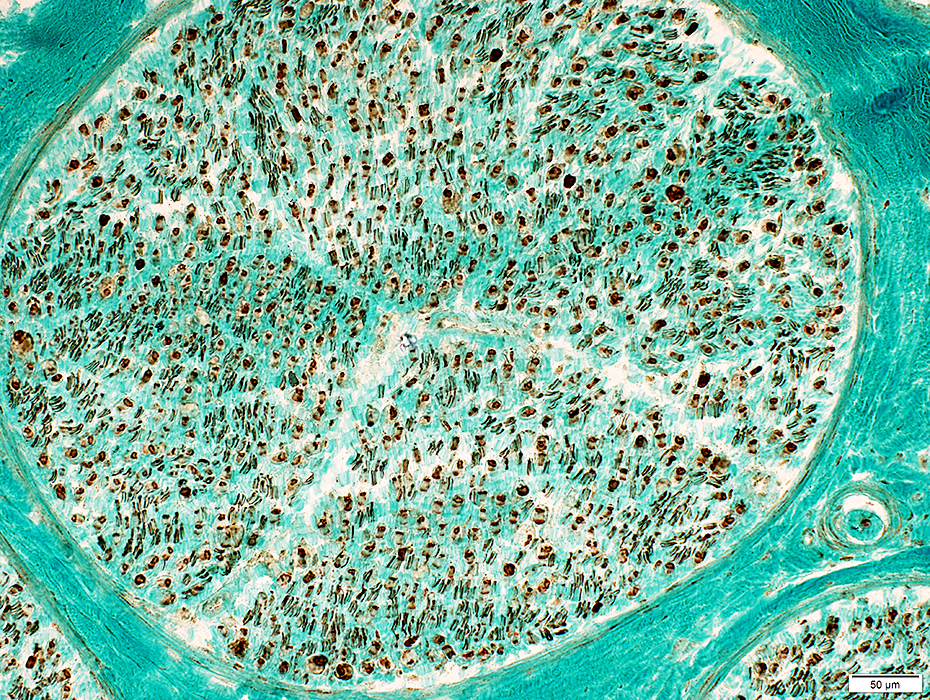
Schwann Cells & Myelin
Contains patches of NCAM stain (Yellow) Bungner Bands (Few) (Yellow) Contain both P0 & NCAM Scattered in endoneurium Small Axons (Yellow) Nearly normal numbers
MBP stains myelin around both large and small axons Normals mainly have MBP in mnyelin around larger axons
Near normal numbers of myelinated (Yellow) and unmyelinated (Green) axons
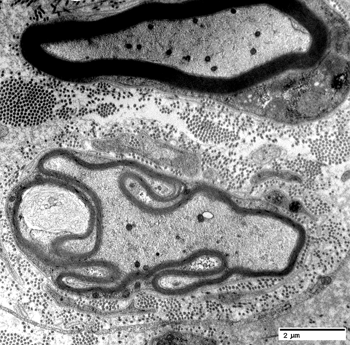
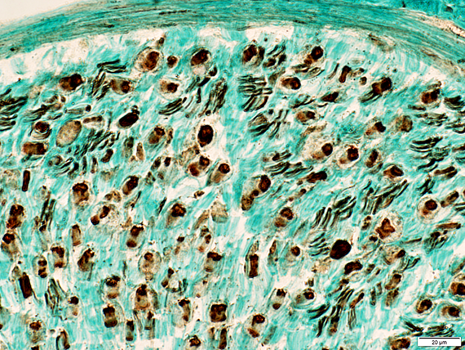
Most myelin, even around smaller axons contains both P0 and MBP (Yellow) Some myelin sheaths have only MBP (Red) MLD: Reduced or Absent Myelin Sheath
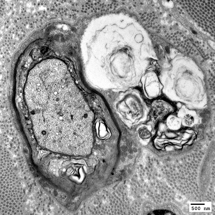
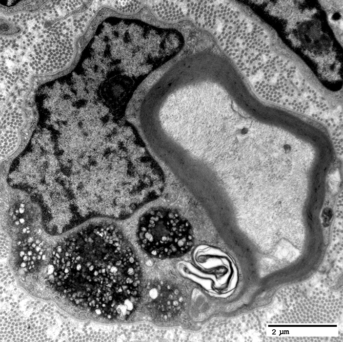
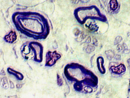
Axons with thin, or folded, myelin sheath
Small, single layer of basal lamina & Schwann cell processes surrounds axons & Schwann cells
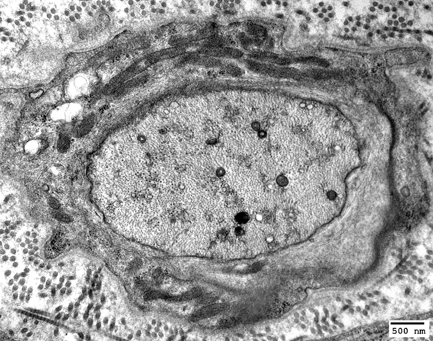
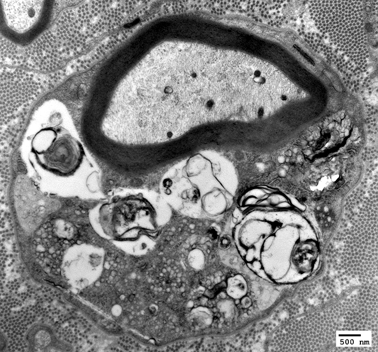
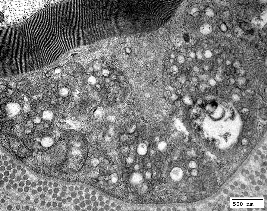
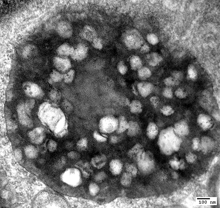
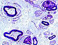
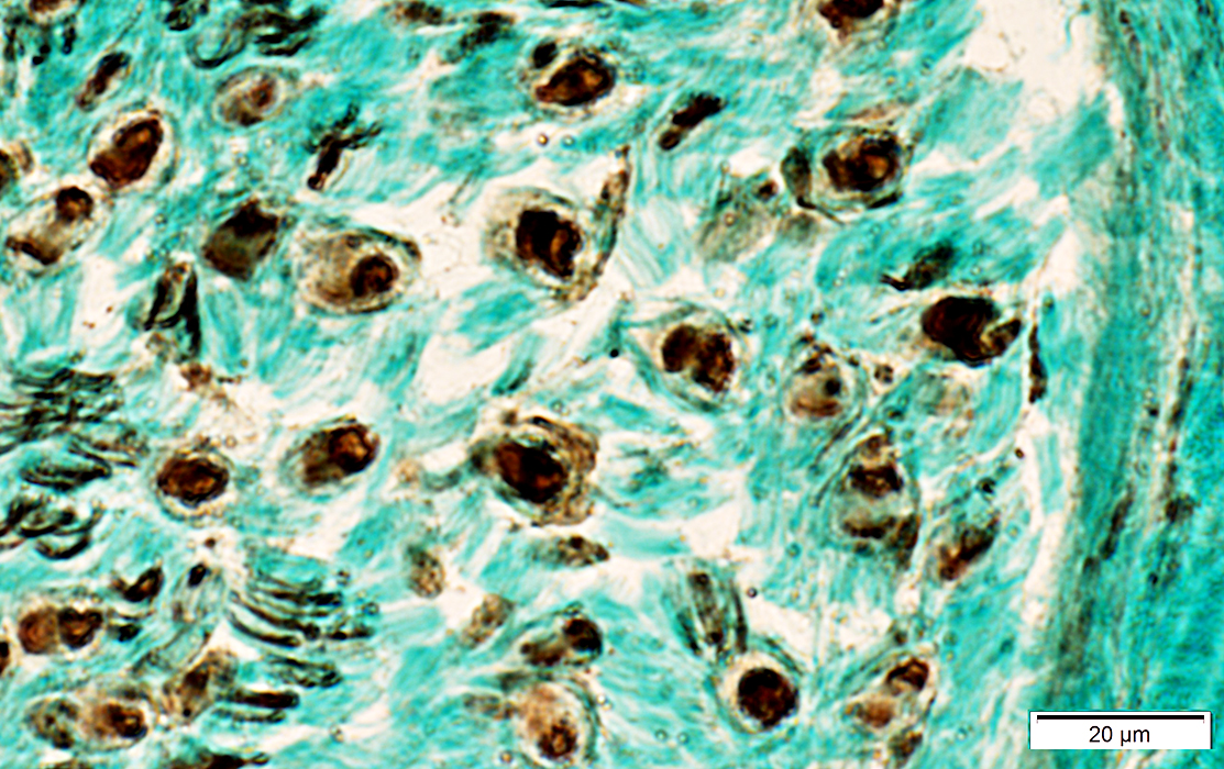
MLD: Schwann cell inclusions
Axons: Moderately or Severely thin myelin sheath Schwann cells: Contain myeloid debris in secondary lysosomes
Schwann cell cytoplasm: Contains myeloid debris in secondary lysosomes
Myelin sheath: Thin
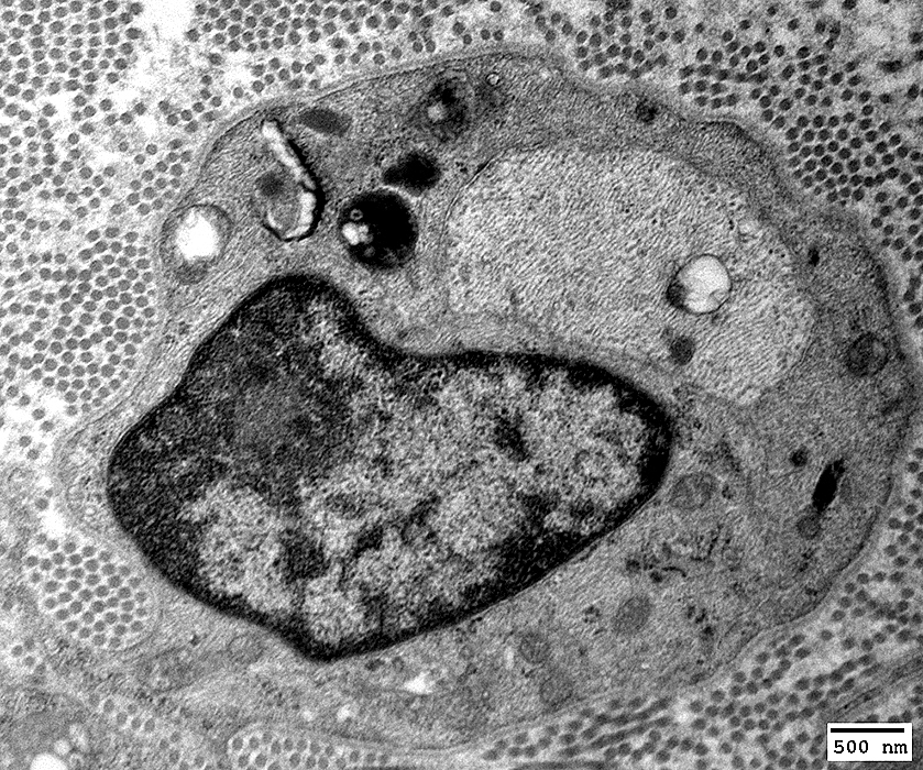
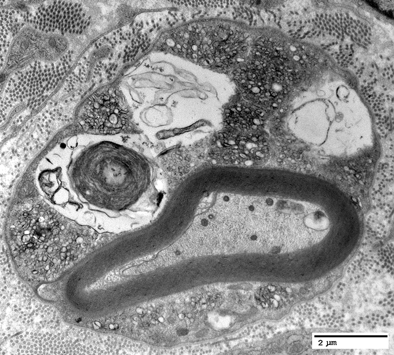
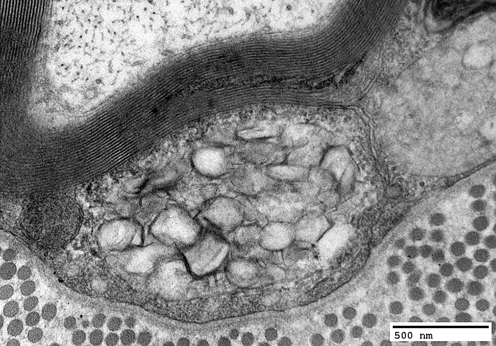
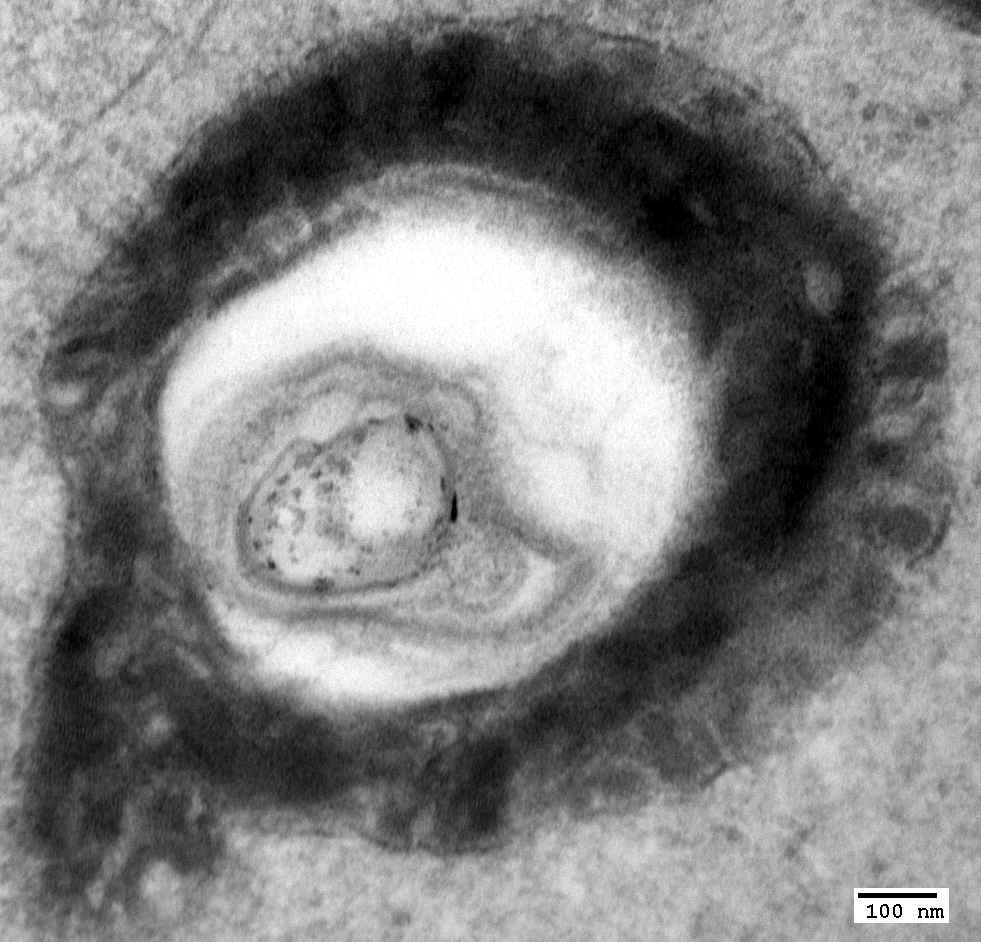
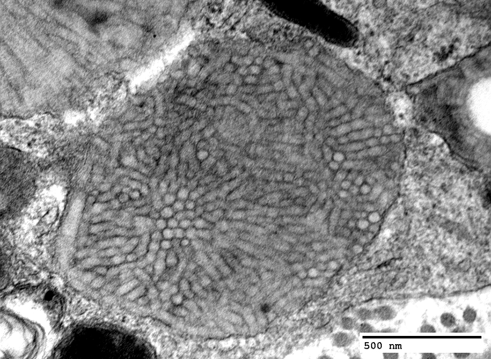
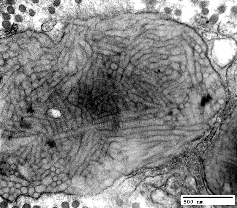
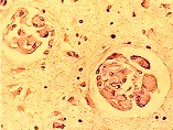
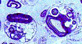
MLD: Tuffstone Bodies
Electron dense: Sharply demarcated Most common in Schwann cells of myelinated & unmyelinated axons Contain Irregularly oriented granular material Areas with 5.8 nm periodicity Peripheral lamellae oriented perpendicular to surface Vacuoles: Electon lucent
Tuffstone bodies
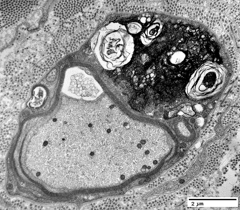
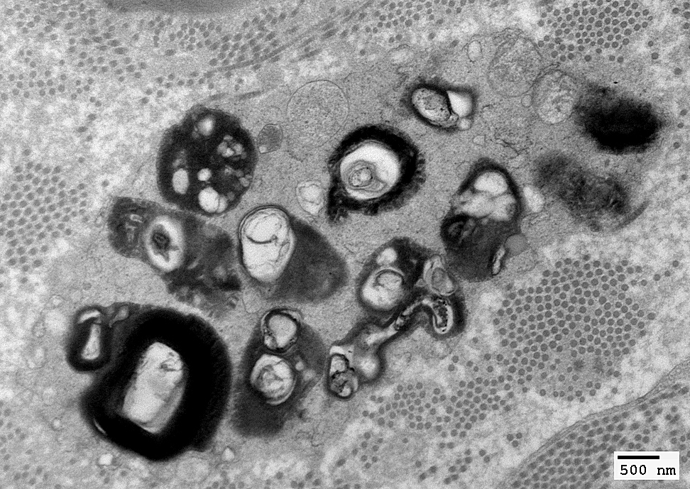
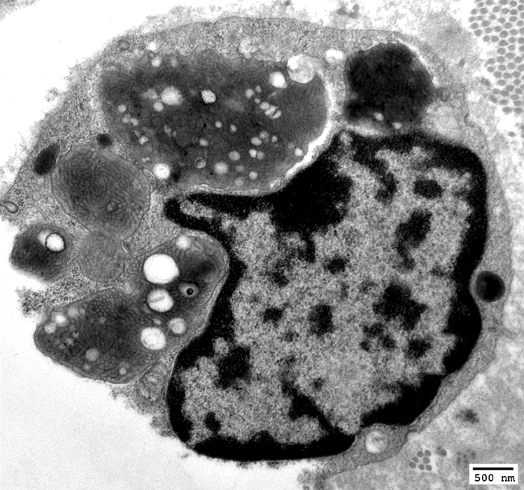
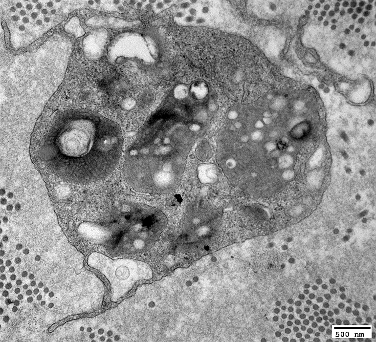
MLD: Macrophages
Commonly in endoneurial macrophages Stacks of lamellar disks
Commonly in endoneurial macrophages Stacks of lamellar disks
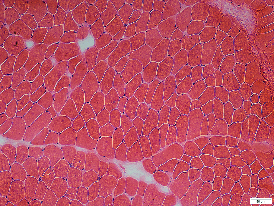
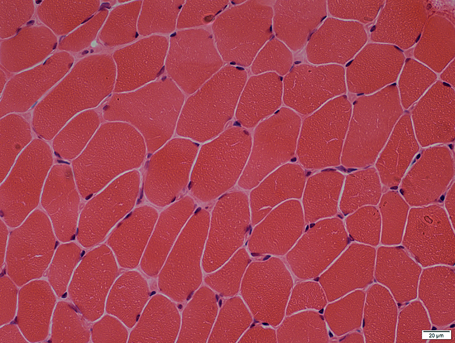
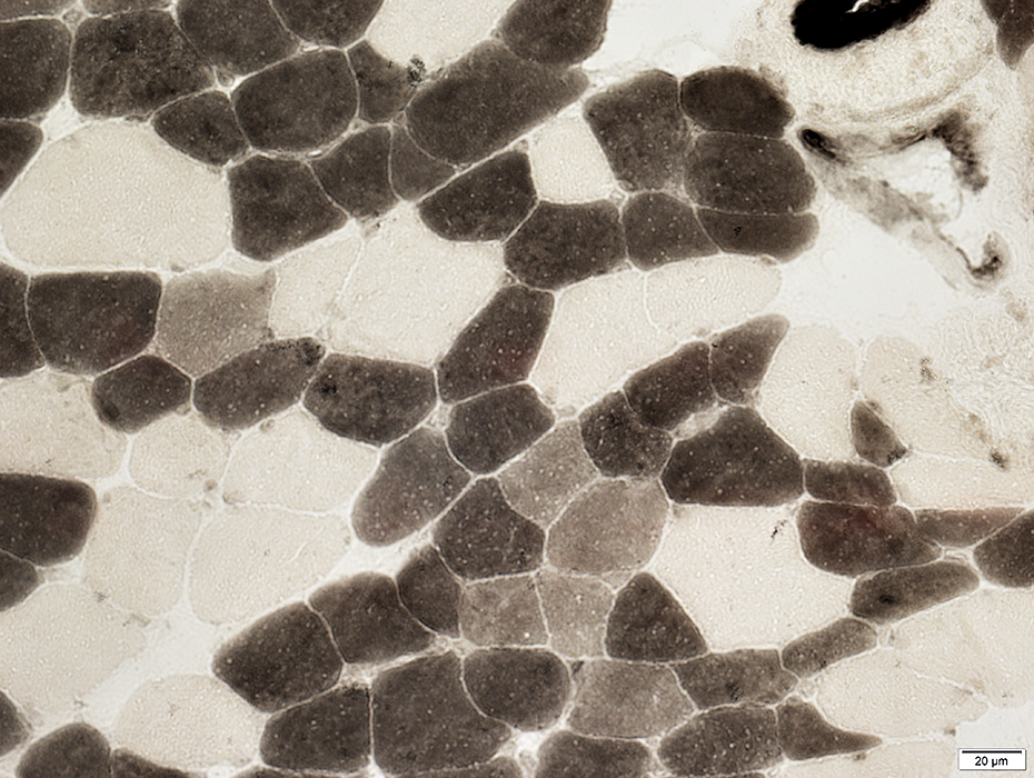
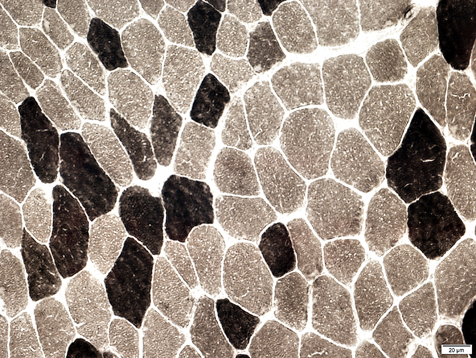
MLD: Muscle
Fiber type grouping: Mild Type 2C: Increased numbers
Return to Normal nerve biosies Return to Biopsy illustrations Return to Neuromuscular Home Page Return to Nerve biopsy Return to Demyelinating neuropathies 8/11/2025 |
P0(r).jpg)
NCAM(r).jpg)
MBP(r).jpg)
P0(r).jpg)
MBP(r).jpg)