mtRNA Leu (MTTL1) A3243G mutations: Pathology
MELAS Syndromes
|
Patients Ages: 2nd & 3rd decades Brain Muscle Age: 67 year old |
Muscle Pathology: Young Adult
|
Muscle fibers Perimysial vessels |
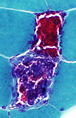
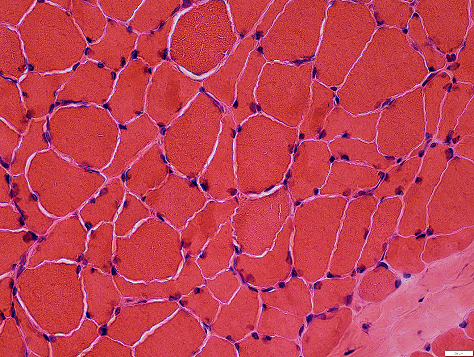
Ragged Red Fibers: Scattered
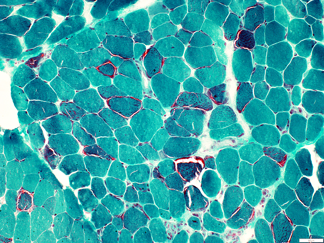 Gomori trichrome stain |
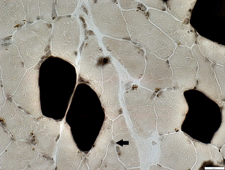
Scattered fibers with clear rim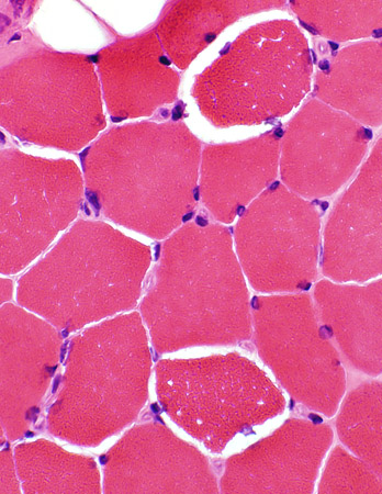 H&E stain |
"Ragged red" fibers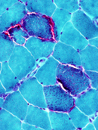 Gomori trichrome stain |
 Gomori trichrome stain |
 Gomori trichrome stain |
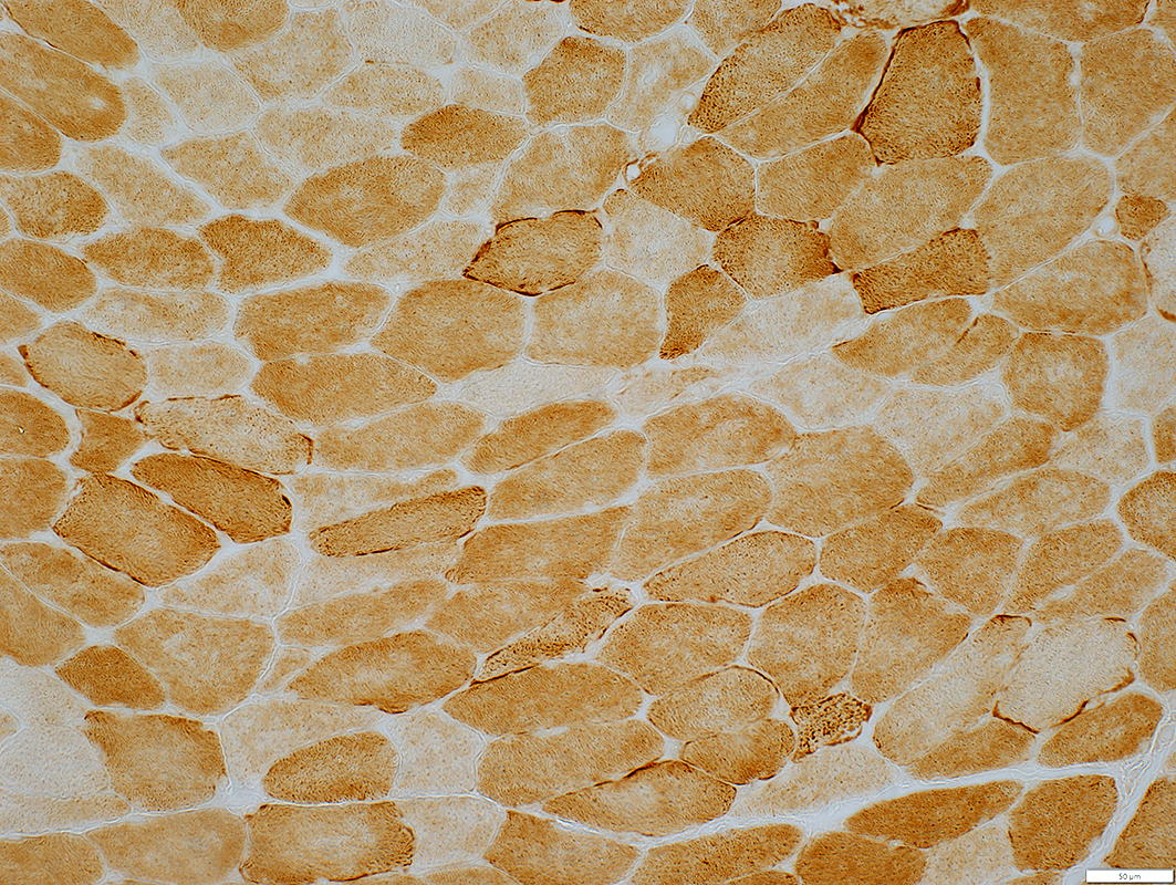
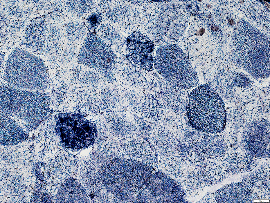
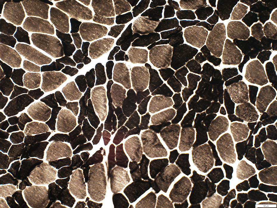
Mitochondrial Proliferation: Scattered muscle fibers with Increased SDH staining
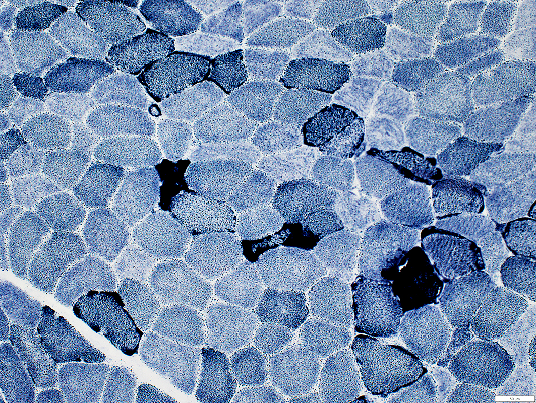 SDH stain |
|
Muscle Fibers: Increased SDH stain Scattered through muscle Variable degrees 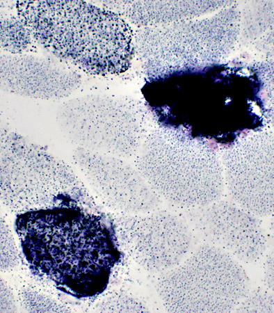 |
Muscle fiber: Increased SDH stain: Individual mitochondria within muscle fiber are enlarged compared to surrounding fibers 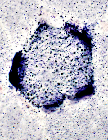 |
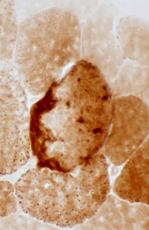
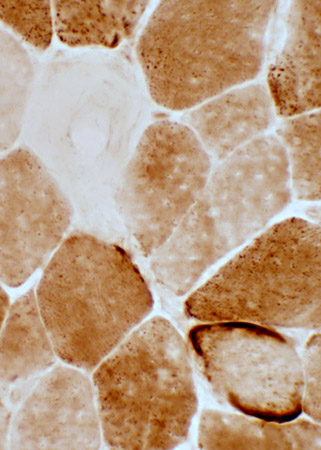
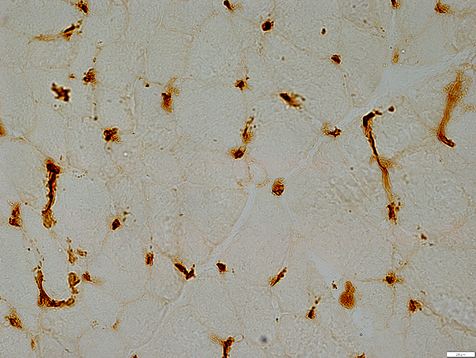
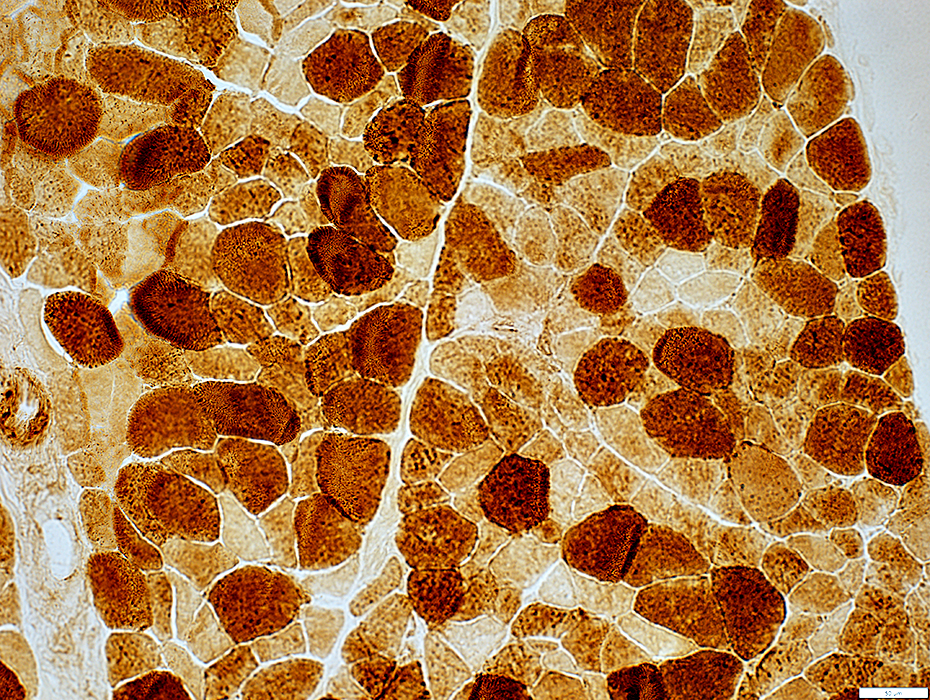
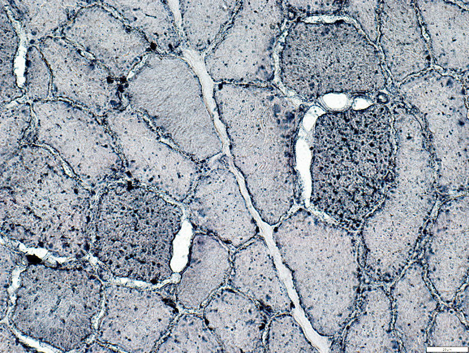
Mitochondrial Proliferation: Scattered muscle fibers with Increased COX staining
 Cytochrome oxidase (COX) stain |
|
|
 Succinate dehydrogenase (SDH) stain 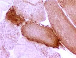 Cytochrome oxidase (COX) stain |
Muscle fibers with increased SDH stain may also have increased COX stain
 Succinic dehydrogenase (SDH) stain |
 COX stain |
|
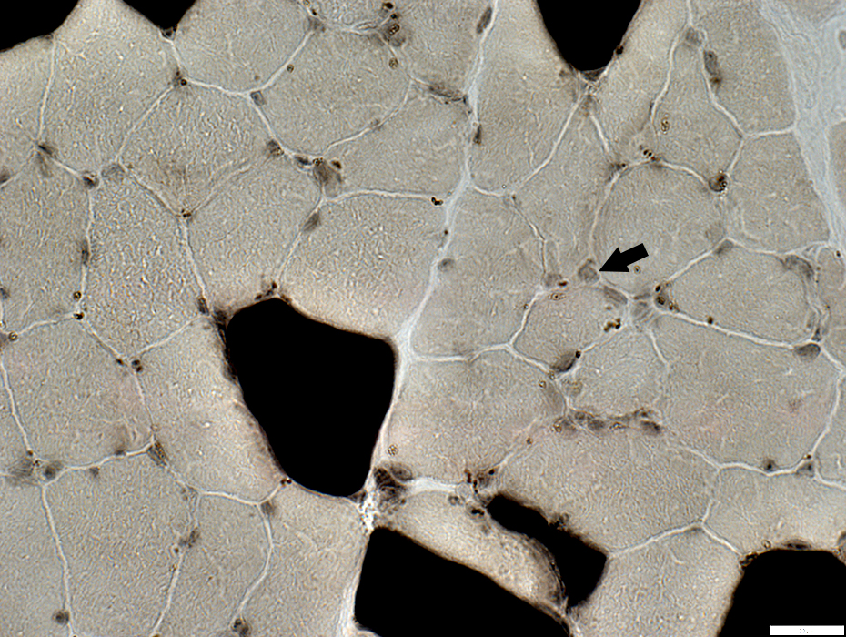
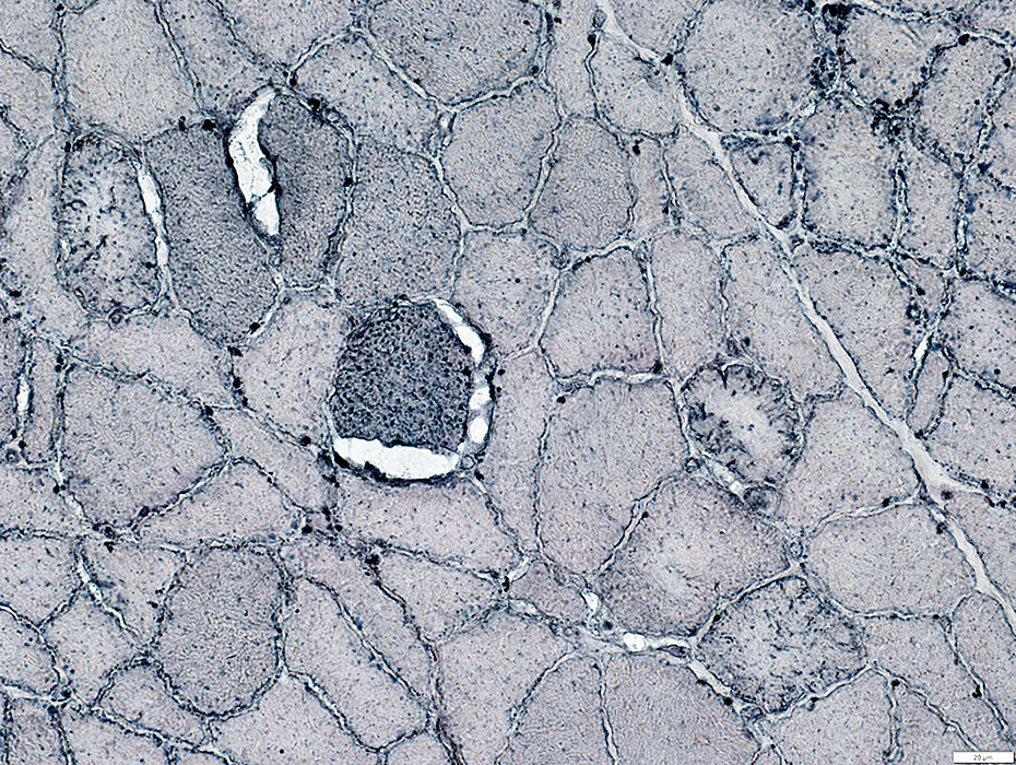
Cytochrome Oxidase (COX) stain: Reduced or Increased Muscle fibers may have reduced, normal or increased COX staining (Right) 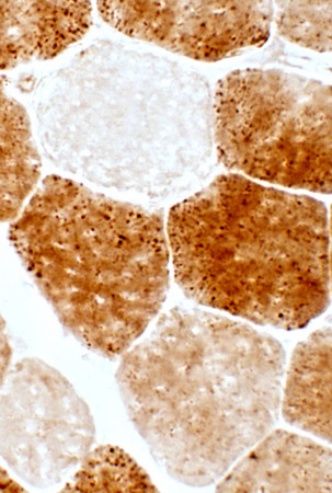
|
Lipid accumulation Increased Sudan black stain in muscle fiber with mitochondrial proliferation 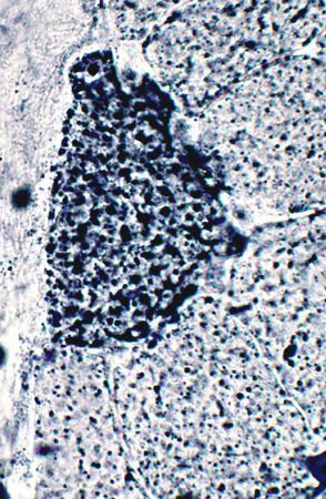 |
|
COX stain |
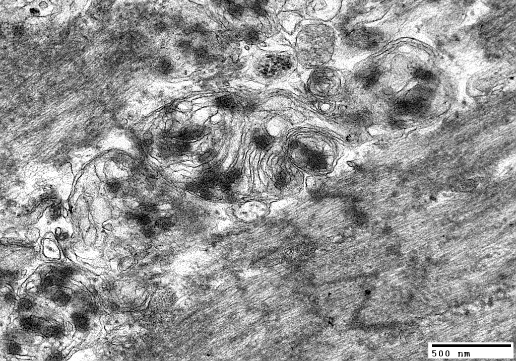
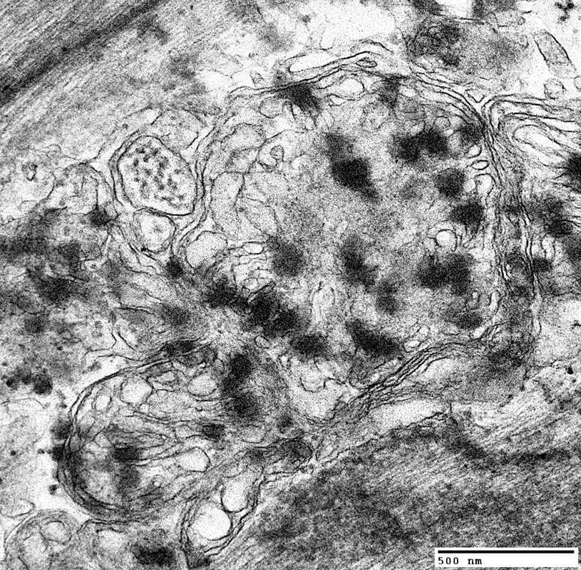
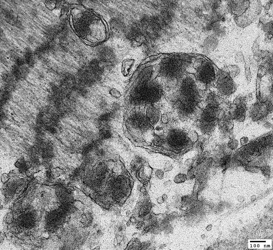
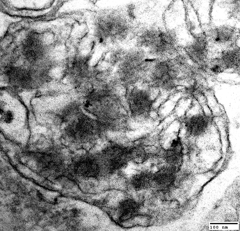
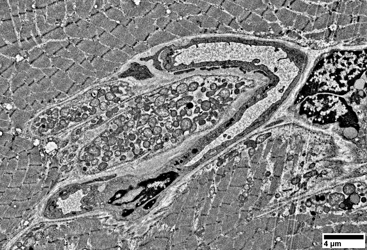
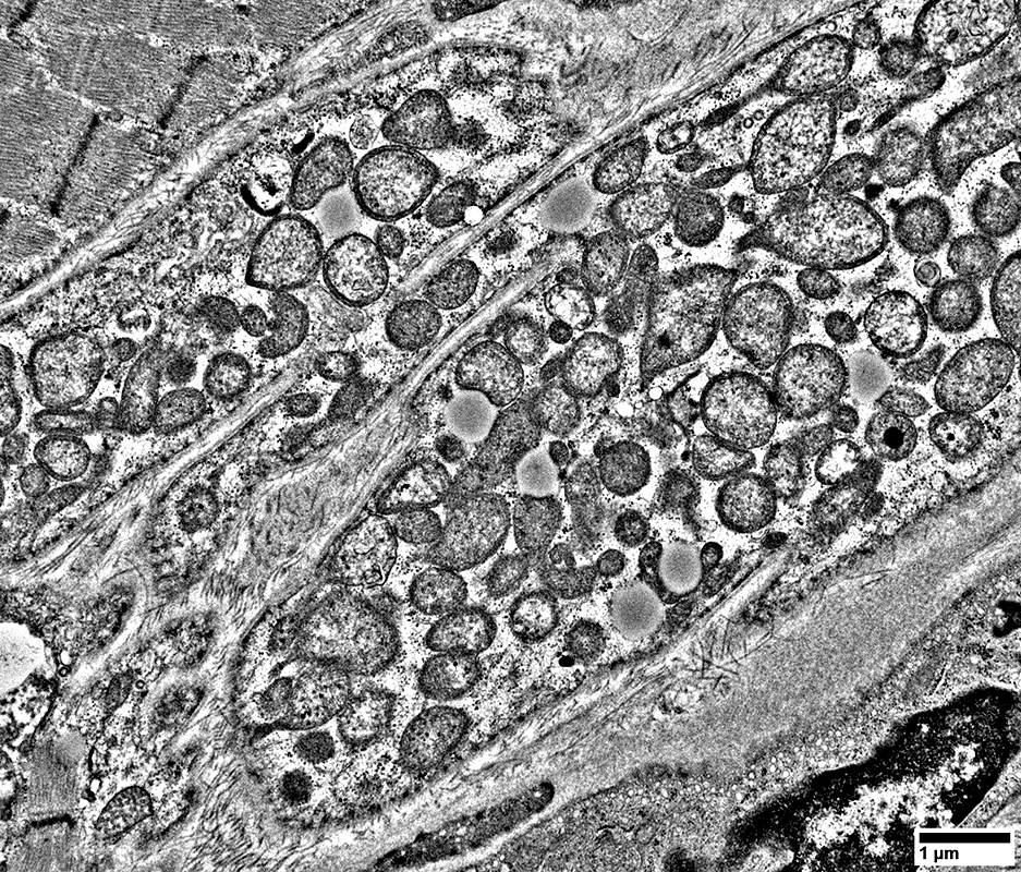
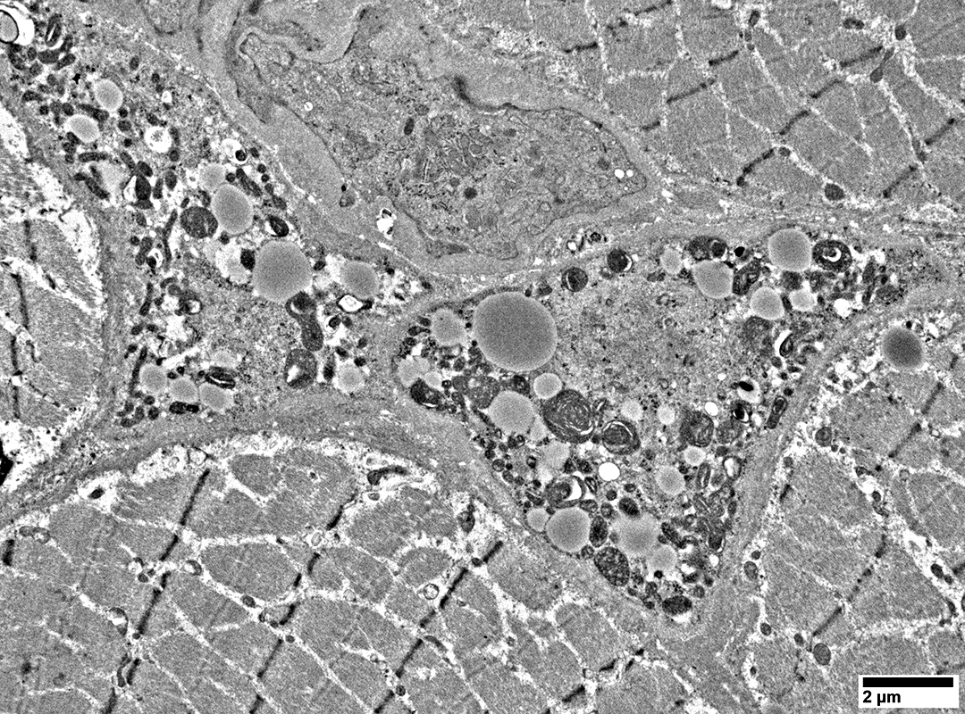
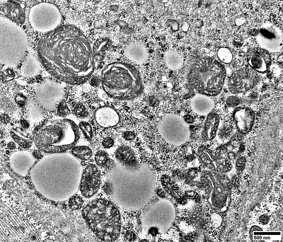
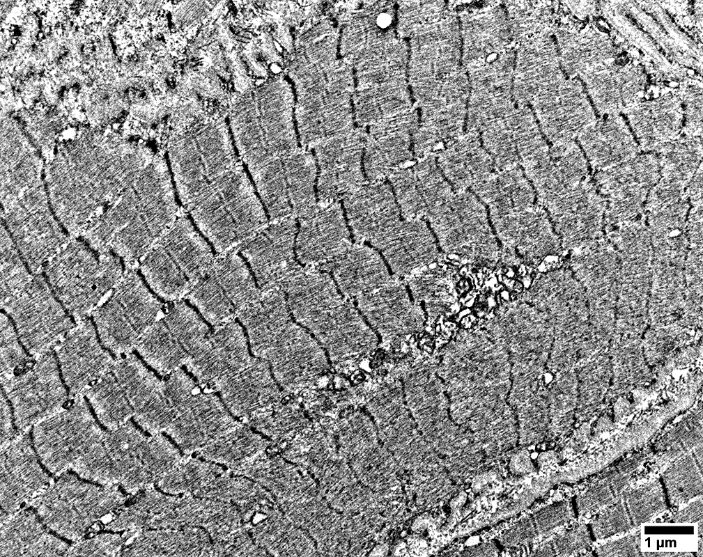
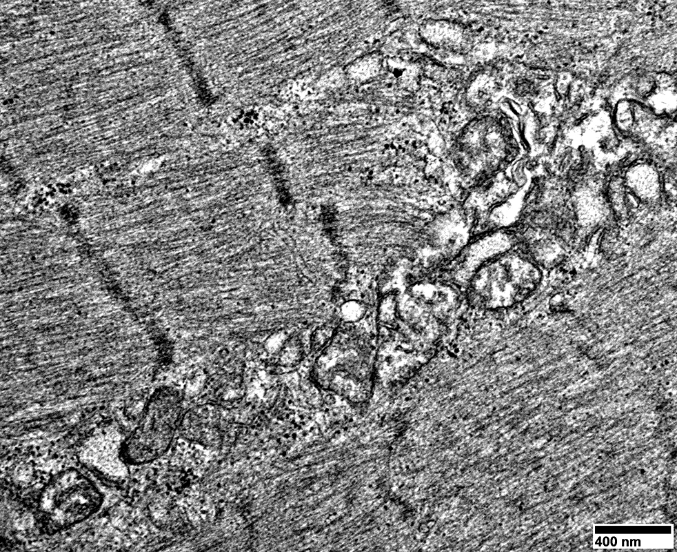
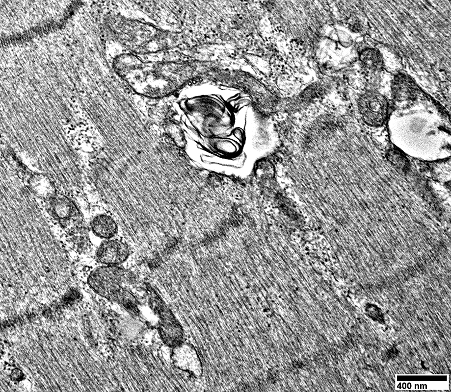
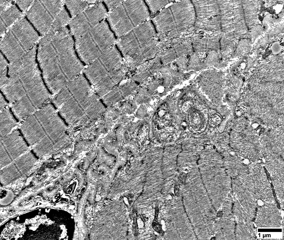
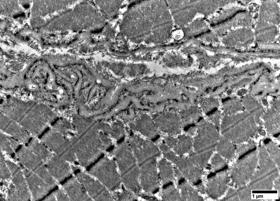
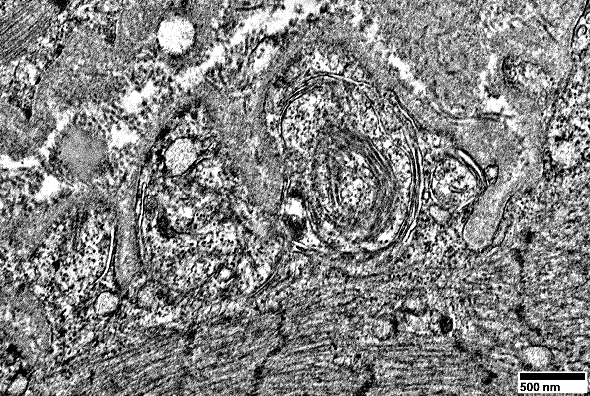
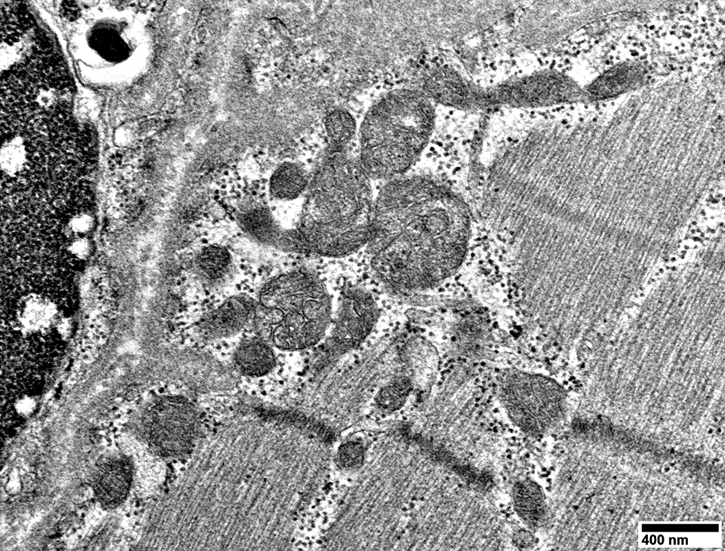
MELAS: Mitochondrial Ultrastructure
 From: R Schmidt |
 From: R Schmidt |
 From: R Schmidt |
 From: R Schmidt |
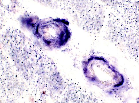
MELAS Vessels
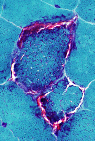
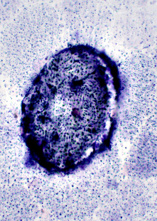
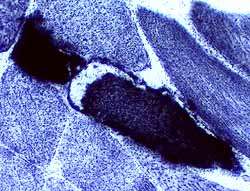
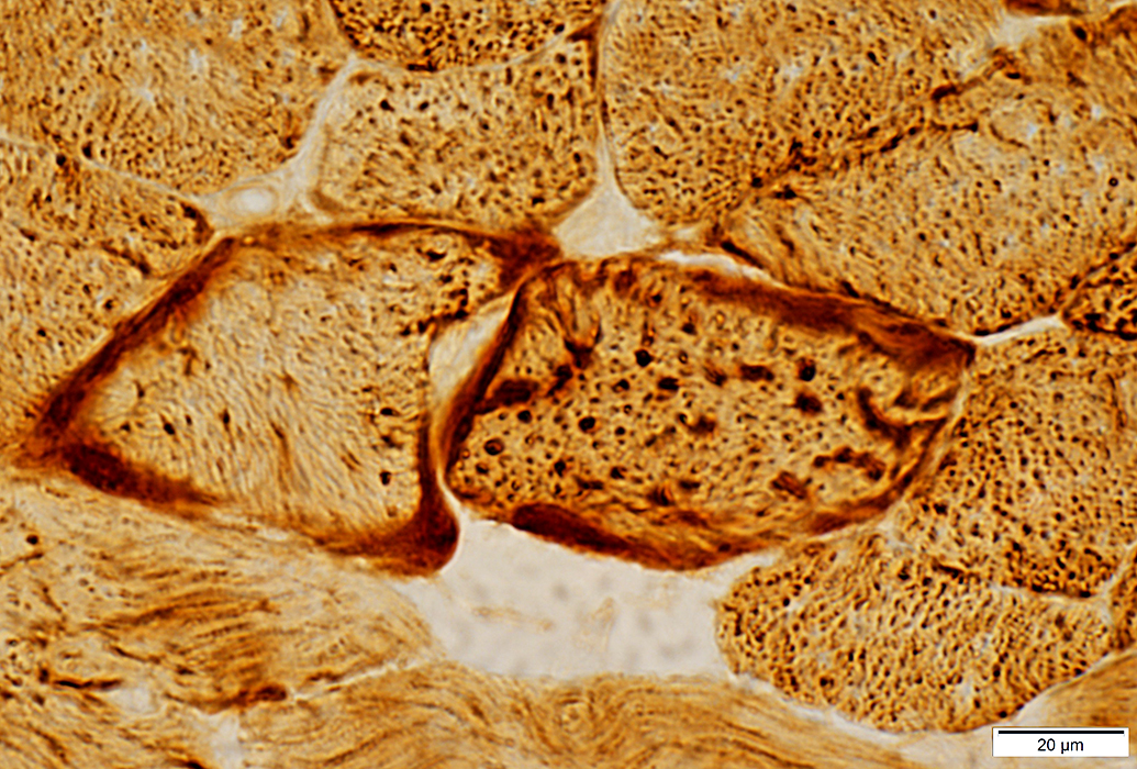
SDH staining of intramuscular vessels is Increased
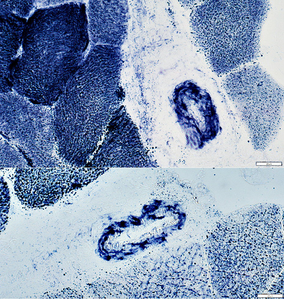 Succinic dehydrogenase (SDH) stain |
Medium-sized perimysial vessels have increased SDH staining
Contrast with normal staining in MNGIE & Control
 Succinic dehydrogenase (SDH) stain 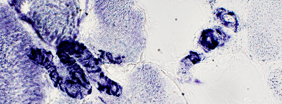
|
Normal: Mild SDH staining of medium sized perimysial vessels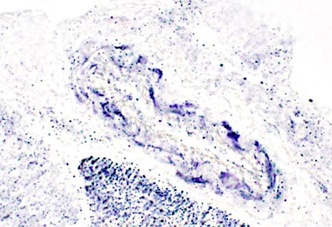
|
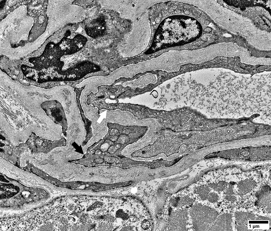
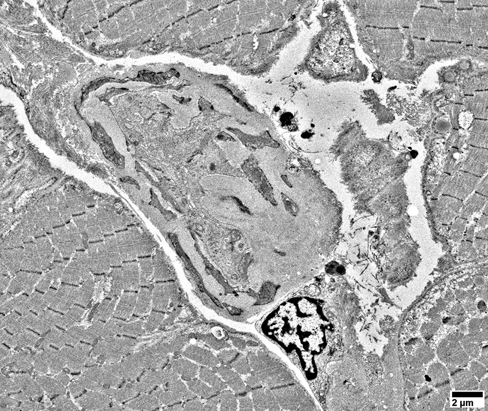
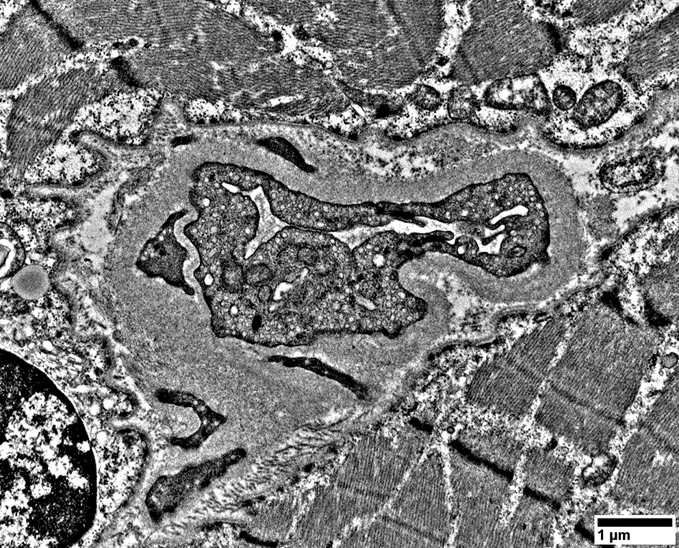
MELAS: Capillaries
 From: R Schmidt |
 From: R Schmidt |
 From: R Schmidt |
 From: R Schmidt |
Brain pathology in MELAS |
MRIs during "stroke-like" episodes
| |
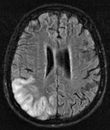 FLAIR image |
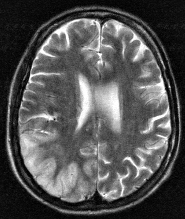 T2 weighted image |
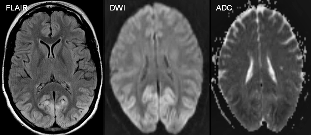 From: Cindy Ly |
| CT during episode of homonymous hemianopia | |||
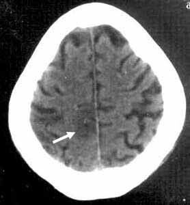 Medial occipital lesion (Arrow) |
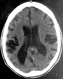 Enlarged ventricles |
Late in disease course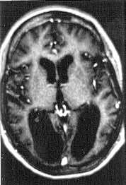 MRI: Severe involvement of occipital cortex |
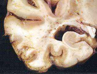 Temporal gray matter: Severely abnormal Temporal horn of Ventricles: Enlarged. |
MELAS: 67 year-old female
mtDNA A3243G 66% heteroplasmyOther disorders: Diabetes; Renal disease, End-stage
|
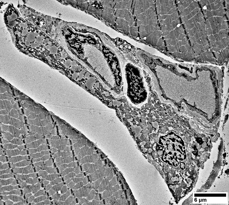
Capillaries Larger vessels Muscle fibers |
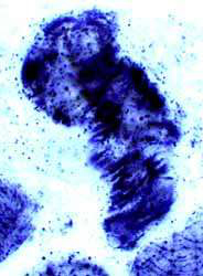
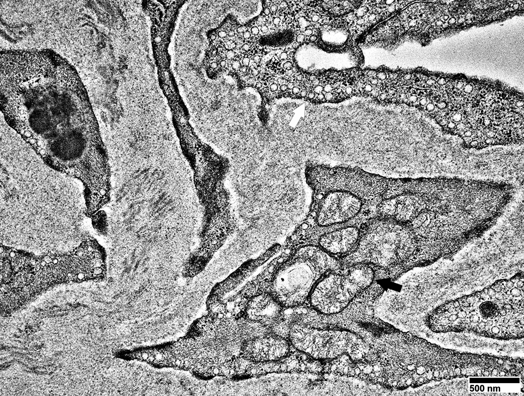
Vessel Pathology
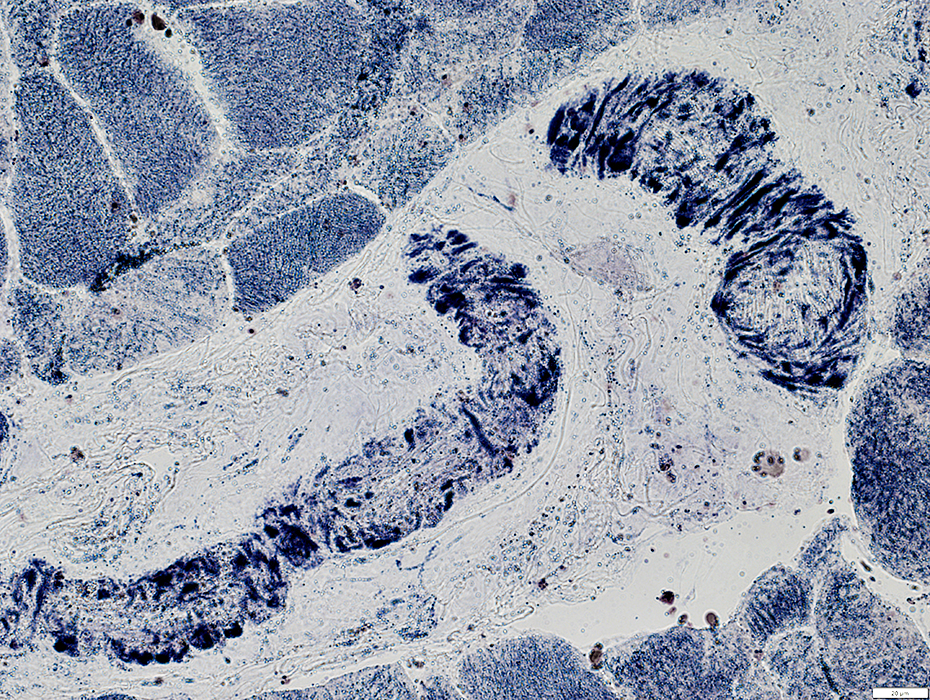 SDH stain |
Punctate dark stainin in walls
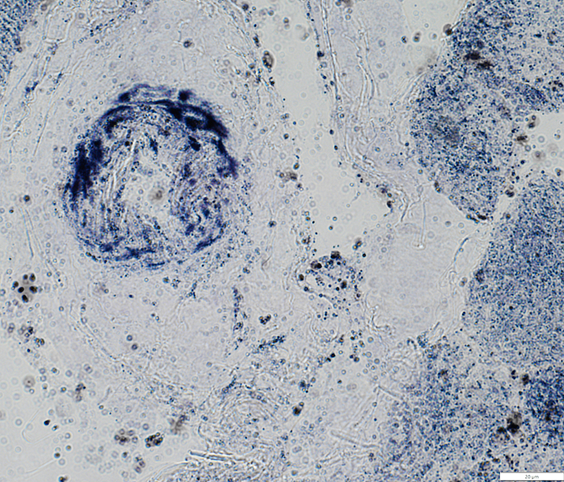 SDH stain |
 From: R Schmidt |
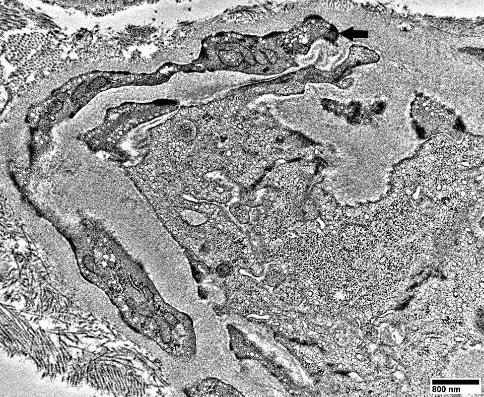
Endothelial cells Multiple small vacuoles (White arrows)
 From: R Schmidt |
 From: R Schmidt |
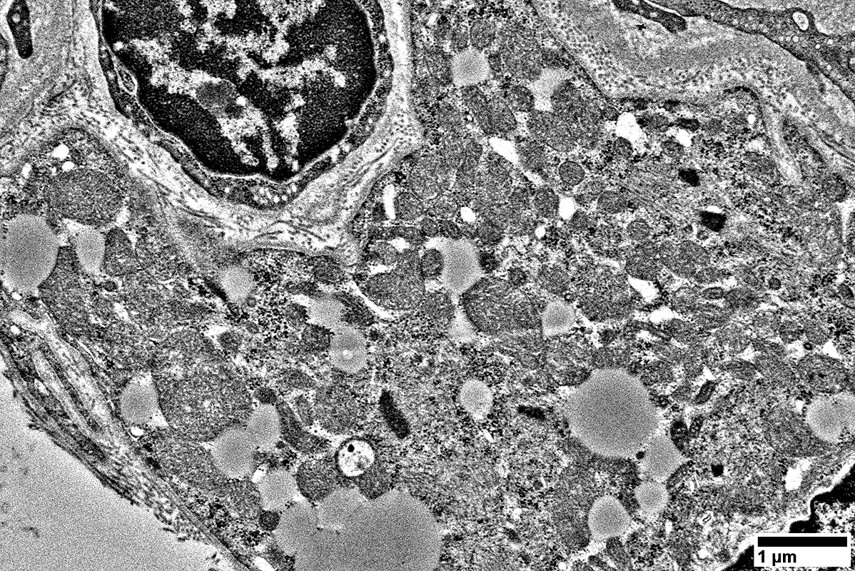
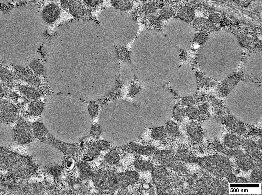
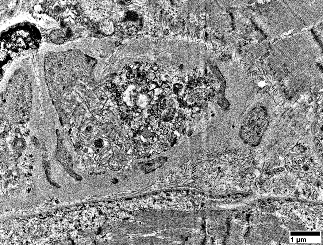
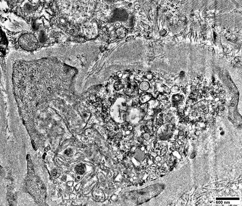
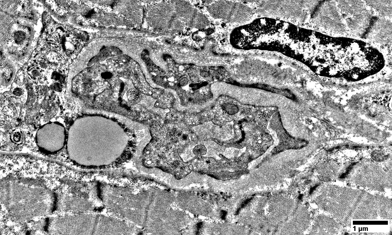
Mitochondrial proliferation & enlargement
Lipid droplets
 From: R Schmidt |
 From: R Schmidt |
 From: R Schmidt |
Mitochondrial proliferation & enlargement
Lipid droplets
 From: R Schmidt |
 From: R Schmidt |
Mitochondrial proliferation & enlargement
Lipid droplets
 From: R Schmidt |
 From: R Schmidt |
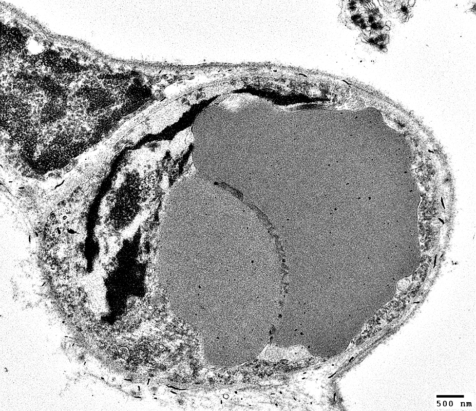
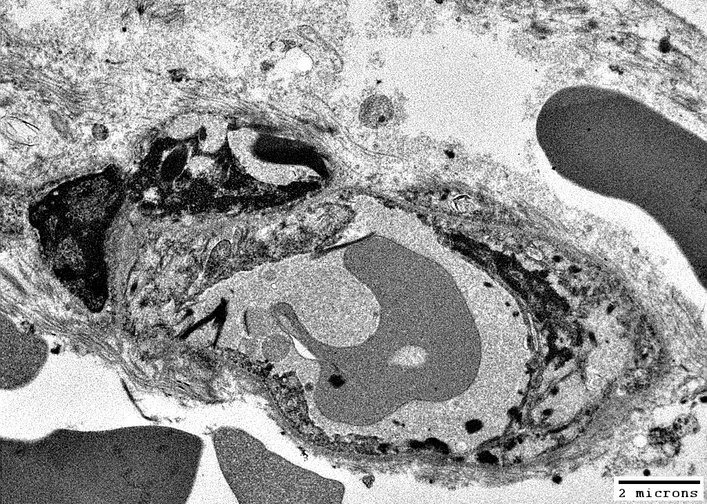
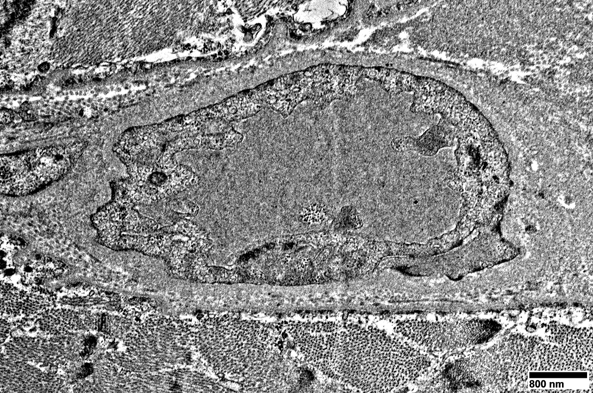
Large endothelial cells with many vesicles
No lumen
Many smooth muscle cells in wall, some with mitochondrial proliferation (Arrow)
Thick wall
 From: R Schmidt |
 From: R Schmidt |
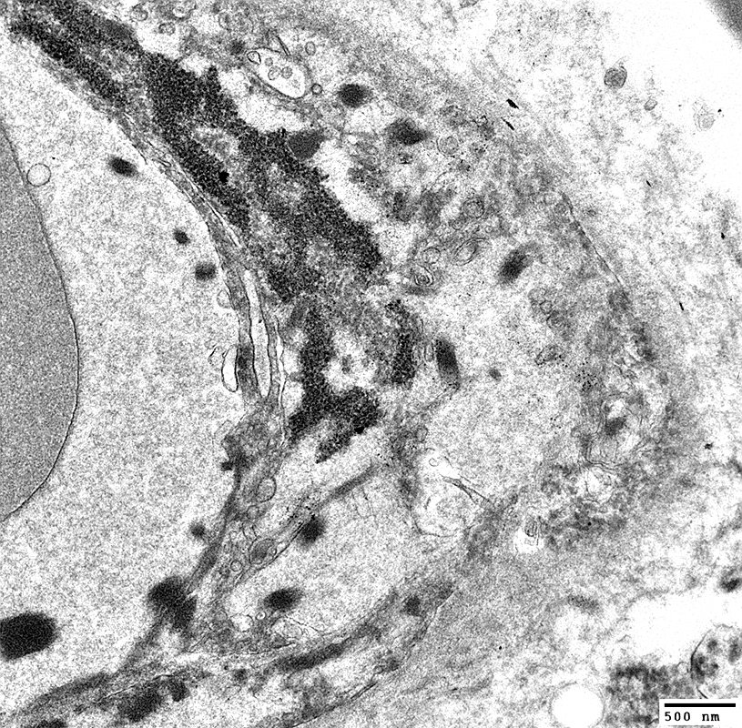
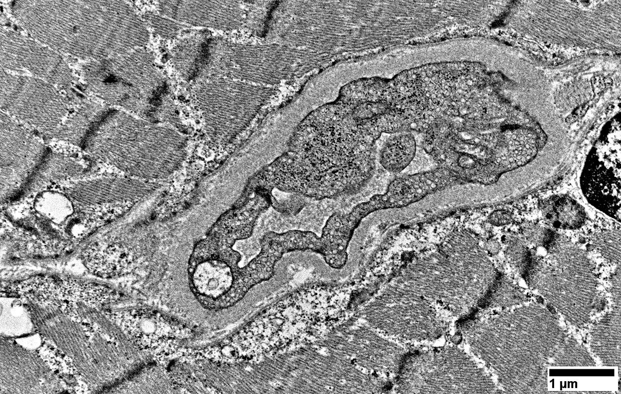
Large endothelial cell with many vesicles
No lumen
Scattered smooth muscle cells in wall
Thick wall
 From: R Schmidt |
 ATPase pH 4.3 |
Pale staining by ATPase pH 4.3 (Arrows)
Large size
 ATPase pH 4.3 |
 UEA-I |
Large size
 From: R Schmidt |
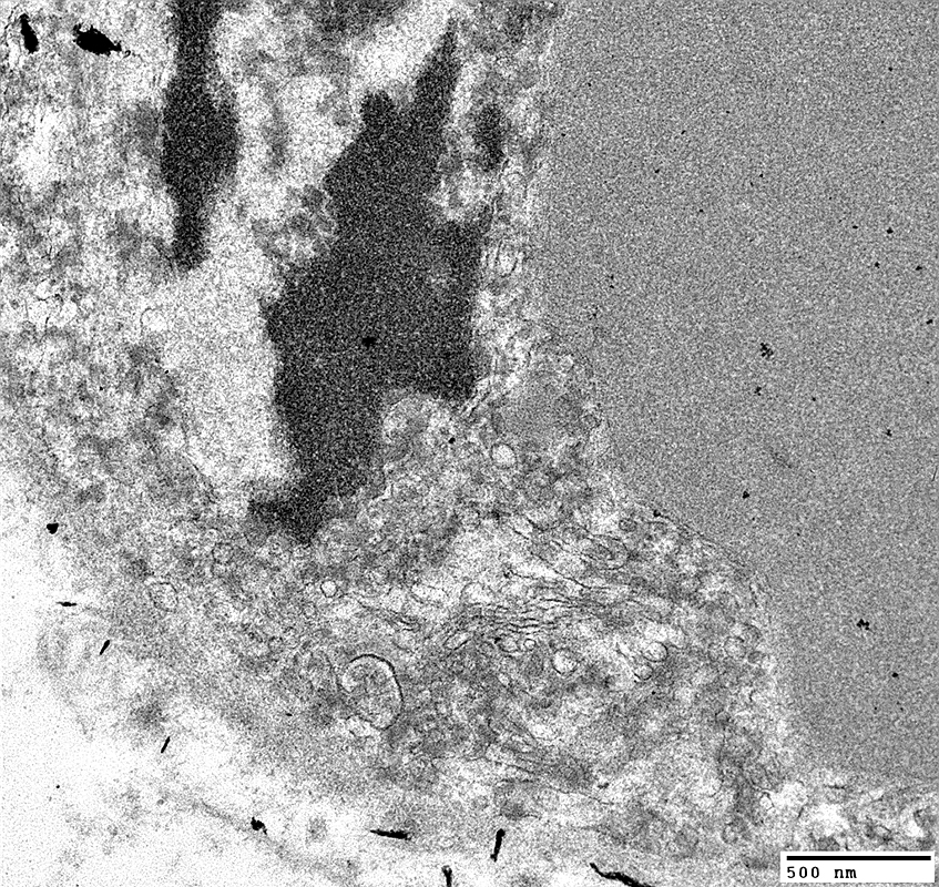
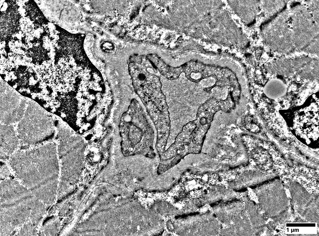
Endothelial cells with many vesicles
Large size
Thick wall
 From: R Schmidt |
 From: R Schmidt |
Endothelial cells with many vesicles
Large size
Thick wall
 From: R Schmidt |
 From: R Schmidt |
MELAS: Muscle Fibers
 H&E stain |
Bimodal variation
 SDH stain |
Dark stained cytoplasm
 COX stain |
Pale stained cytoplasm
 Sudan black stain |
 Sudan black stain |
 ATPase pH 9.4 stain |
 From: R Schmidt |
 From: R Schmidt |
 From: R Schmidt |
 From: R Schmidt |
Thick
Undulating
 From: R Schmidt |
 From: R Schmidt |
 From: R Schmidt |
Return to Mitochondrial pathology
Return to Mitochondrial syndromes
Return to Muscle biopsies
12/1/2025