MNGIE: Muscle Pathology
|
Histochemistry Ultrastructure |
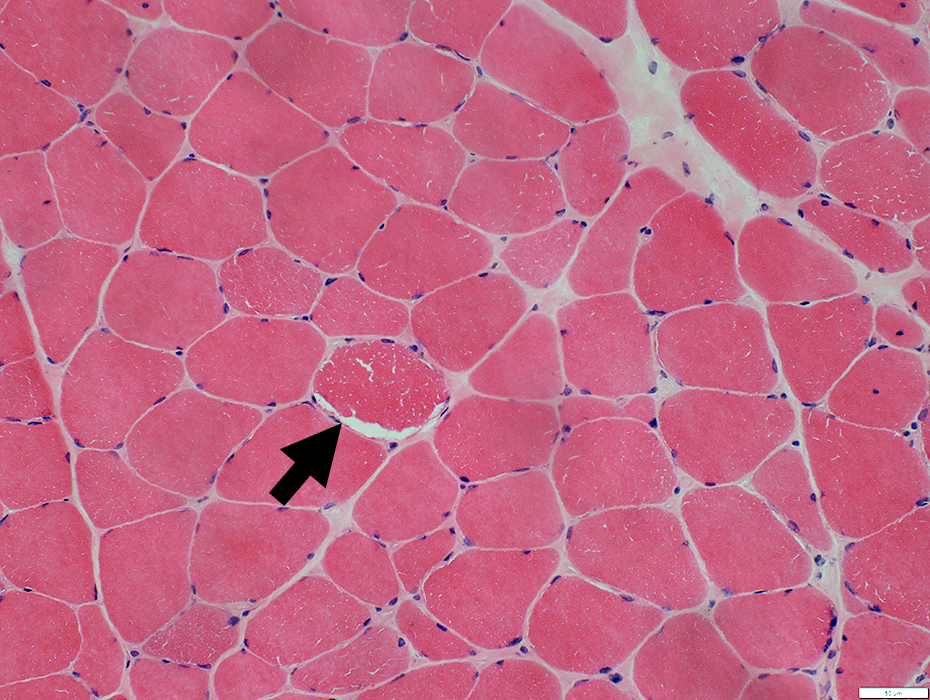
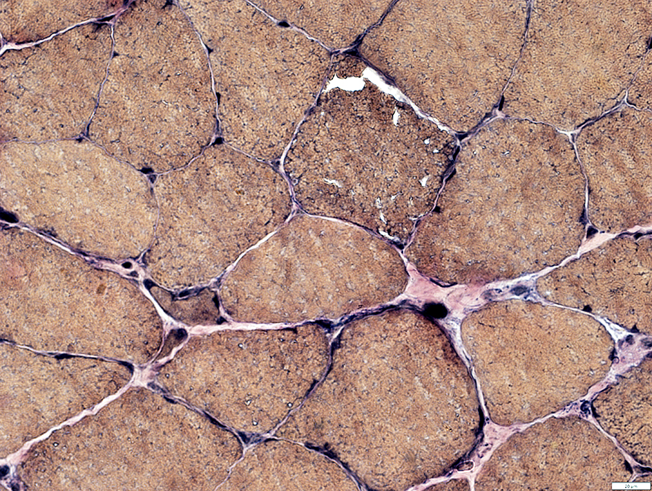
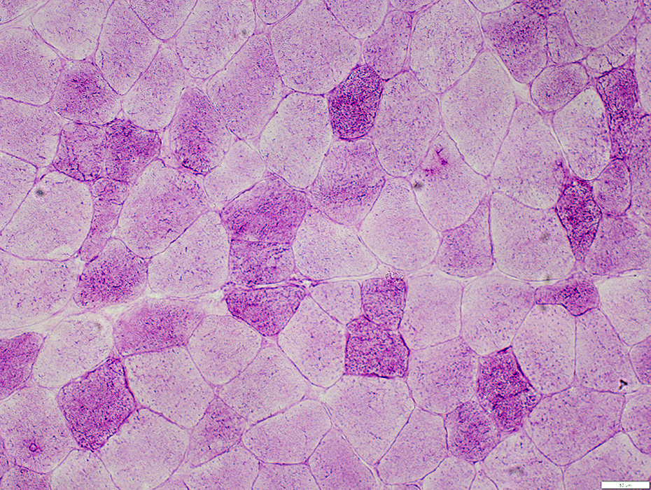
Muscle fibers with Mitochondrial Proliferation
Irregular red stain (Ragged red)
 Gomori trichrome stain |
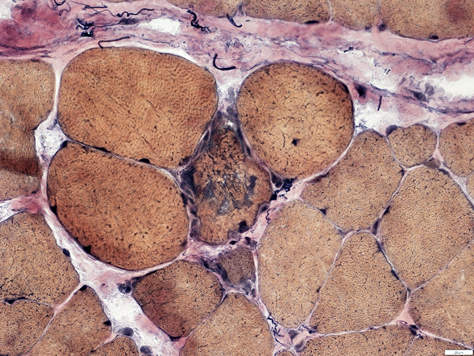
 H&E stain |
Internal architecture: Irregular in some muscle fibers (Arrow)
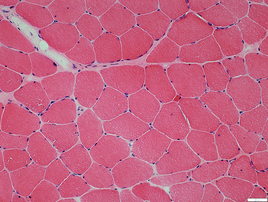 H&E stain |
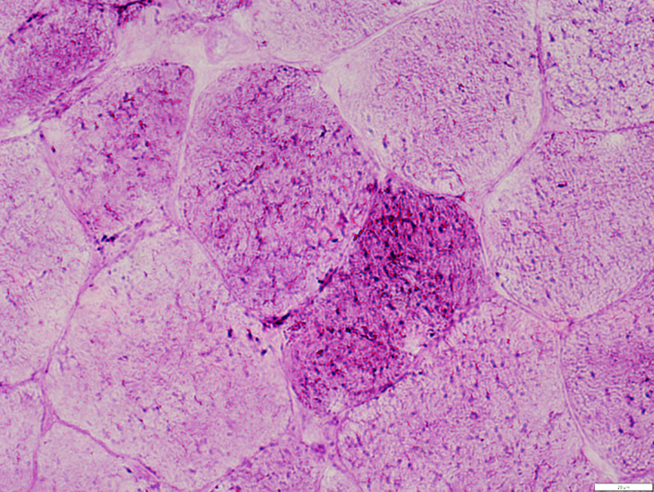
Muscle fibers with Mitochondrial Proliferation
Internal architecture: Irregular
 H&E stain |
 VvG stain |
Internal architecture: Irregular
 VvG stain |
Lipid Droplets
Mildly/Moderately increased in muscle fibers
 Sudan black stain |
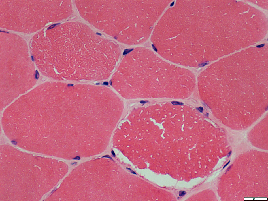
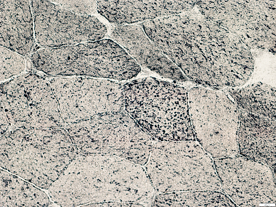
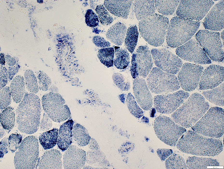
 COX stain |
Reduced staining in scattered muscle fibers
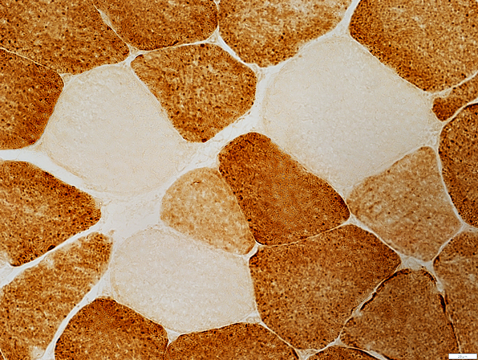 COX stain |
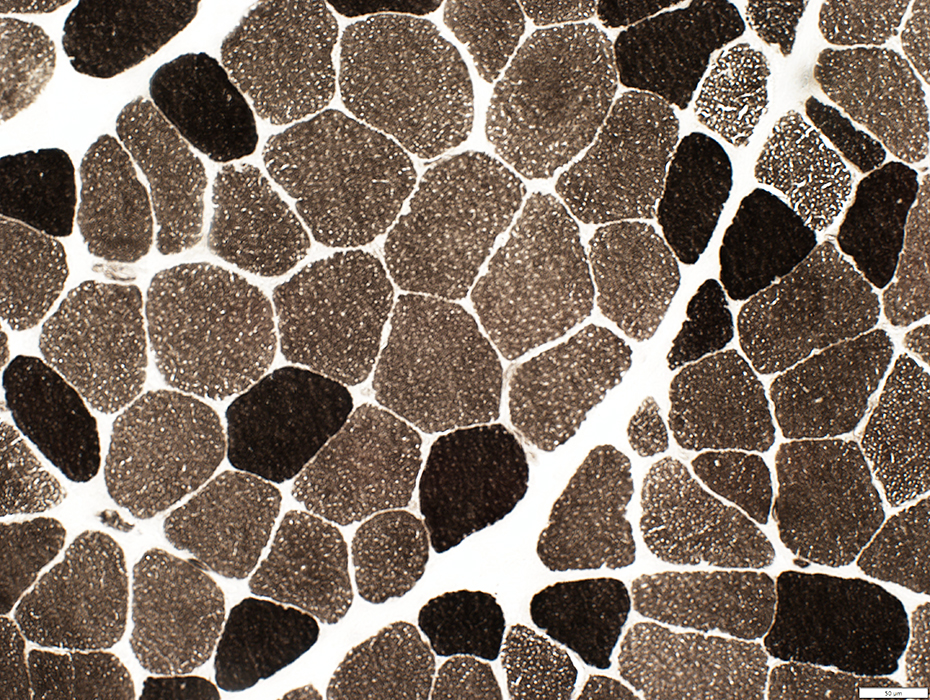
COX stain
Muscle fibers with reduced staining
Mitochondria not visible
 COX stain |
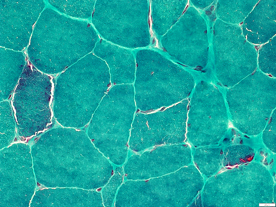
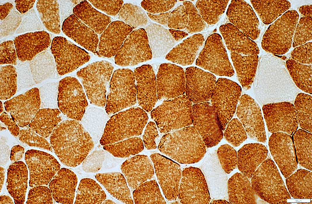
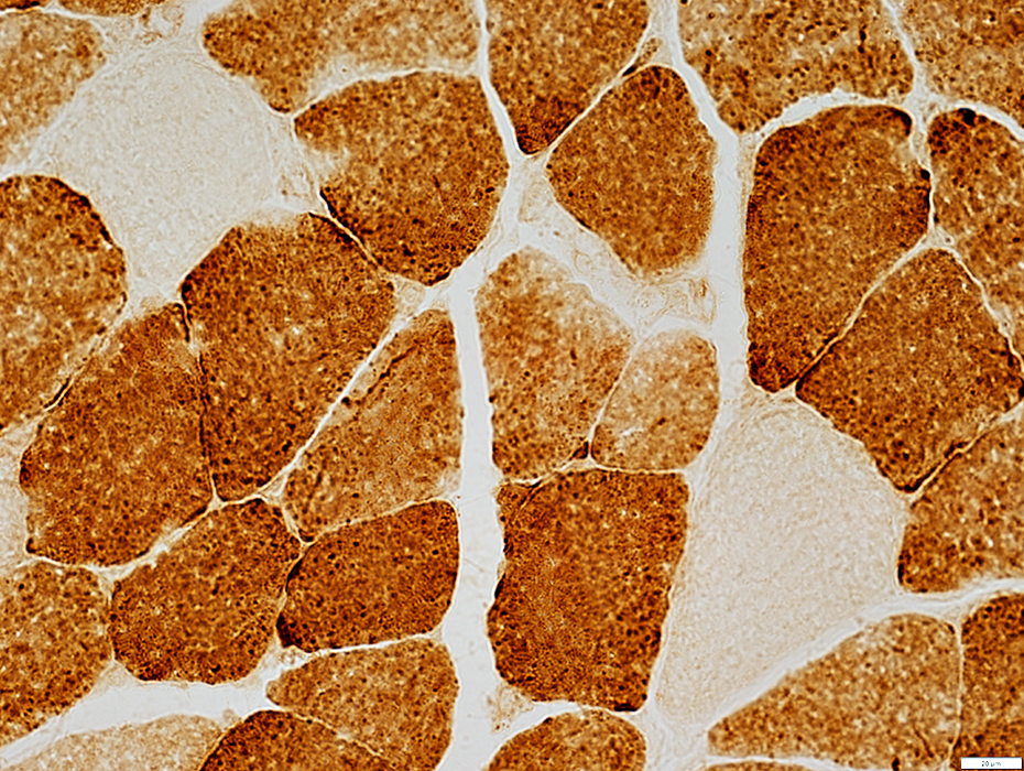
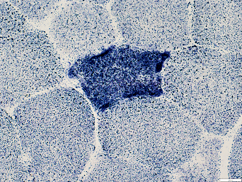
Succcinate Dehydrogenase (SDH)
Scattered fibers with increased staining
 SDH stain |
 SDH stain |
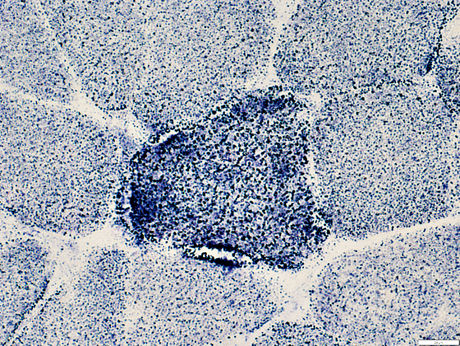
Muscle fibers have increased staining
Mitochondria appear large
 SDH stain |
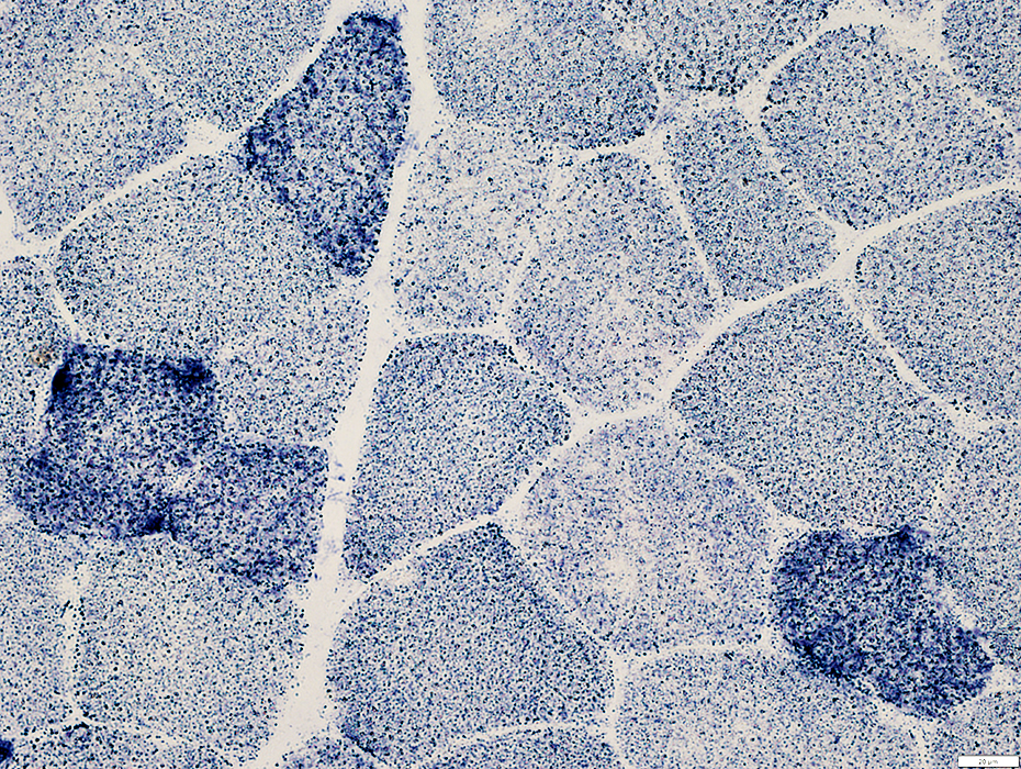
Scattered muscle fibers with increased staining
Mitochondria appear large
 SDH stain |
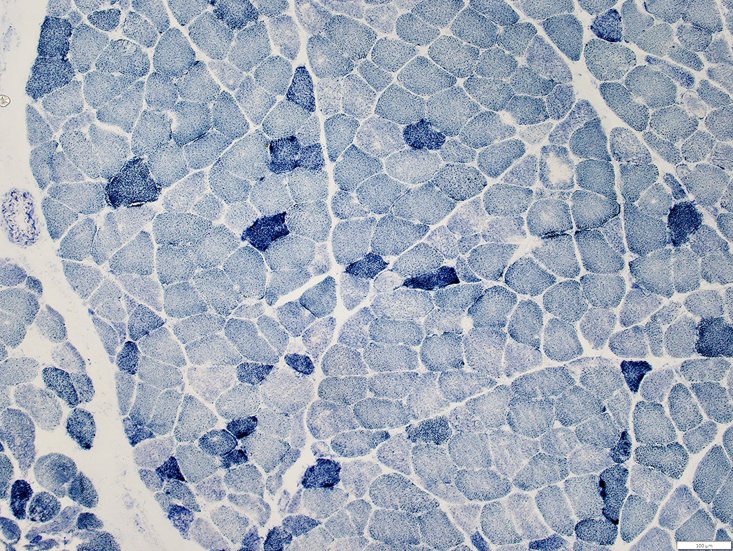
Succcinate Dehydrogenase (SDH)
Perimysial vessels: Normal staining
Differs from increased staining in MELAS
 SDH stain |
MNGIE: Muscle fiber types
Type I muscle fibers: Mild predominance
Lipid droplets: Increased in a few type 1 (Intermediate-stained) muscle fibers
 ATPase pH 9.4 stain |
 PAS stain |
 PAS stain |
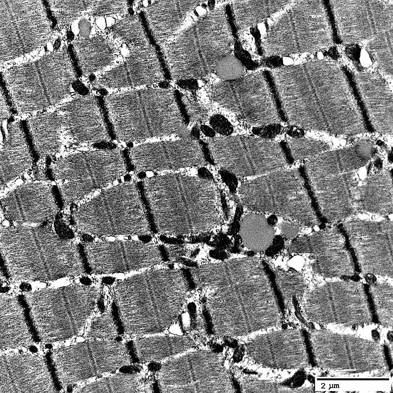
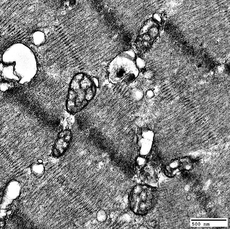
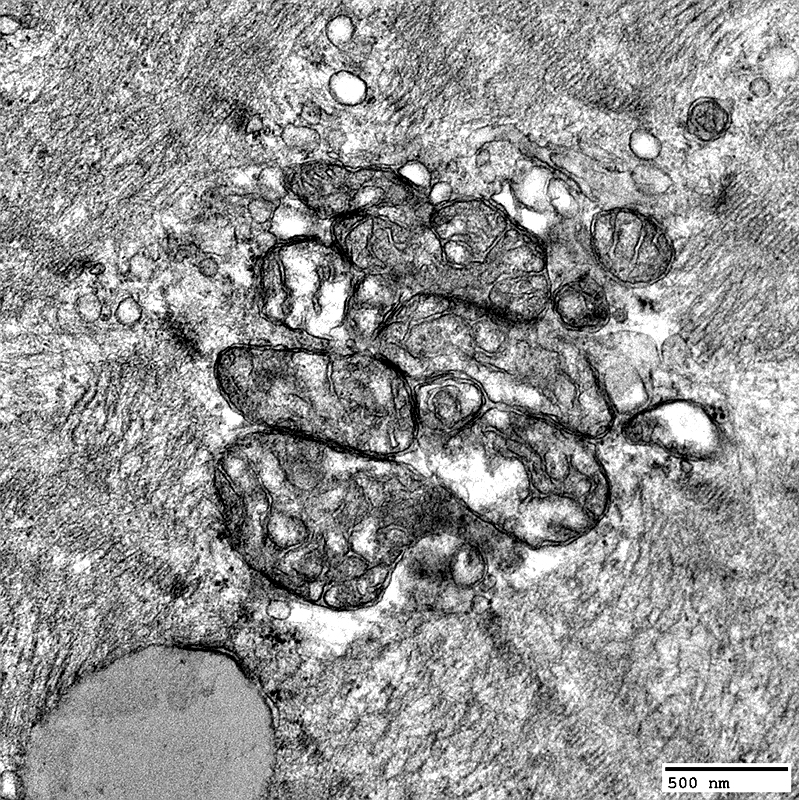
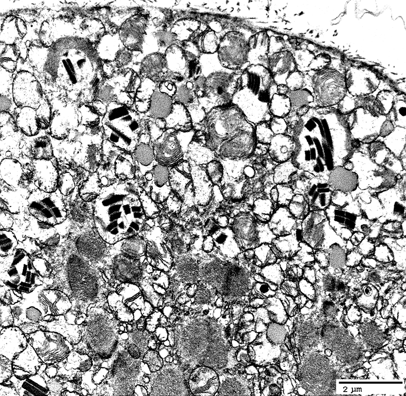
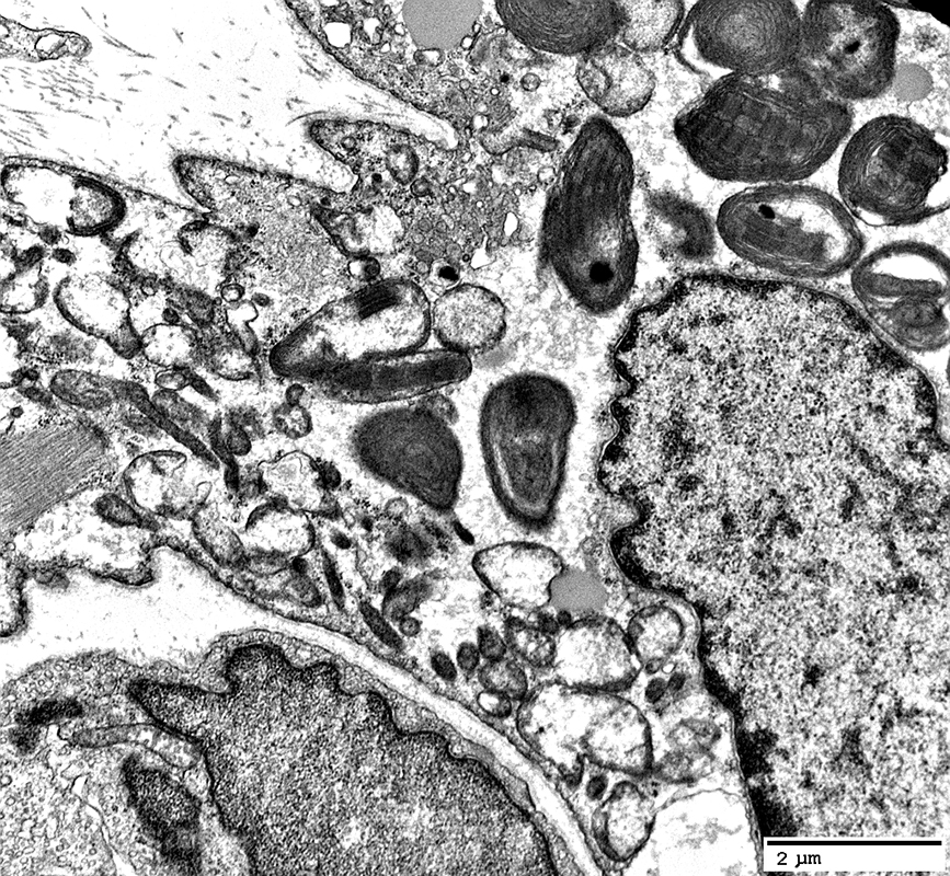
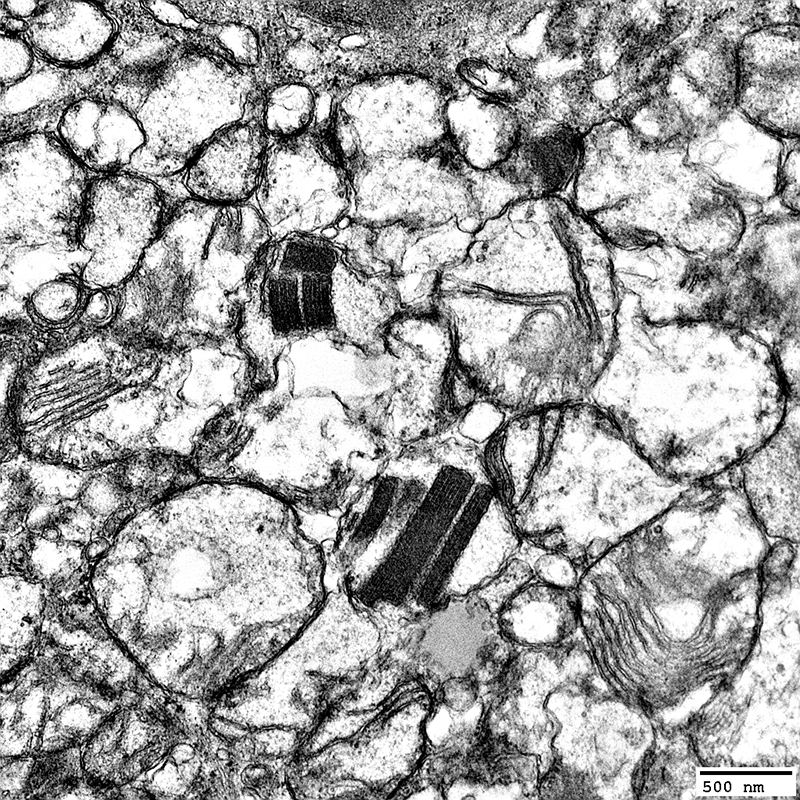
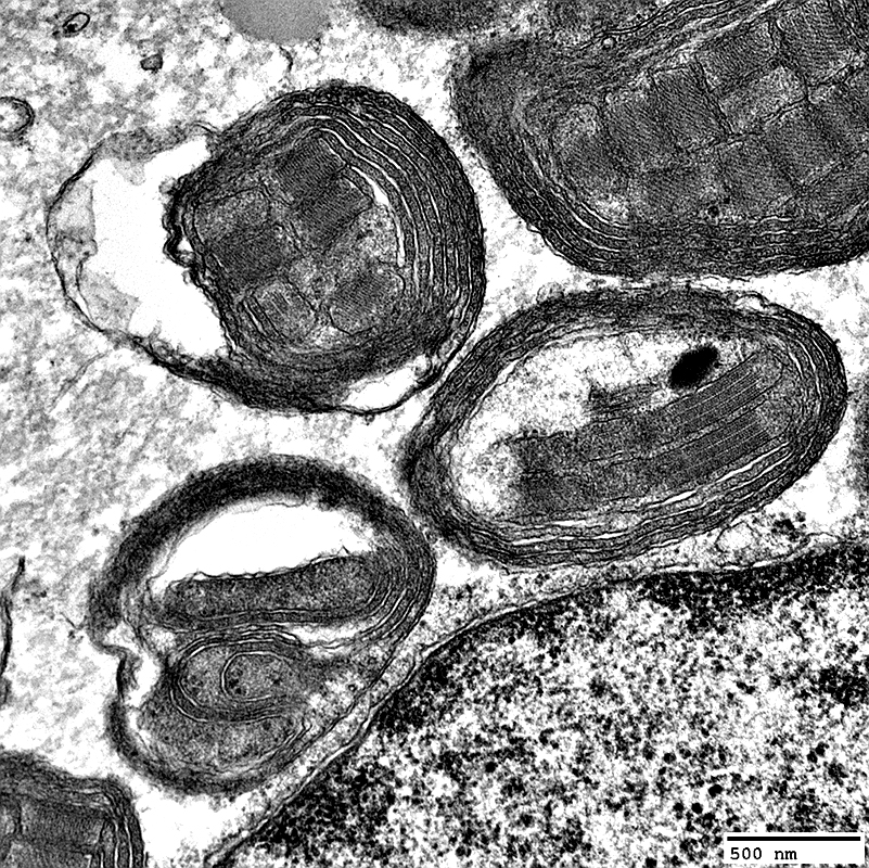
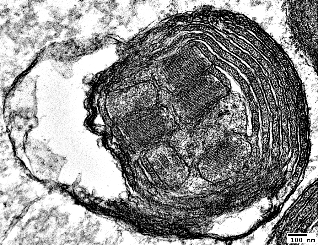
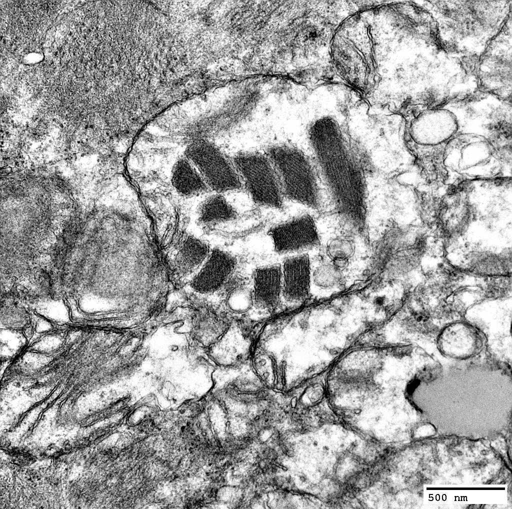
MNGIE: Muscle Ultrastructure
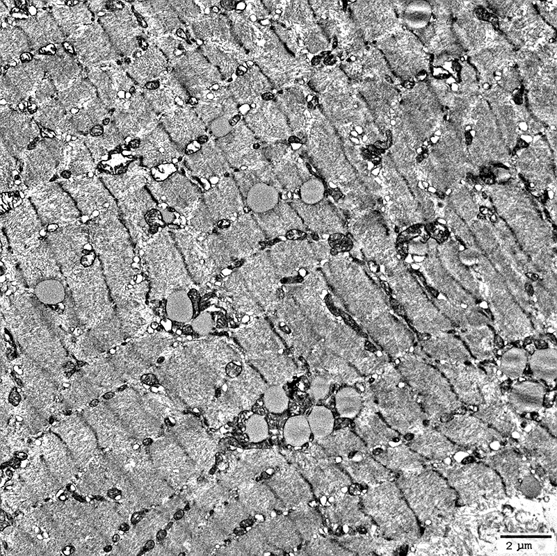 From: R Schmidt |
Mitochondria: Moderate proliferation associated with lipid droplets
 From: R Schmidt |
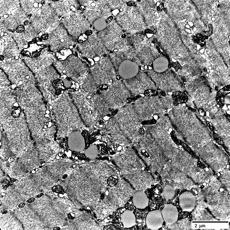
 From: R Schmidt |
 From: R Schmidt |
Mitochondria: Proliferation; Mildly large; Round shape
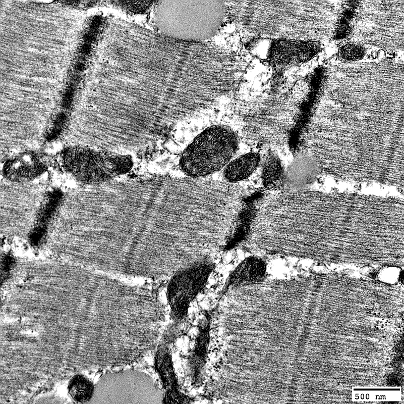 From: R Schmidt |
 From: R Schmidt |
Mitochondria: Proliferation; Mildly large; Round shape, Clustered
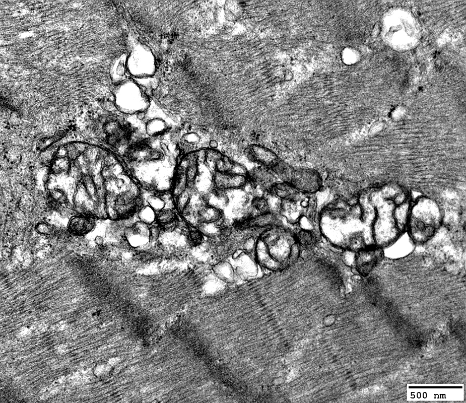 From: R Schmidt |
 From: R Schmidt |
Mitochondria: Marked proliferation; Contain paracrystalline arrays (Parking lot inclusions)
 From: R Schmidt |
 From: R Schmidt |
Mitochondria: Marked proliferation; Contain paracrystalline arrays (Parking lot inclusions); Whorls
 From: R Schmidt |
Mitochondria: Whorled internal architecture with parking lot inclusions
 From: R Schmidt |
 From: R Schmidt |
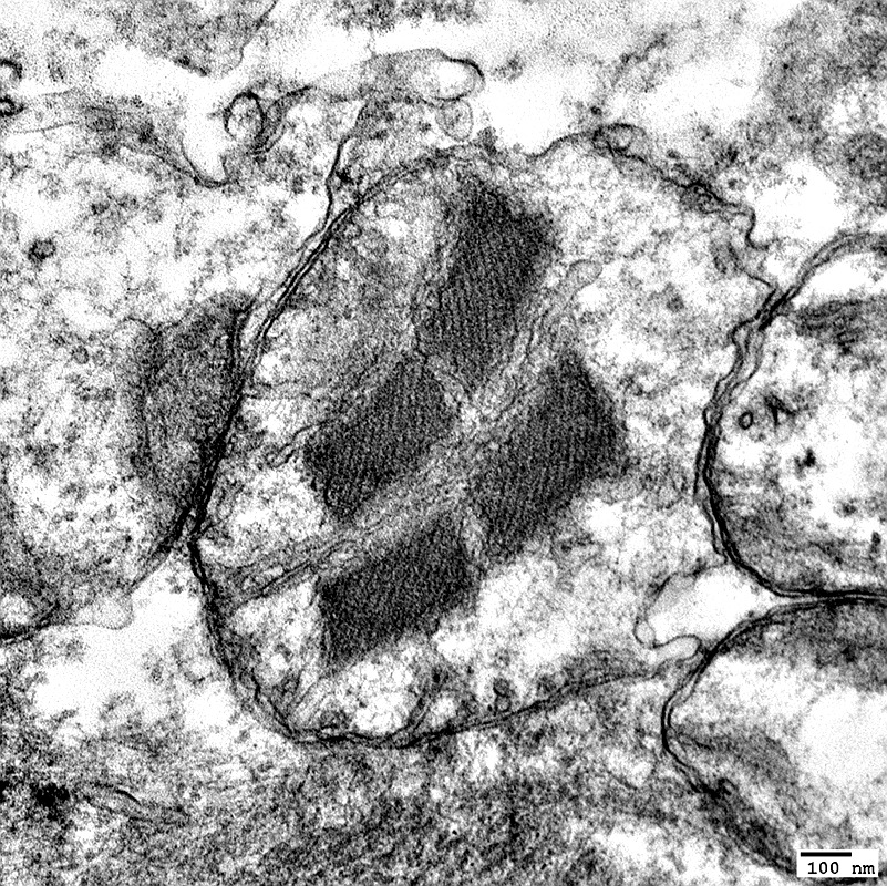
Mitochondria: Contain paracrystalline arrays (Parking lot inclusions)
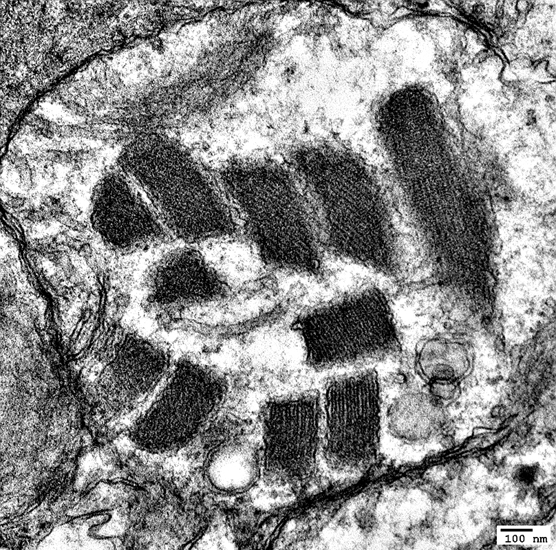 From: R Schmidt |
 From: R Schmidt |
Return to: MNGIE
5/26/2026