
LEPROSY
|
Skin Hands MRI: Nerve Roots Nerve Pathology Ultrastructure |
Leprosy: Skin
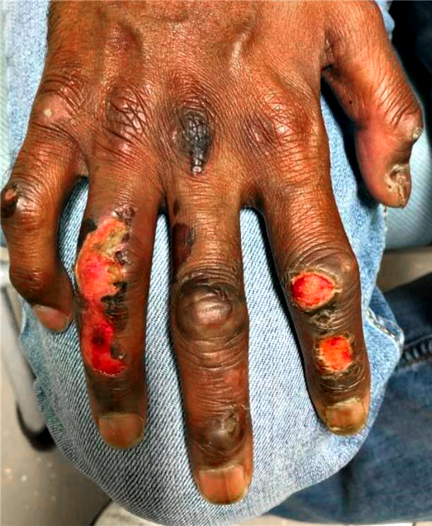 From: A Sabouri Pre-Treatment |
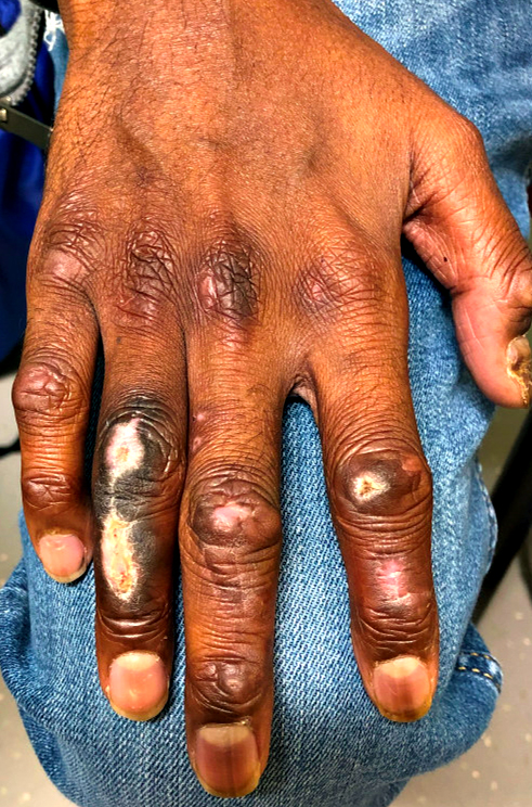 From: A Sabouri Post-Treatment |
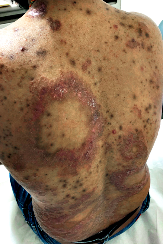 From: A Sabouri |
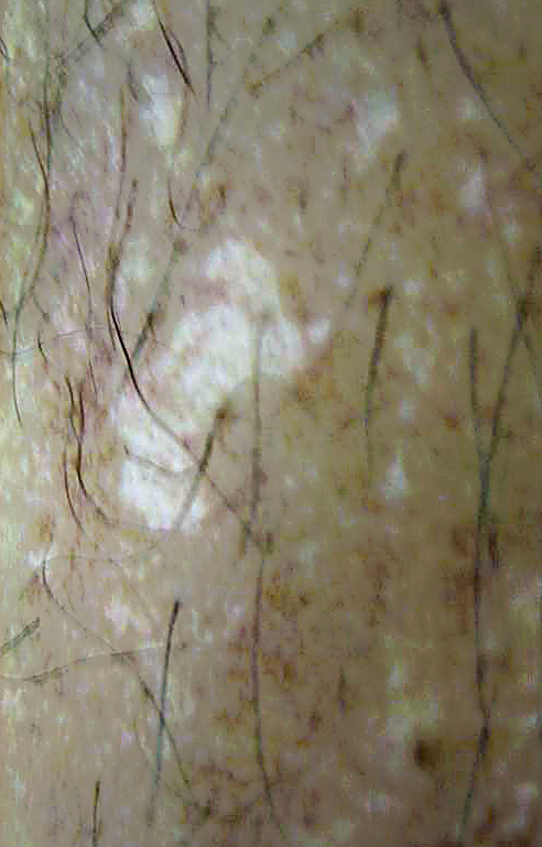 From: M Al-Lozi Skin: Hypopigmented lesion |
Leprosy: Hands
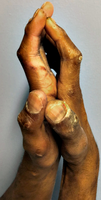 From: A Sabouri |
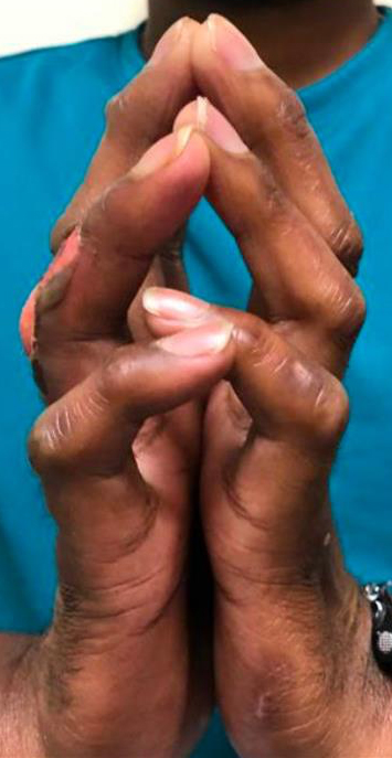 From: A Sabouri |
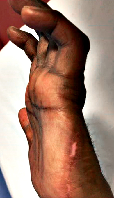 From: A Sabouri |
Leprosy: Nerve Root Enlargement
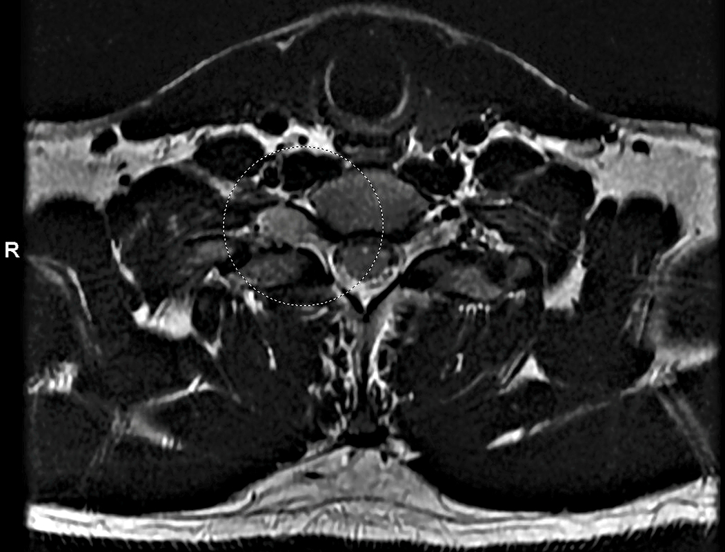 From: A Sabouri |
C8 & T1 nerve roots: Large on right
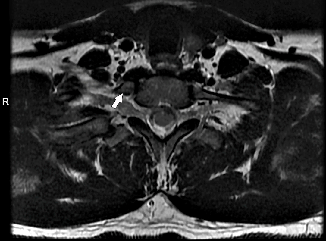 From: A Sabouri |
Leprosy: Nerve Pathology
- Nerves often biopsied
- Ulnar: Cutaneous dorsalis branch
- Sural
- Patterns of involvement
- Asymmetric: Differential involvement among fascicles
- Endoneuritis, mononuclear cell
- Granulomas, epithelioid
- Fibrosis
- Fascicular: Tuberculoid leprosy; Chronic disease
- Perineurial: Chronic disease
- Lepromatous leprosy
- Diagnosis
- M leprae detected (10%)
- qPCR+ (34%)
- qPCR on adjacent skin (25%)
Tuberculoid Leprosy: Nerve Pathology
Multiple fascicles: Endoneurial structure is lost
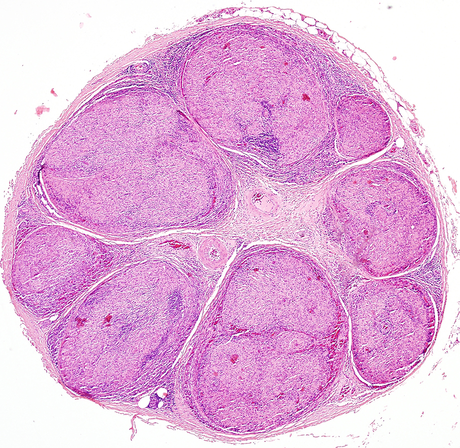 From: R Schmidt |
Fascicle replaced by granulomatous structure (Arrow)
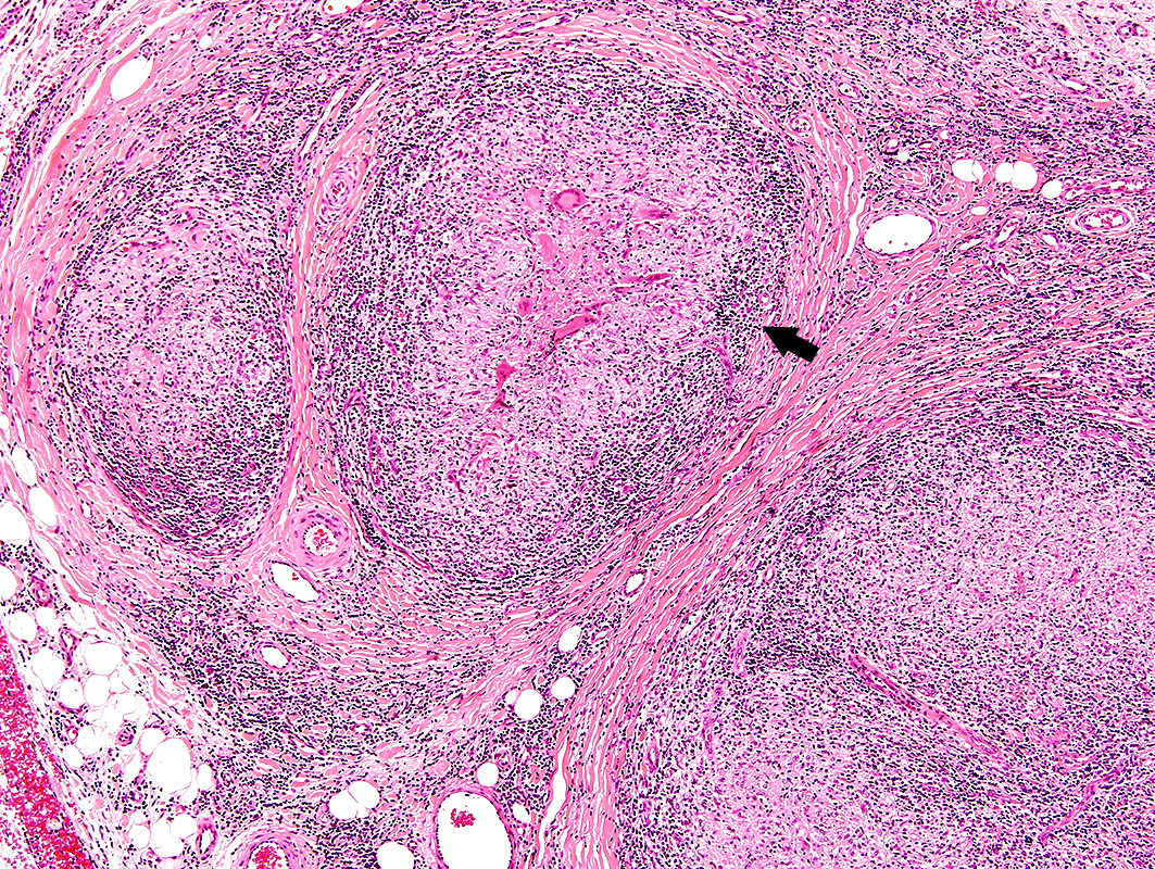 From: R Schmidt |
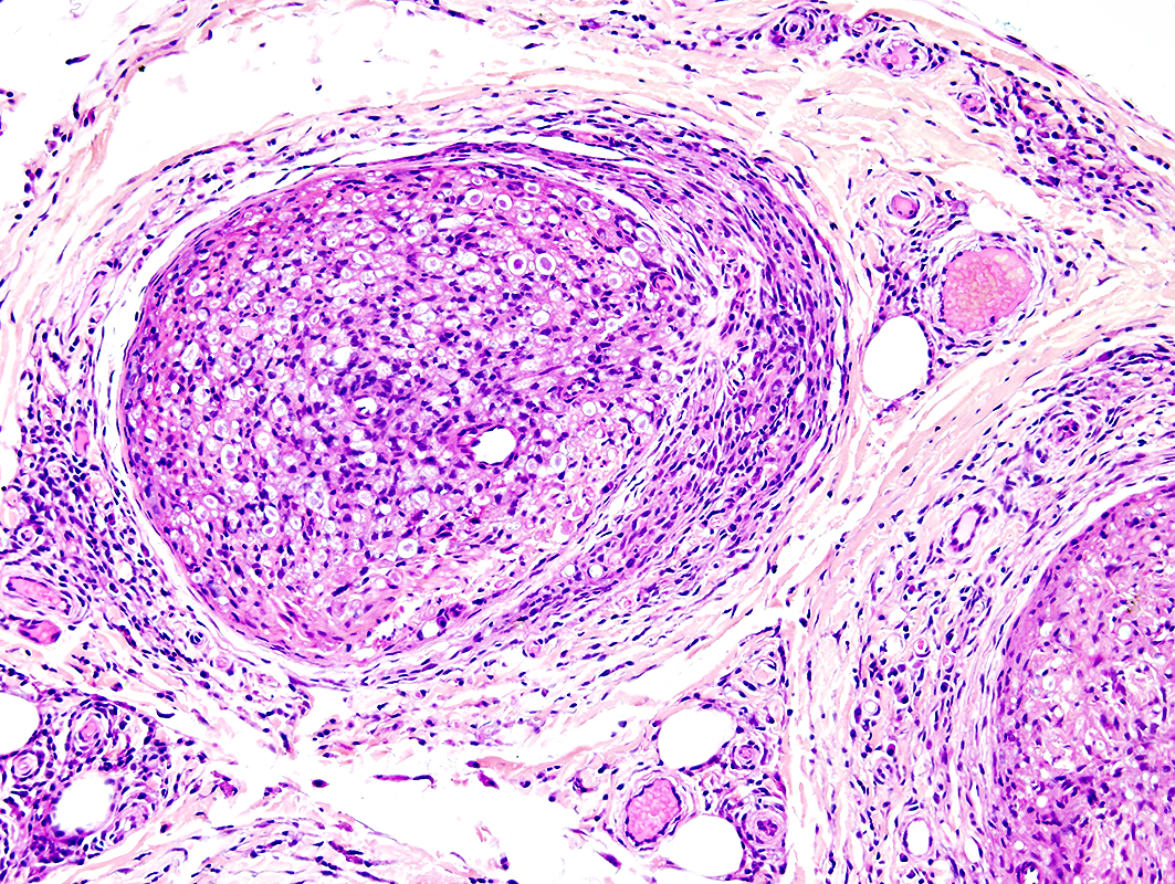 From: R Schmidt |
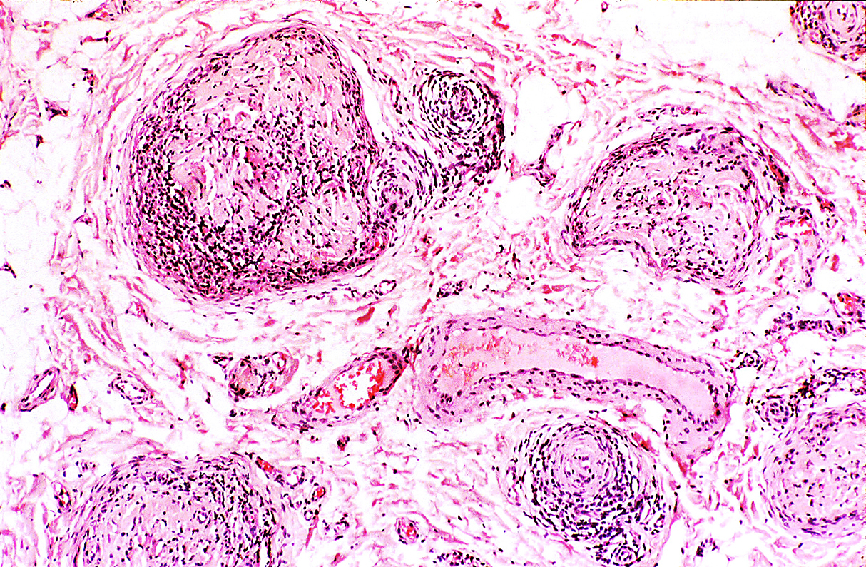
Lepromatous Leprosy: Nerve Pathology
Inflammation: Perineurial & Perivascular
 From: R Schmidt |
Locations: Perineurium; Perivascular
Leprosy: Nerve Pathology
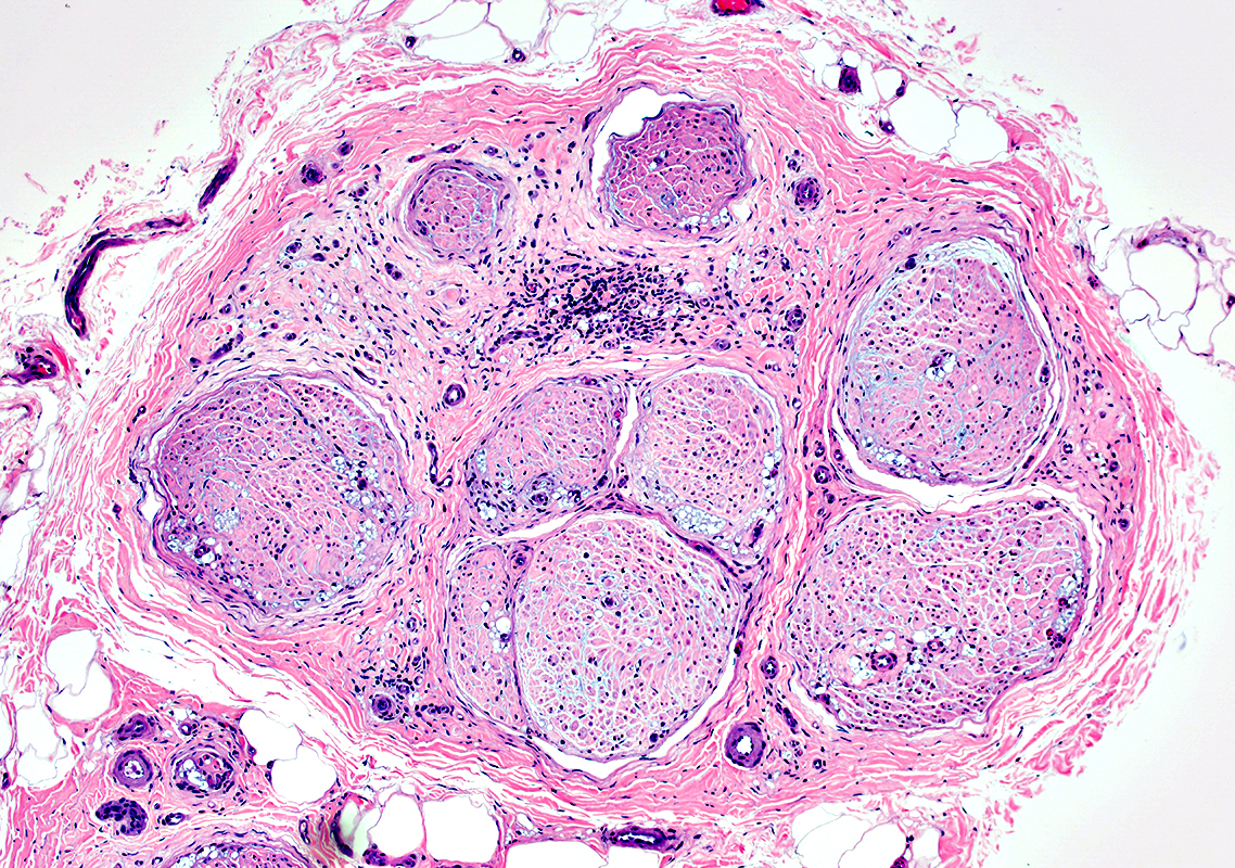 H&E stain |
Multifocal leprosy lesions in endoneurium & epineurium
Inflammatory cell focus in epineurium
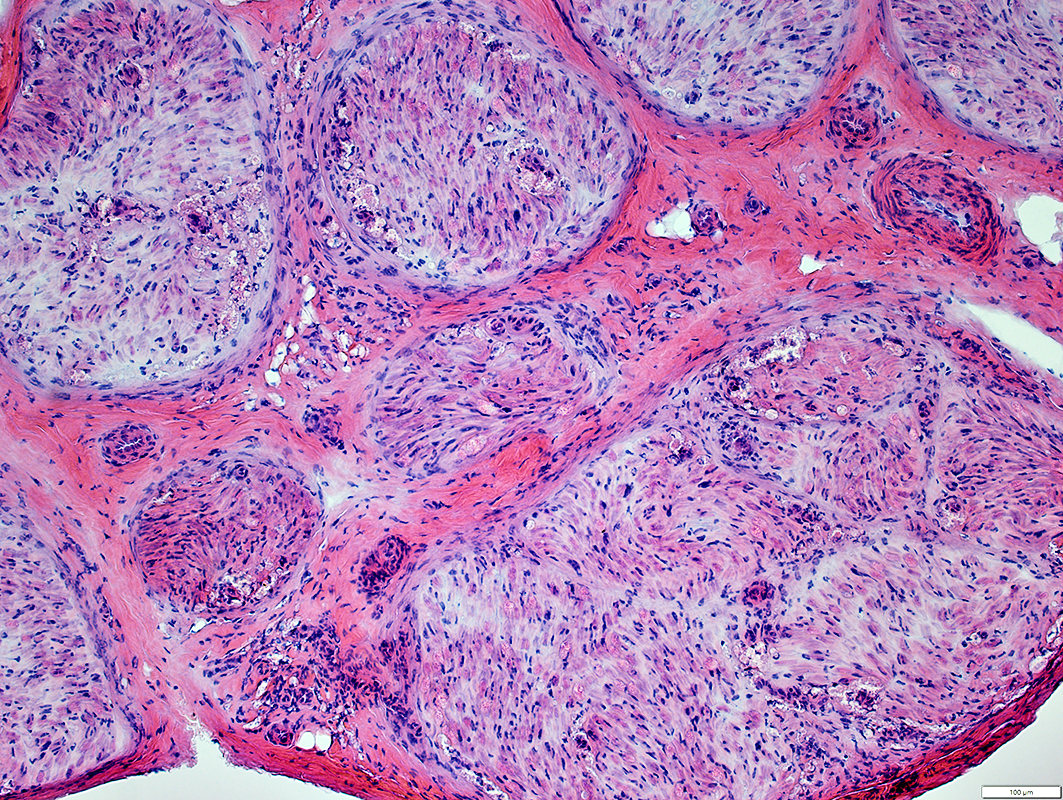 H&E stain |
Multifocal lesions in endoneurium & epineurium
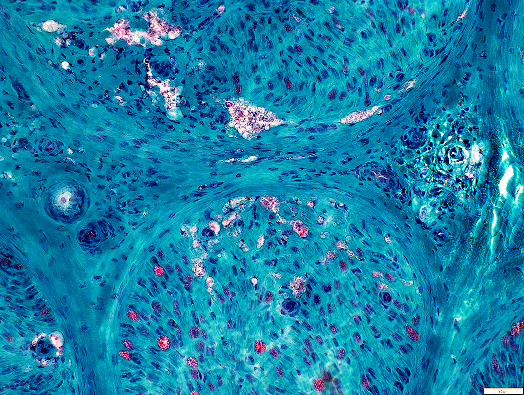 Gomori trichrome stain |
Multifocal lesions in endoneurium & epineurium
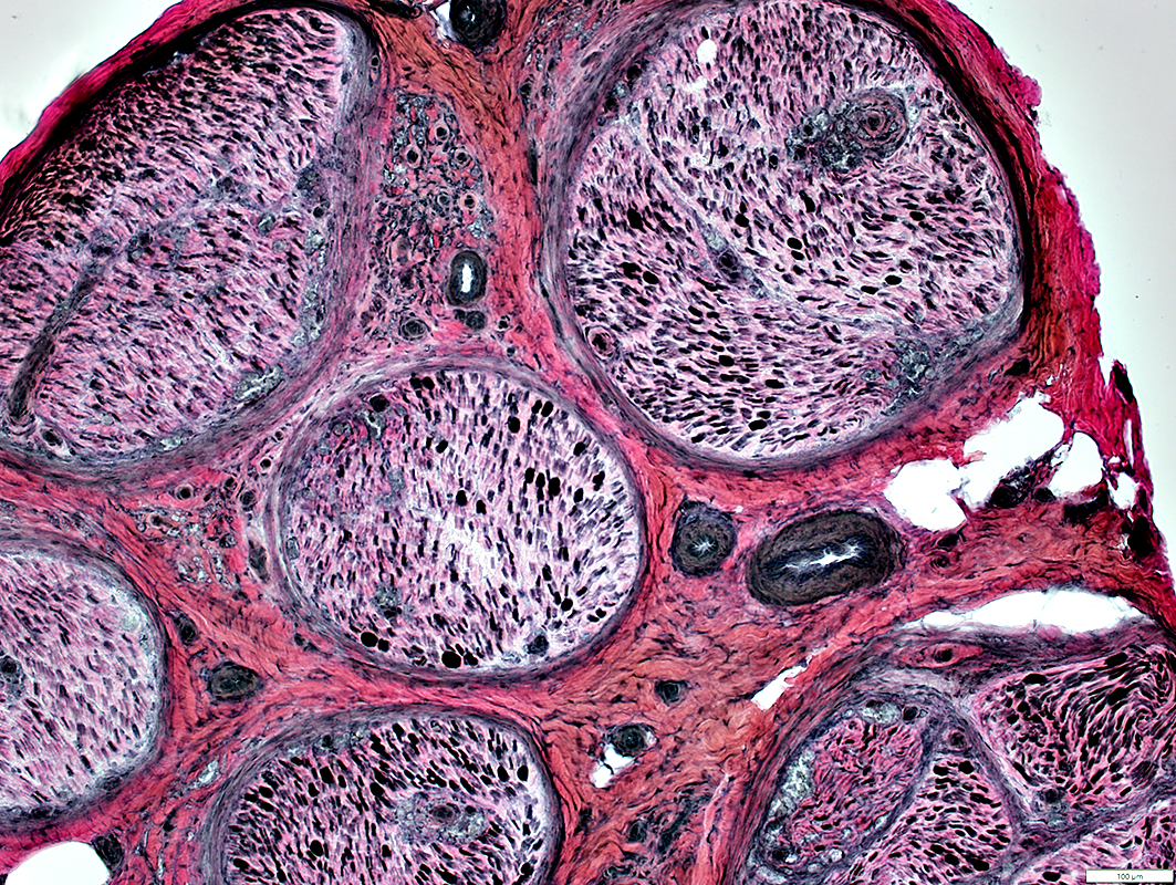 VvG stain |
Multifocal lesions in endoneurium & epineurium
Loss of large myelinated axons: Severe; Varied within & among fascicles
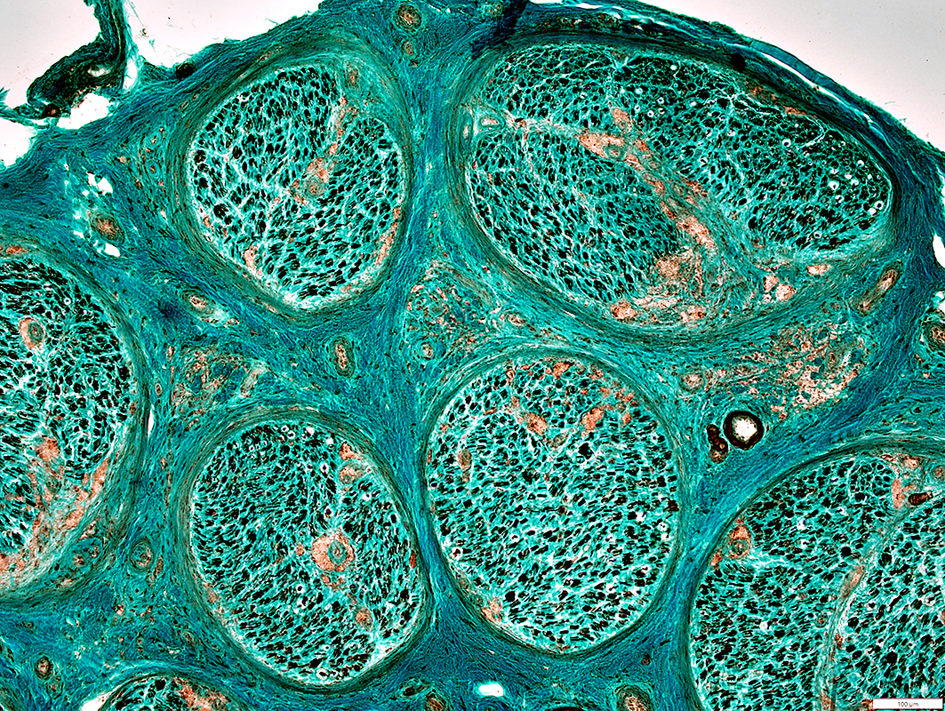 Neurofilament stain |
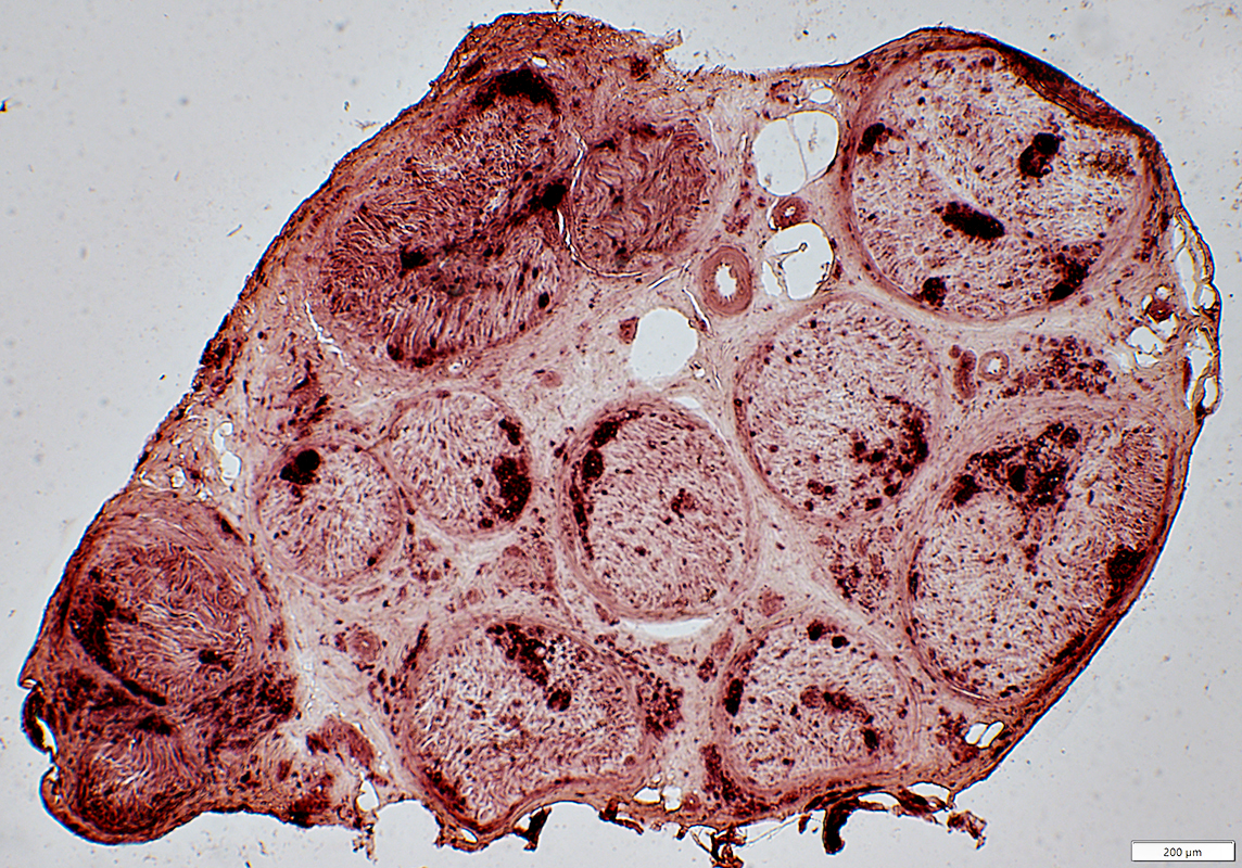 Acid Phosphatase stain |
Multifocal lesions in endoneurium, subperineurial regions & epineurium
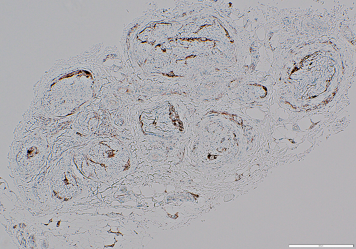 CD68 stain |
Leprosy: Focal Lesions
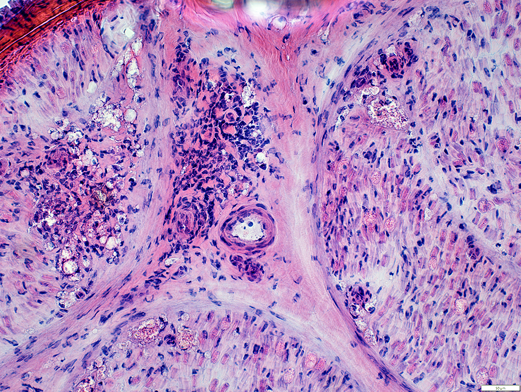 H&E stain |
Multifocal lesions in endoneurium & epineurium
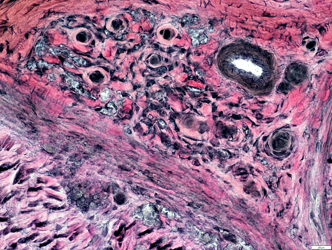 VvG stain |
Multifocal lesions in endoneurium & epineurium
Leprosy bacilli in lesions are dark-stained
Lesions often contain small vessels with dark-stained endothelium
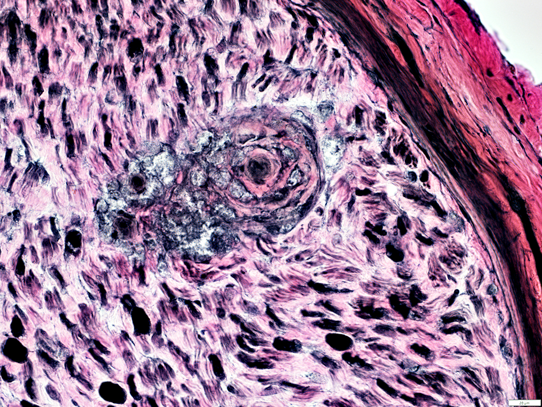 VvG stain |
Focal lesion in endoneurium
Lesions often contain small thick-walled vessels with abnormal structure
Axons
Large Myuelinated axons: severe loss
Remaining myelinated axons: Mostly thin myelin sheaths; Likely regenerated
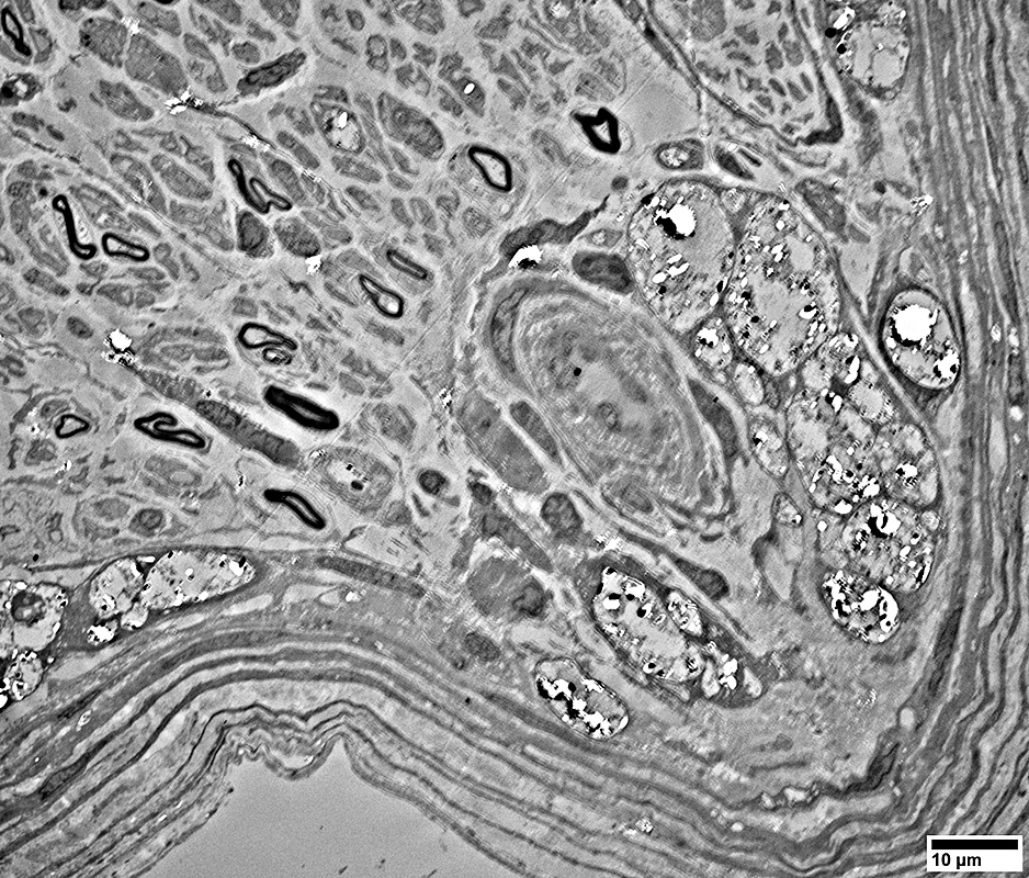 From: R Schmidt |
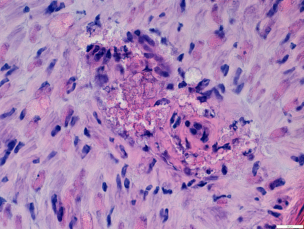 H&E stain |
Focal lesion in endoneurium
Lesions often contain small thick-walled vessels
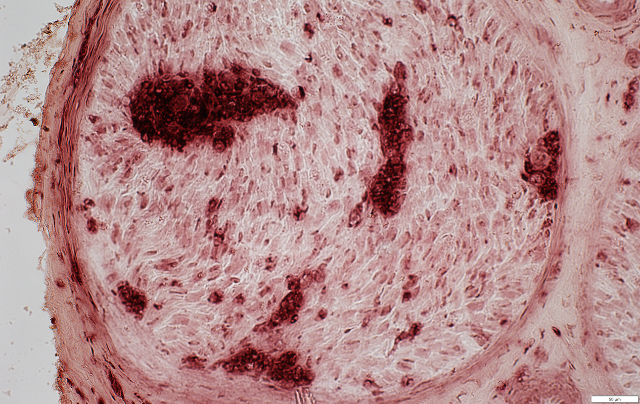 Acid phosphatase stain |
Multifocal lesions in endoneurium
Lesions stain strongly for acid phosphatase
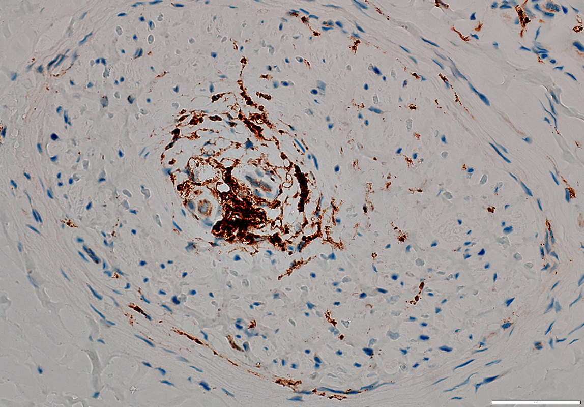 CD68 stain |
Leprous bacilli
Stained for: Acid fast; Fite; Gomori trichrome
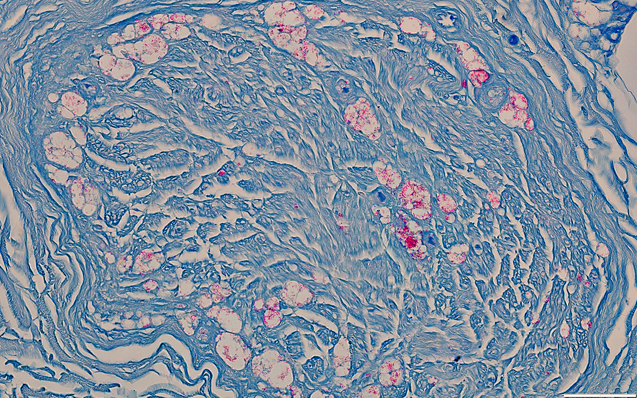 Acid Fast stain |
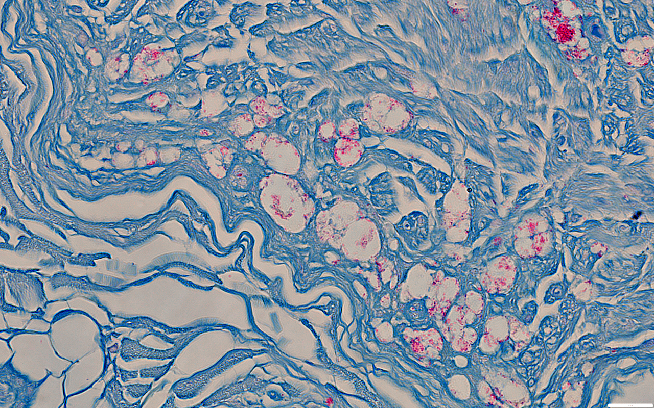 Acid Fast stain |
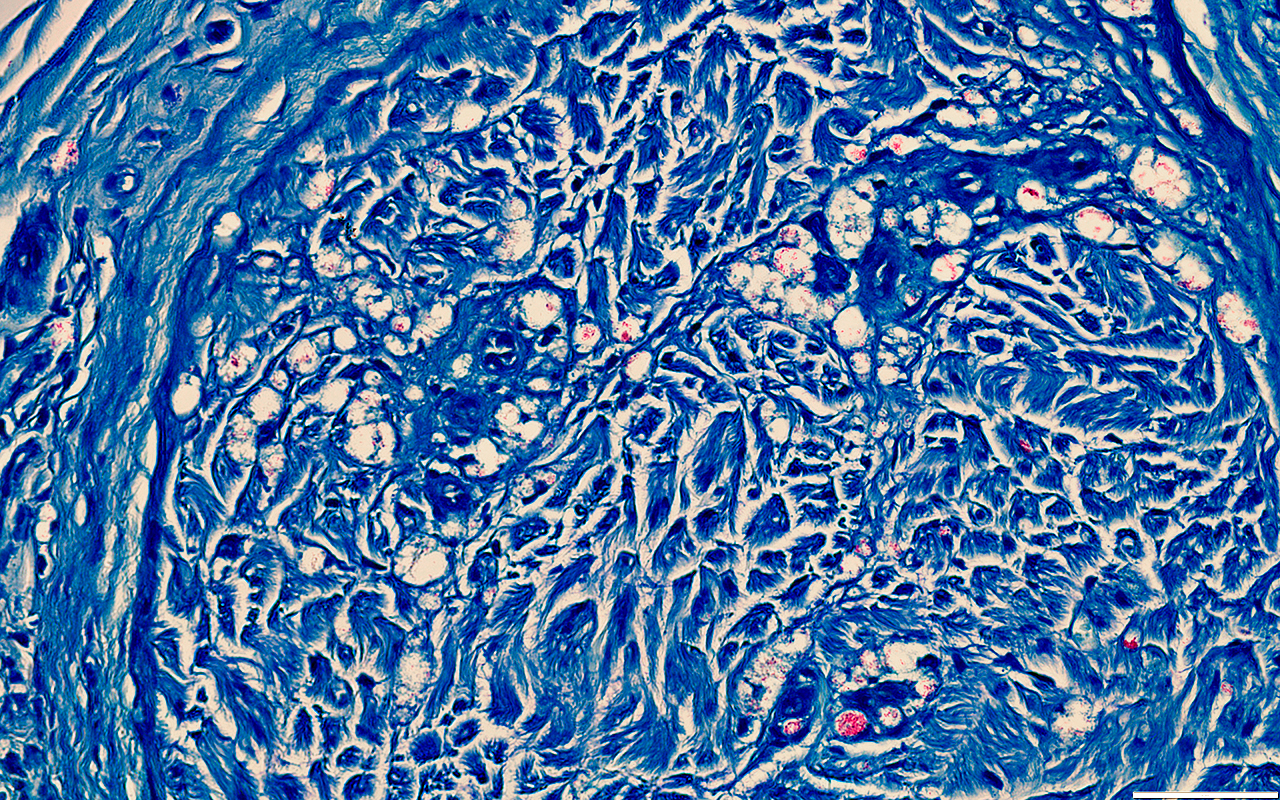 Fite stain |
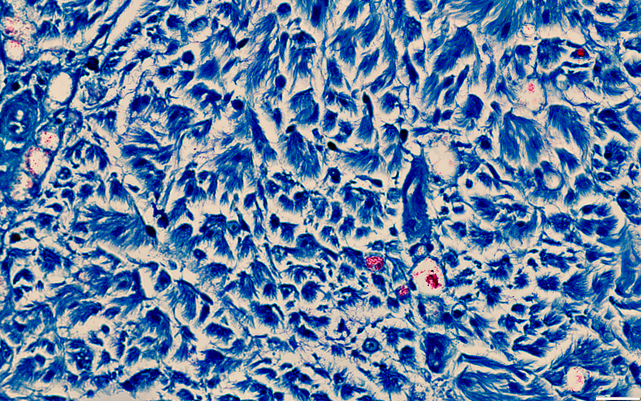 Fite stain |
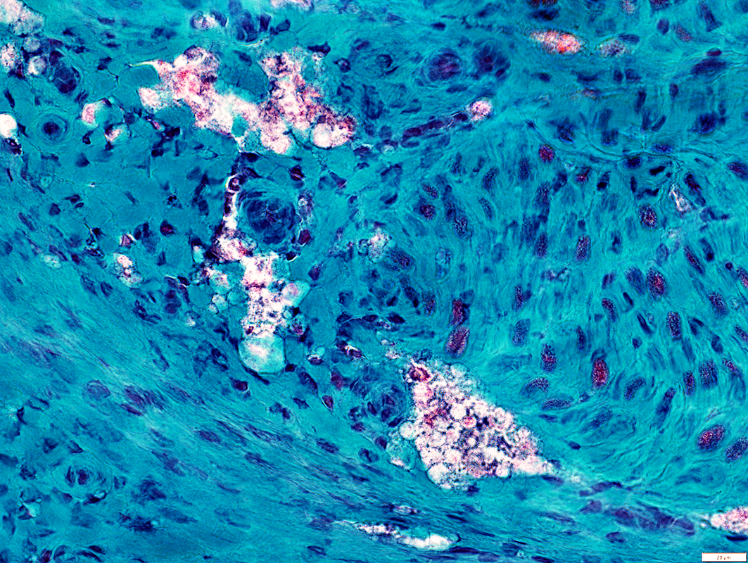 Gomori trichrome stain |
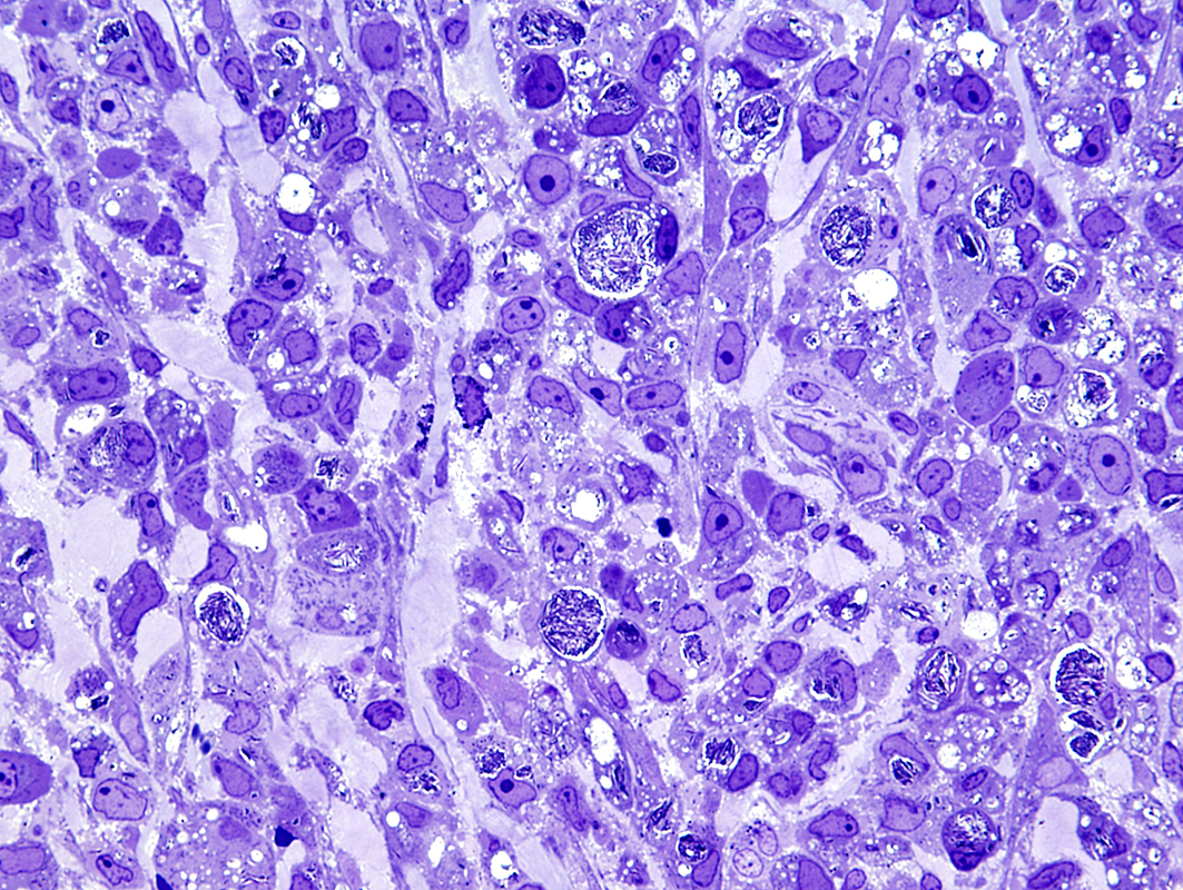 From: R Schmidt |
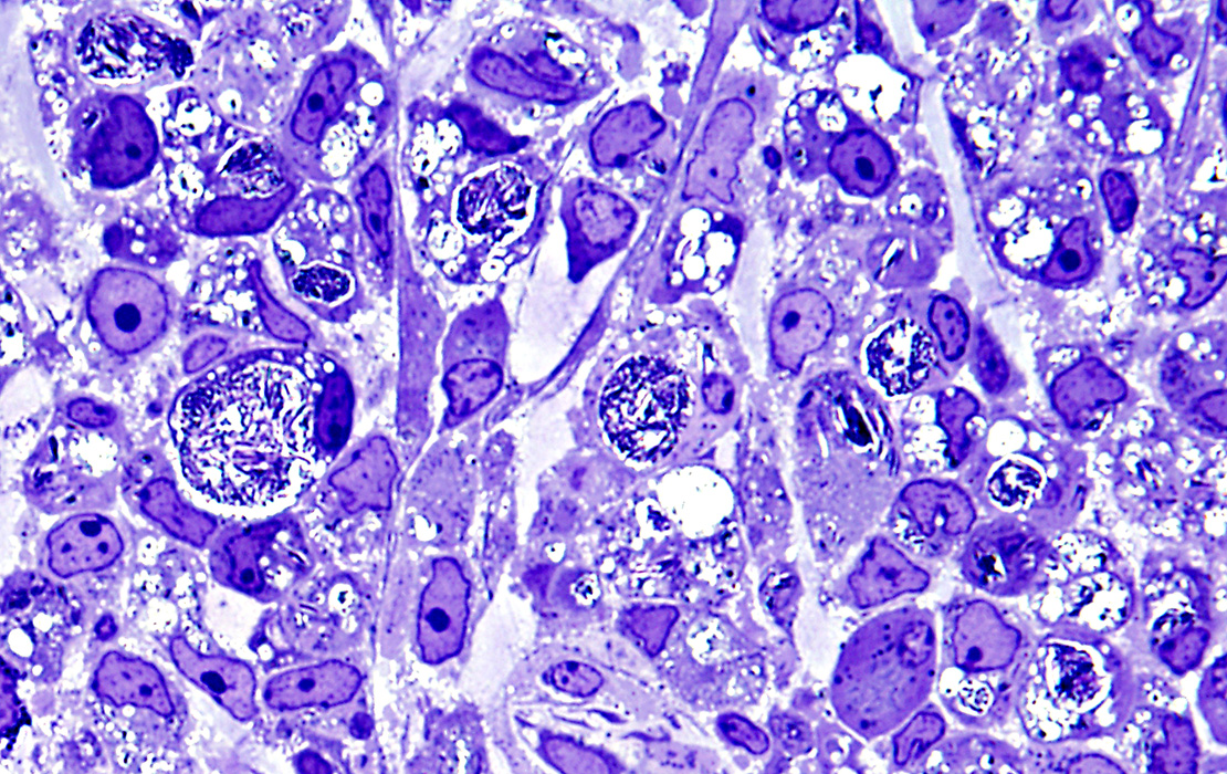 From: R Schmidt |
Leprosy Nerve: Intracellular M. leprae
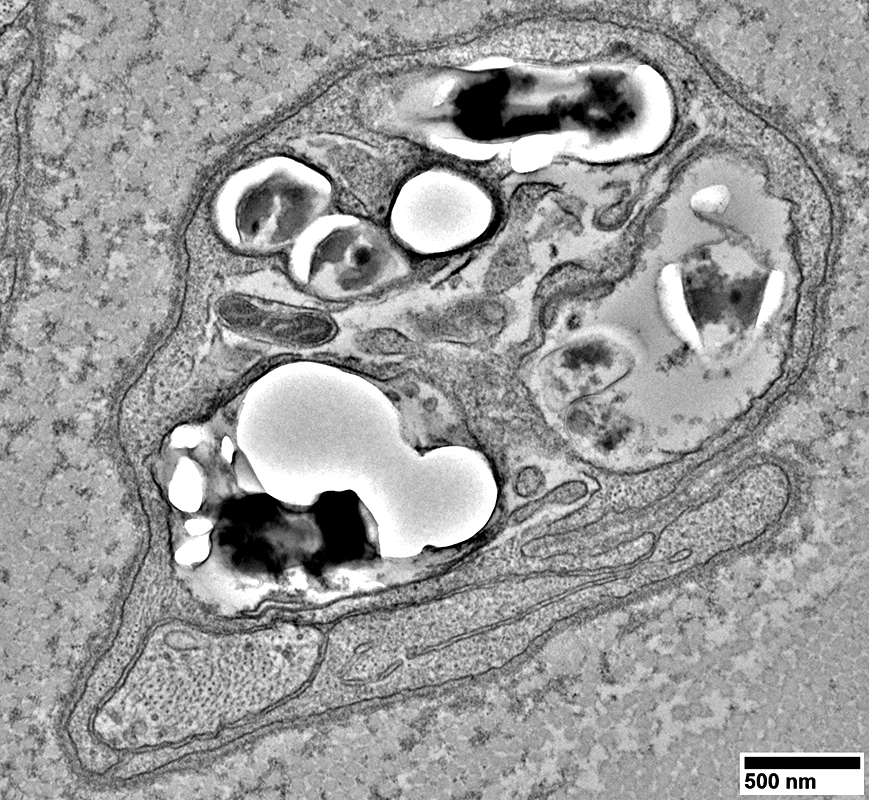 From: R Schmidt |
Leprosy bacilli: Within a non myelinating Schwann cell (Identified by basal lamina outside surface mambrane)
Schwann cell: Surrounds an unmyelinated axon (Lower left; Above)
Nerve (Below)
Other neighboring non-myelinating Schwann cells
Have no intracelluylar bacilli
Surround unmyelinated axons
Myelinated axons are lost
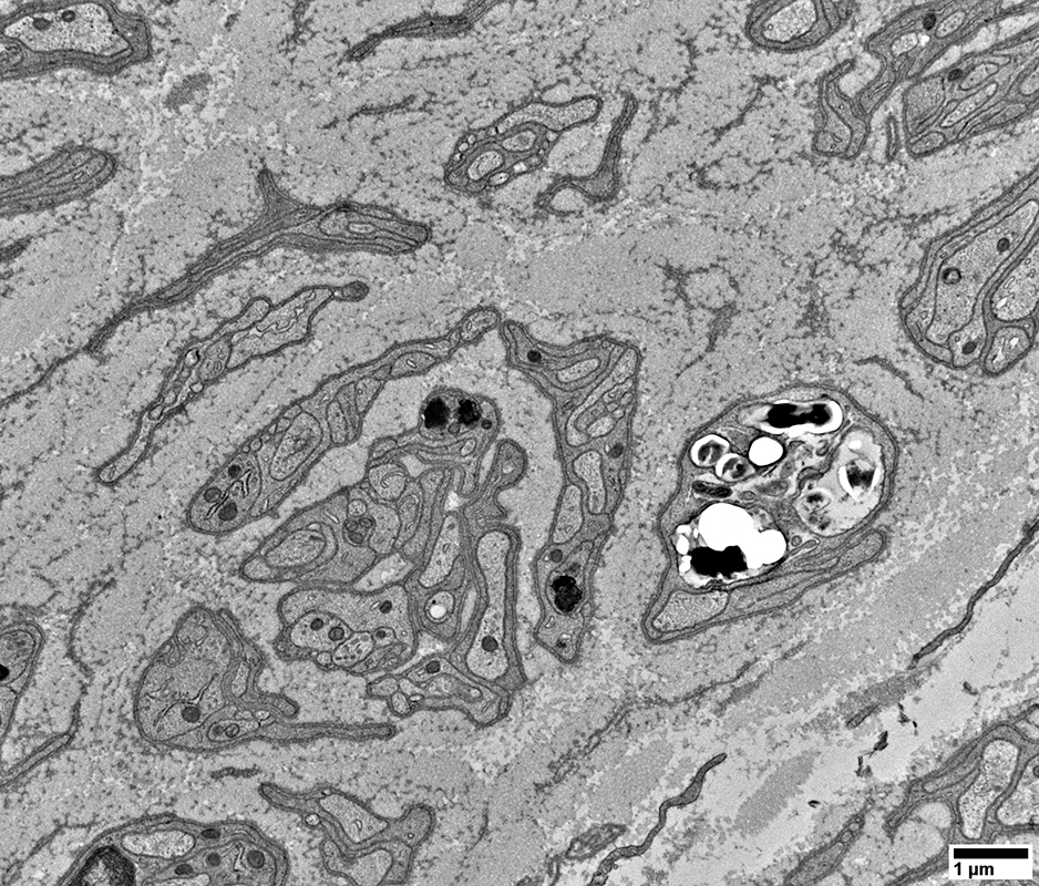 From: R Schmidt |
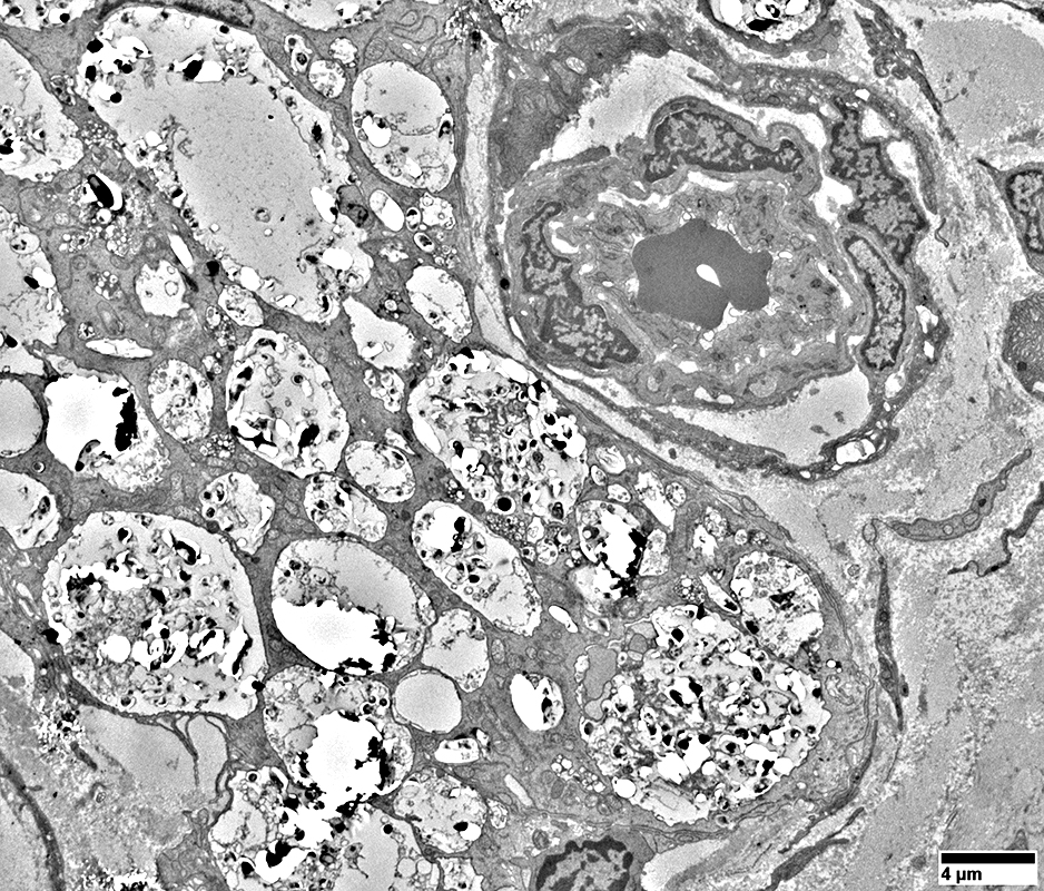 From: R Schmidt |
Many leprosy bacilli within a very large (> 40) μM "Lepra cell" (Left)
Damaged endoneurial microvessel (Upper right)
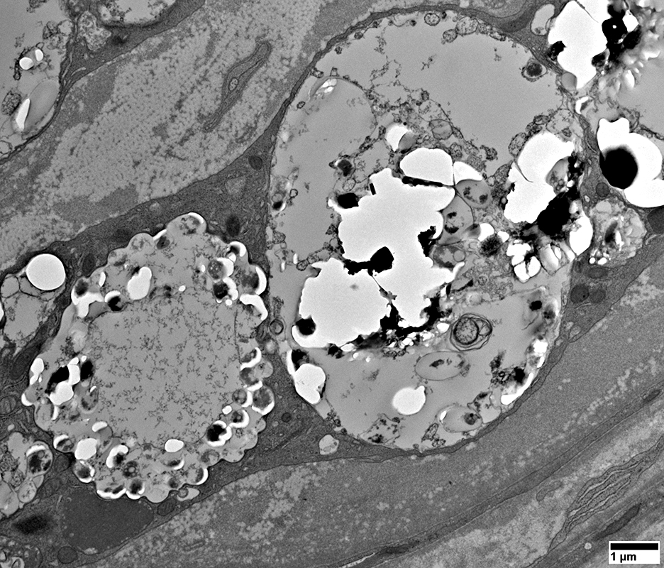 From: R Schmidt |
Many leprosy bacilli within a large "Lepra cell"
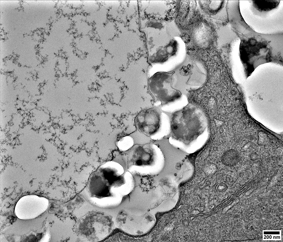 From: R Schmidt |
Leprosy: Intracellular organisms
Leprosy bacilli within a cell" near the perineurium
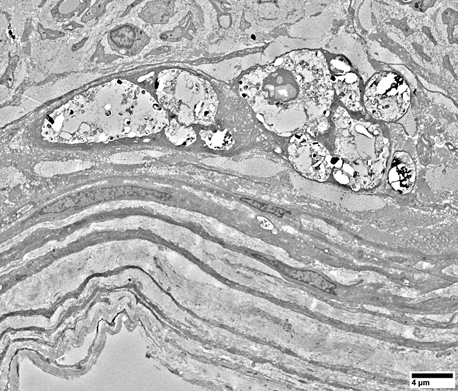 From: R Schmidt |
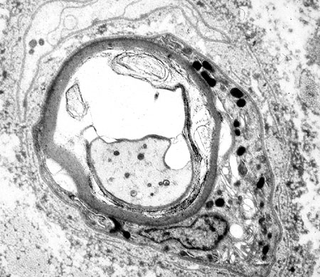 |
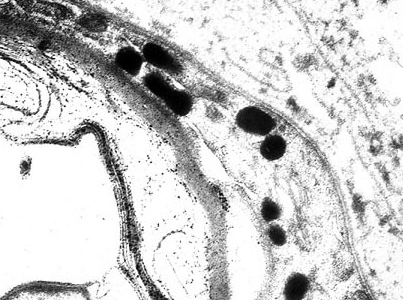 From Dennis K Burns MD Leprosy: Intracellular organisms Leprosy bacilli: Within a myelinating Schwann cell |
Leprosy: Endoneurial Microvessels
 From: R Schmidt |
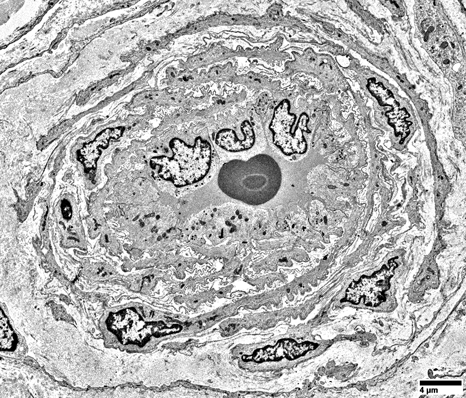
Damaged Wall
Pericyte processes: Multiple; Small; Separated by irregular connective tissue
 From: R Schmidt |
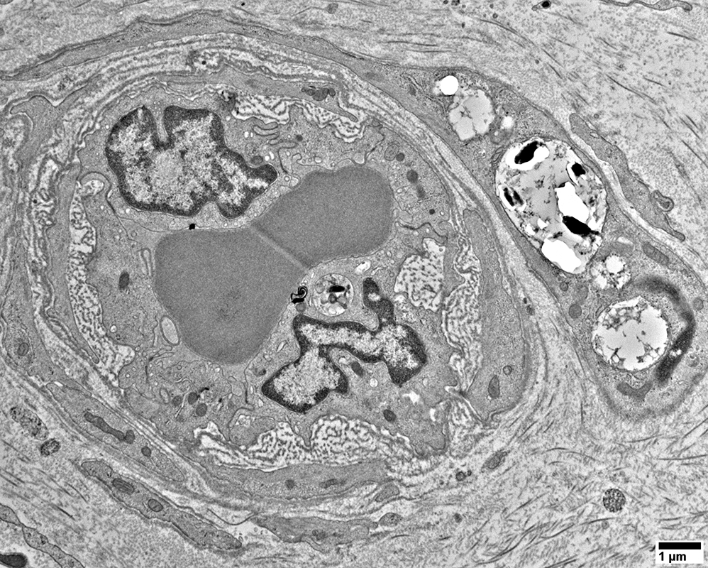
Endoneurial microvessels
Damaged wall with extracellular material between endothelial cells & pericyte processes
Leprosy bacilli: Within a Schwann cell (Upper right)
Schwann cell
Extends process around endoneurial microvessel
Has no associated axon
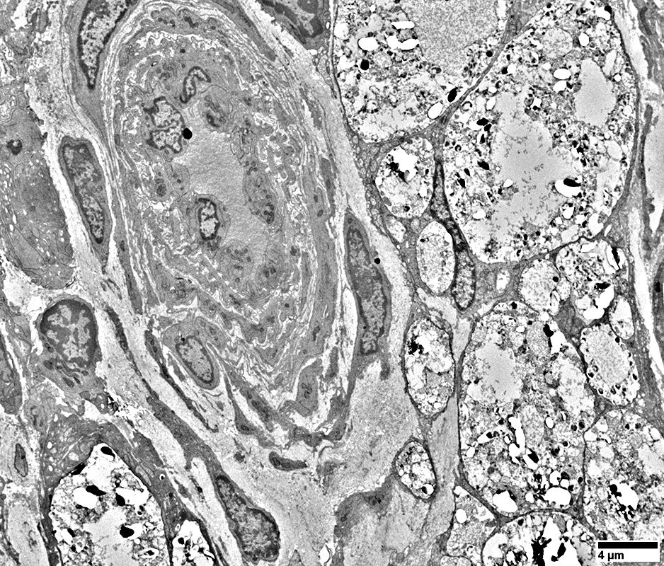 From: R Schmidt |
Damaged Wall
Pericyte processes: Multiple; Small; Separated by irregular connective tissue
Lepra cell
Large
Near vessel wall
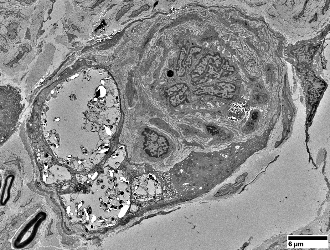 From: R Schmidt |
Damaged Wall
Pericyte processes: Multiple; Small; Separated by irregular connective tissue
Lepra cell
Large
Immediately adjacent to vessel wall
Remaining Myelinated Axons
Thin myelin for Axon size: Consistent with axon regeneration
Leprosy: Other Nerve Constituents
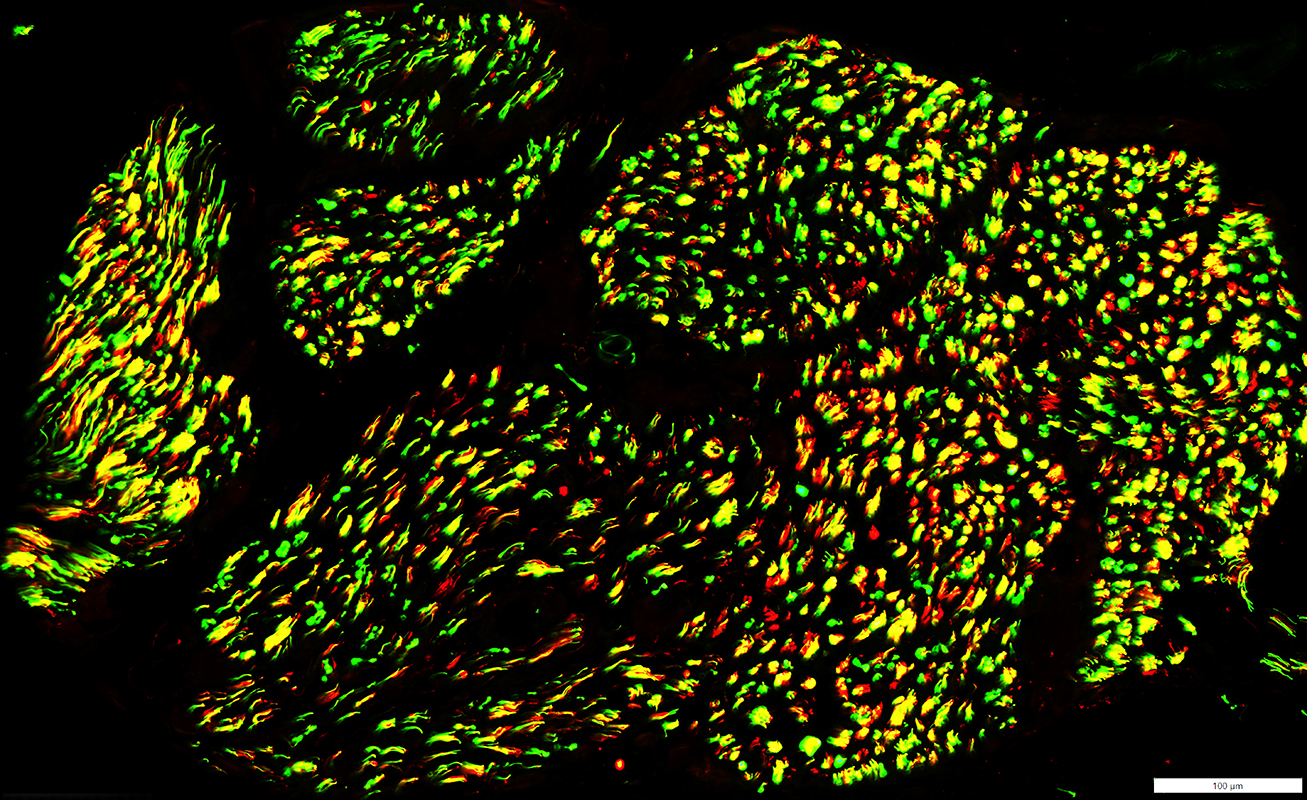
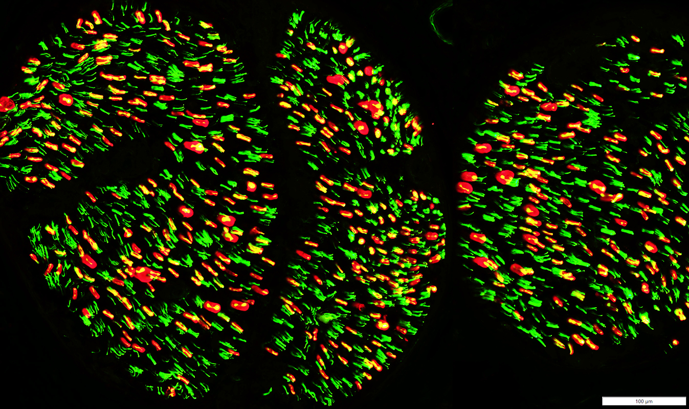 Neurofilament (Green) - P0 protein (P0) (Red) |
Large Myelinated Axons
Most are lost
Small Myelinated Axons
Most remaining myelinated axons are small
Small, Unmyelinated Axons
Most are preserved
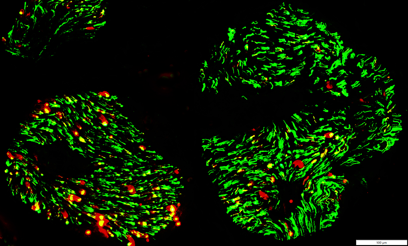 Neurofilament (Green) - Myelin Basic Protein (MBP) (Red) Axons Large Myelinated Axons Most are lost Small Myelinated Axons Most remaining myelinated axons are small & surrounded by MBP myelin: Suggests axon regeneration Differential loss among fascicles Small, Unmyelinated Axons Most are preserved |
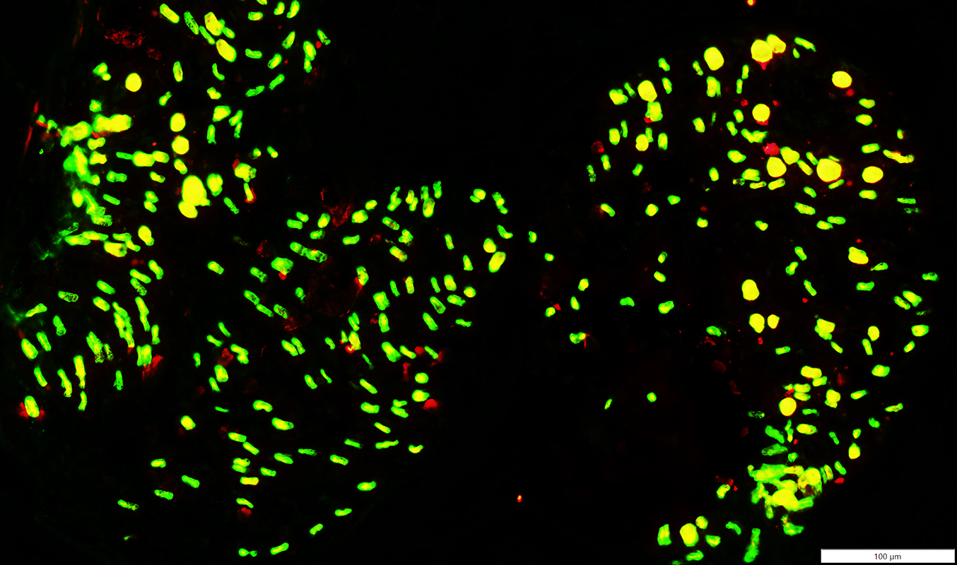
 Neurofilament (Green) - NCAM (Red) |
Larger Axons
Often have co-staining for NCAM: Suggests myelin that forms after axon regeneration
Small Axons
Mostly preserved: Few, scattered empty non-myelinating Schwann cells without axons (Red)
 P0 (Green) - Myelin Basic Protein (MBP) (Red) |
Most small myelin sheaths abnormally stain for MBP and P0: Suggests thin myelin that forms after axon regeneration
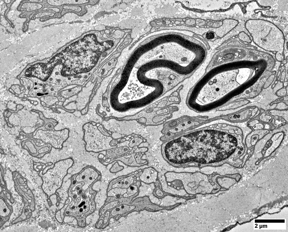 From: R Schmidt |
Myelinated
Myelin thin for axon size; Consistent with previous regeneration
Surrounded by Schwann cell processes consistent with axon regeneration through previous Büngner bands
Unmyelinated: Some small axons still present
Büngner bands: Clusters of Schwann cell processes; Consistent with axon loss
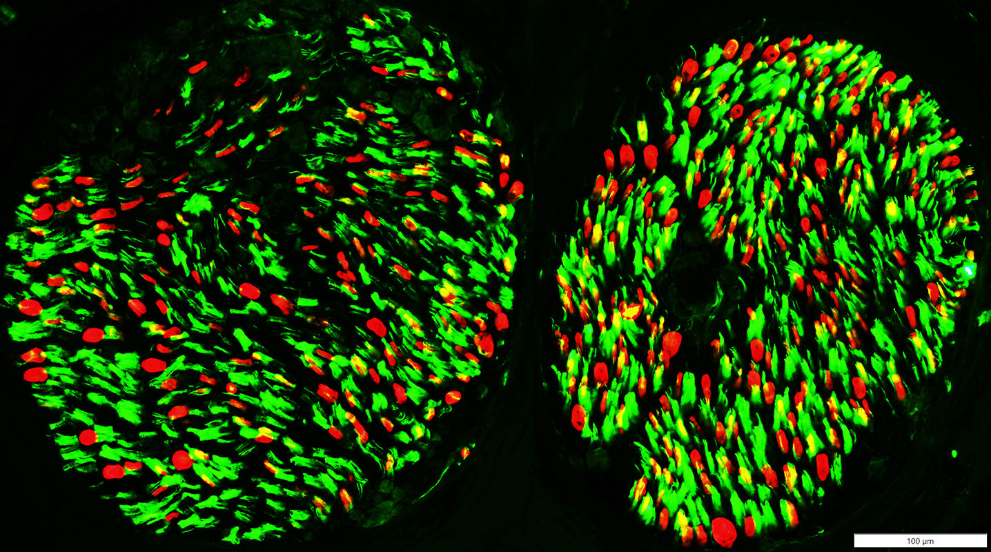 NCAM (Green) - P0 (Red) |
Reduced numbers of Myelin sheaths (Red)
Büngner bands (Yellow) NCAM - P0 costaining): Few
Non-myelinating Schwann Cells (Green): Reduced numbers, or absent, in some focal regions consistent with areas of leprosy lesions
Endoneurial Histiocytes
Dark cytoplasm
No intracellular leprosy bacilli
Near perineurium (Right)
Lepra cell (Bottom)
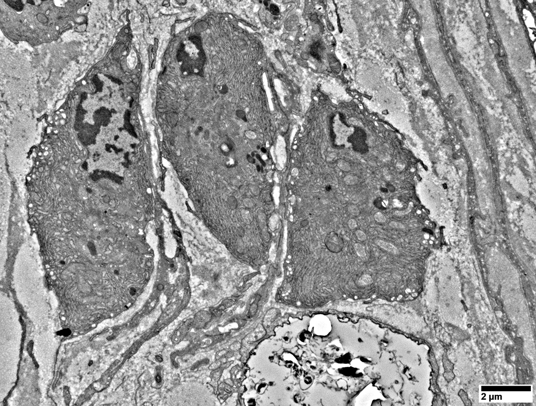 From: R Schmidt |
Return to Leprosy
7/26/2026