Large Histiocyte Immune Myopathy (LHIM) 1
|
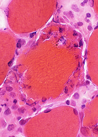
Necrosis Peripheral Histiocytic cells |
Nosology: Macrophage Activation Syndromes (Hemophagocytic Lymphohistiocytosis)
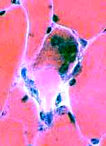
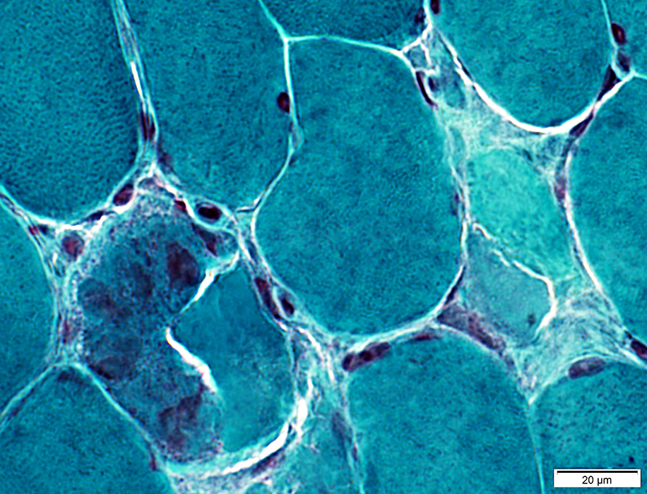
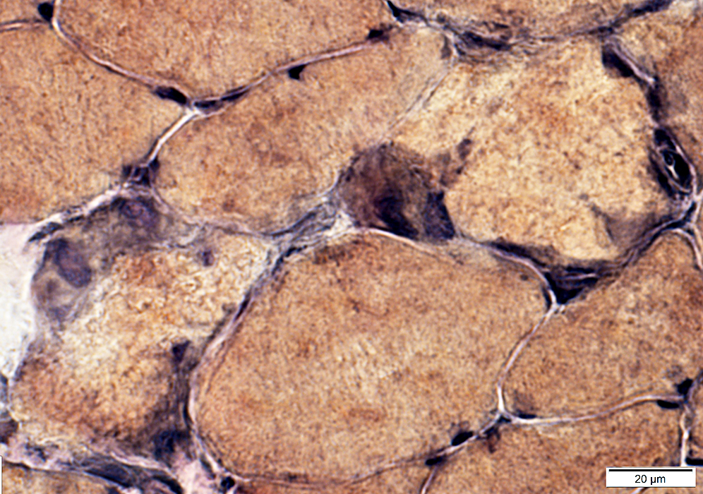
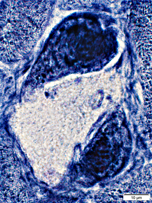
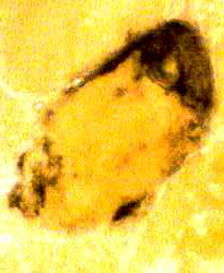
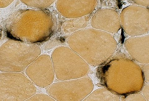
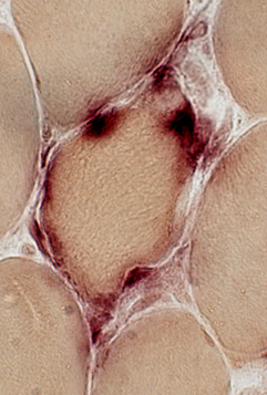
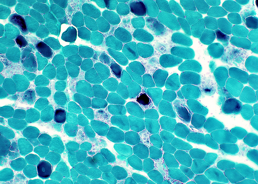 Gomori trichrome stain |
Scattered
Dark-stained or Pale
Some are associated with large cells (Arrow; Histiocyte-like)
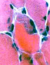
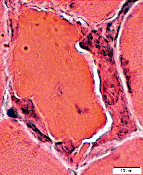
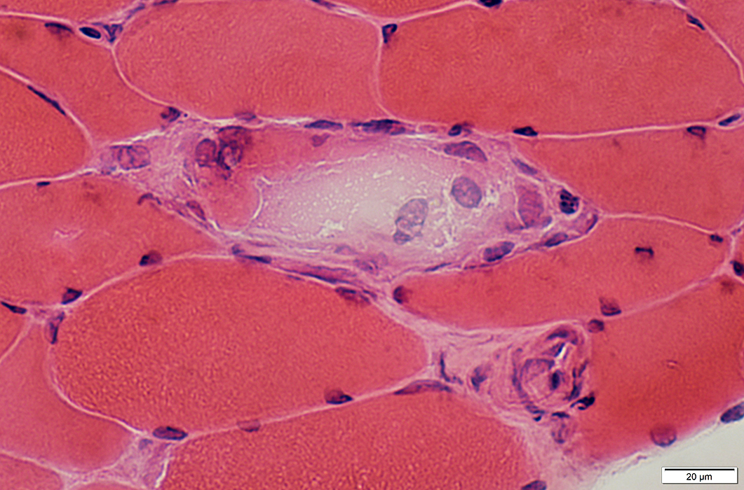
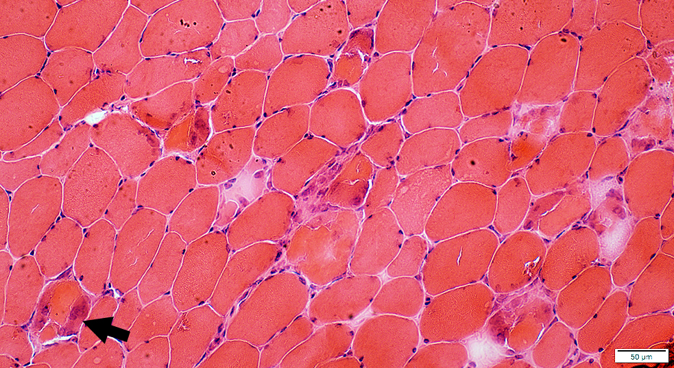 H&E stain |
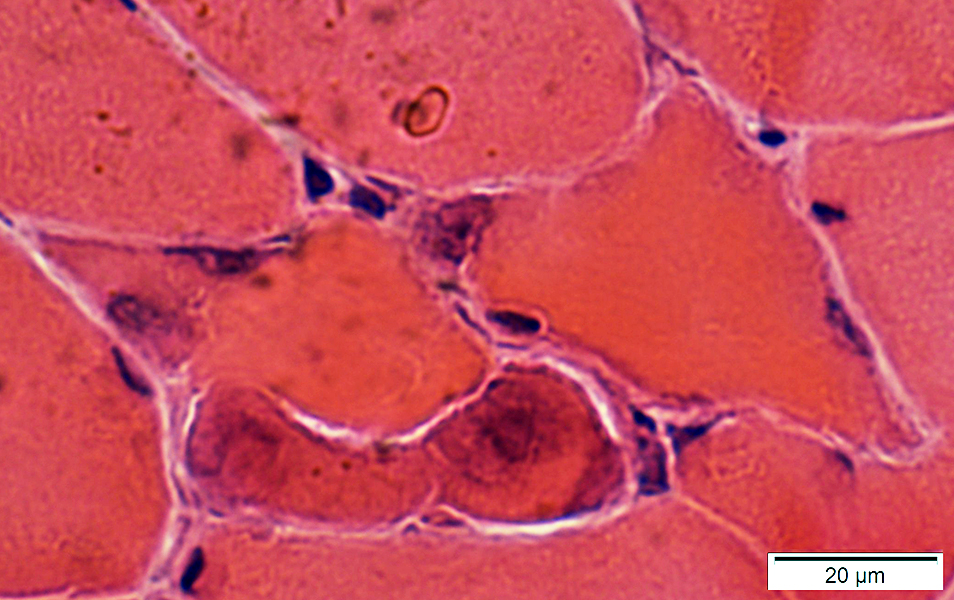
Necrotic muscle fibers
 H&E stain |
Necrosis Pattern: Scattered Muscle Fibers
 C5b-9 stain |
Myophagocytosis
 |
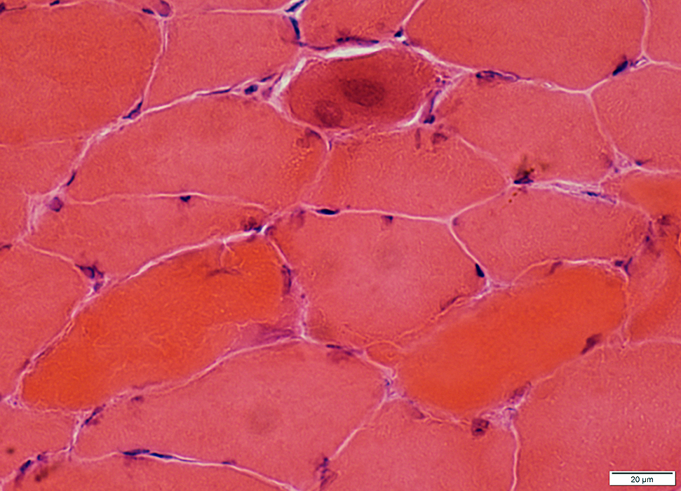
Muscle fibers are necrotic with a hyaline appearance.
 H&E stain |
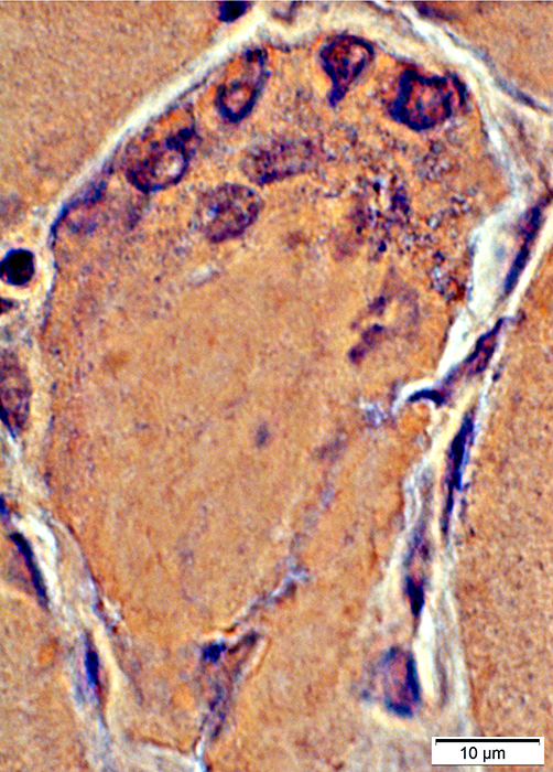
 Congo red stain |
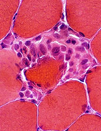
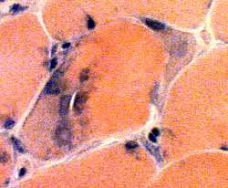
Myophagocytosis: Muscle fiber Pathology
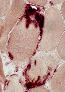
 Gomori trichrome stain |
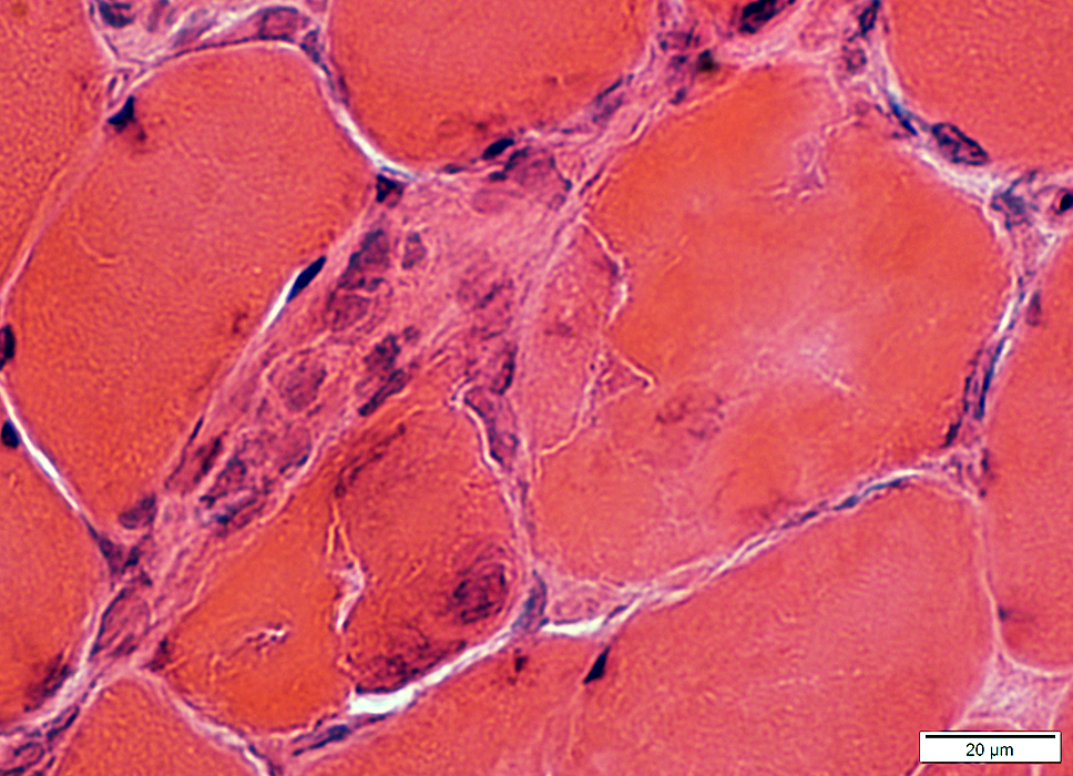
Multinucleated & large
Neighbor necrotic muscle fibers
 H&E stain |
 Congo red stain |
 H&E stain |
 H&E stain |
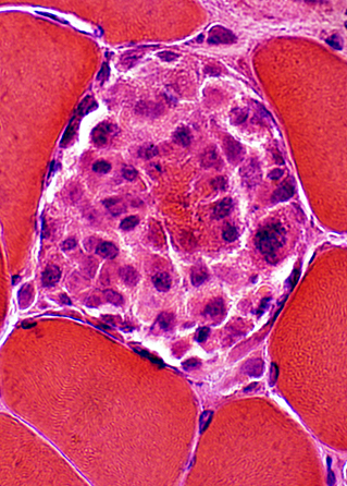
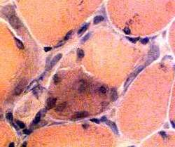
Multinucleated large cells, neighbor necrotic fibers
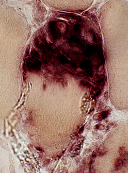
 H&E stain |
 VvG stain |
Multinucleated large cells, neighbor necrotic fibers
VvG stain
VvG stain
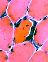
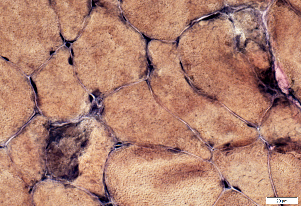
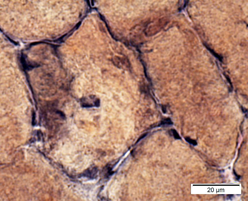
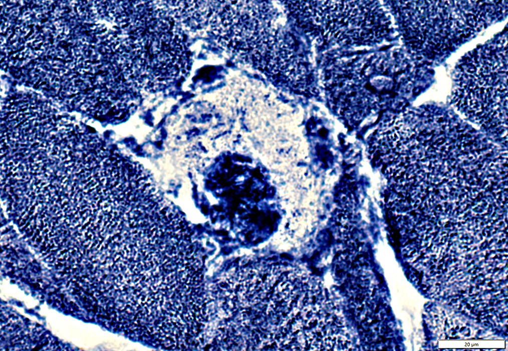
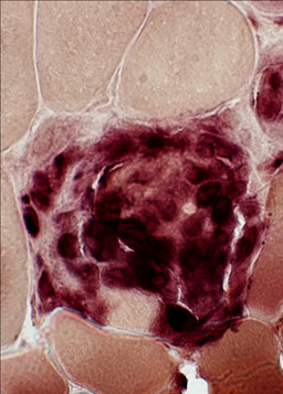
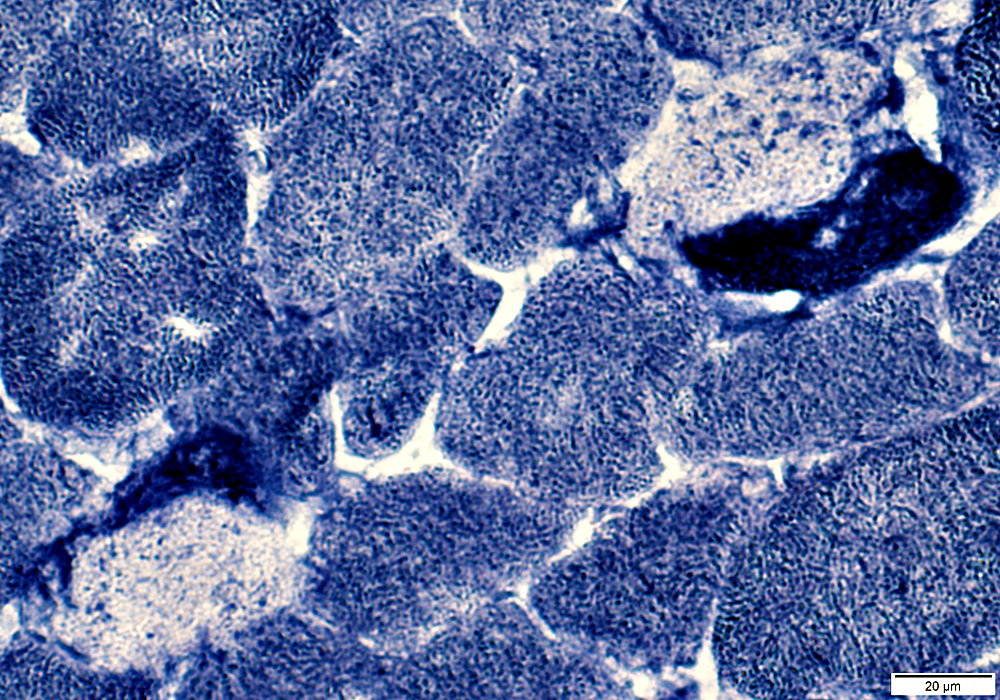 NADH stain |
|
Necrotic muscle fibers Pale Loss of NADH staining Large histiocytic cells Neighbor necrotic fibers May be NADH positive 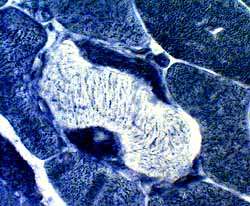 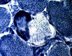 NADH stain |
 NADH stain |
 NADH stain |
Muscle Fiber Necrosis: Patterns & Relation to Histiocytes
LHIM: Necrotic muscle fiber cytoplasm is mainly at periphery of muscle fibers
Typical Necrosis in other disorders: Necrotic muscle fiber cytoplasm diffusely in muscle fibers (Bottom Right)
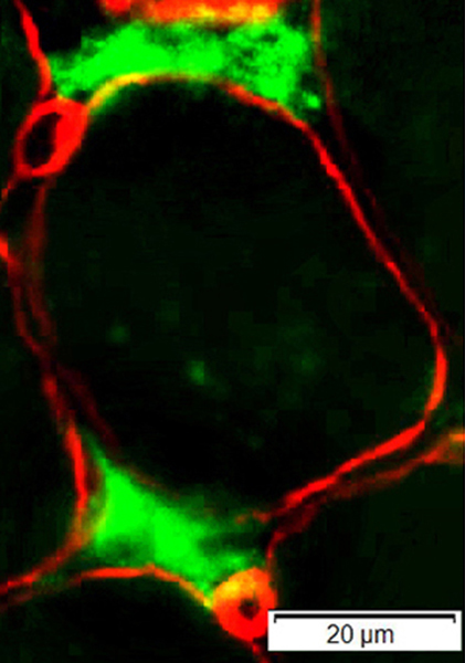
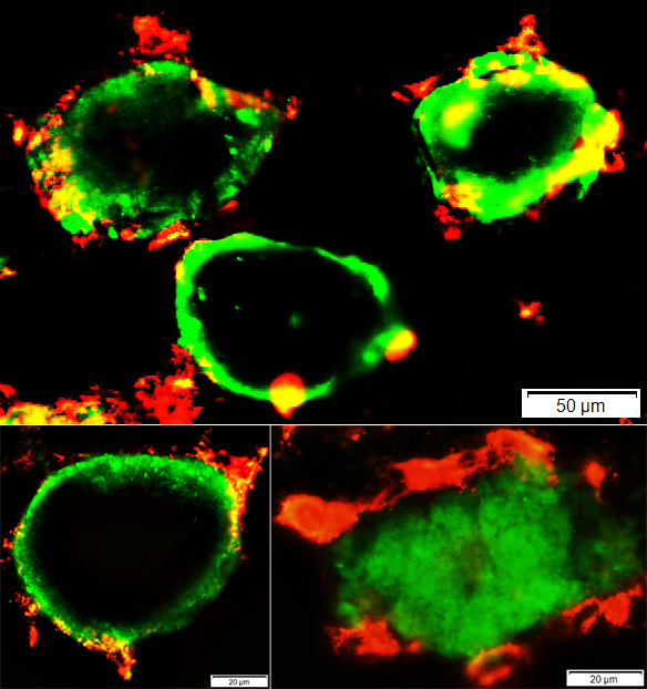 C5b-9 green; HAM56 red |
C5b-9 stained (Green) cytoplasm: In periphery of muscle fibers
Necrosis pattern typical of other disorders (Bottom right): C5b-9 diffusely stains cytoplasm of necrotic muscle fiber
Muscle Fiber Necrosis
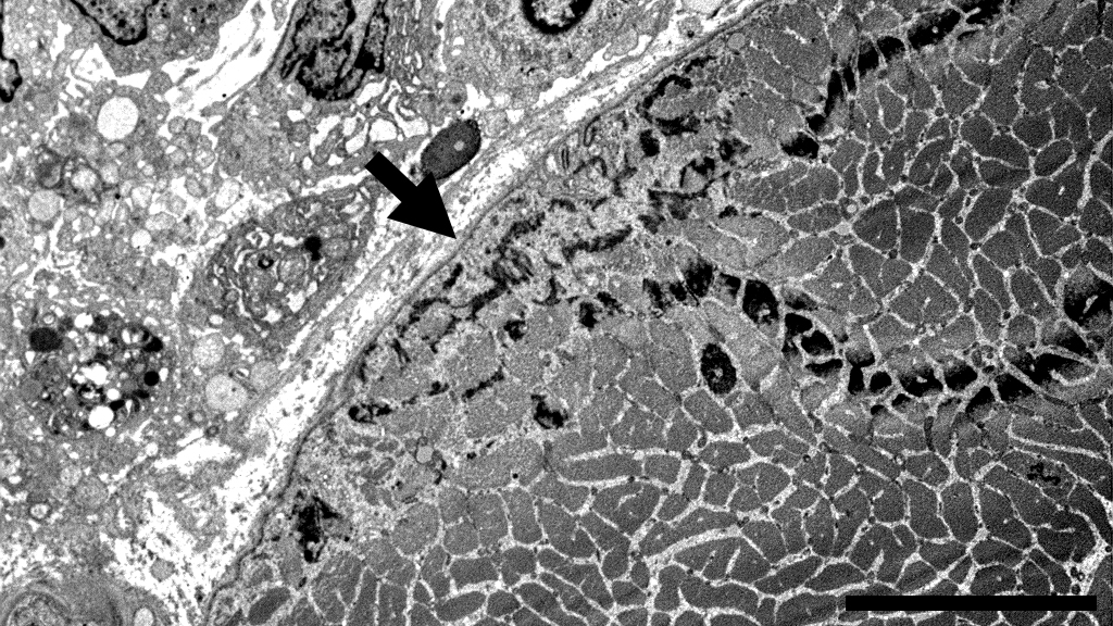 Bar = 10 µM |
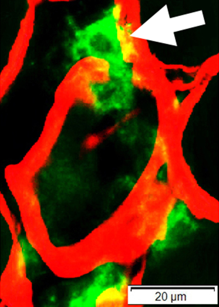
Histiocytic cells: Present in neighboring endomysial connective tissue.
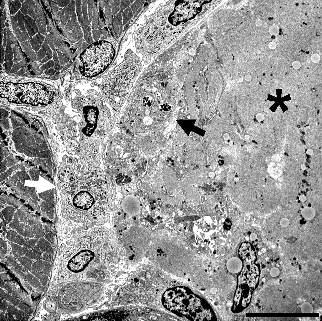 Bar = 10 µM |
Surrounding (White arrow), apposed to, & invading (Black arrow), a necrotic muscle fiber (*)
Necrotic muscle fiber
Pale cytoplasm
Almost complete dissolution of myofibrils and sarcoplasmic organelles
Non-necrotic muscle fibers: Located at left side in image
 Bar = 2 µM |
Surrounded by intact basal lamina
Misoriented sarcomeres with Z-bands (Arrow) & glycogen are scattered, and organelles lost
Amorphous granular material has replaced muscle fiber cytoplasm
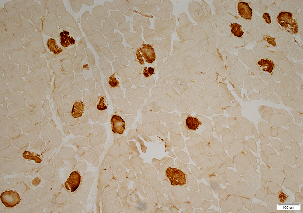
Alkaline phosphatase stain |
Histiocytic (Myophagocytic) cells
 Acid phosphatase stain Myophagocytic cells (red) progressively phagocytose & replace muscle fibers.. |
 CD68 green; Collagen IV red |
 CD163 green; Collagen IV red |
Outside (Above left) & Progressing through muscle fiber basal lamina (red; Arrow)
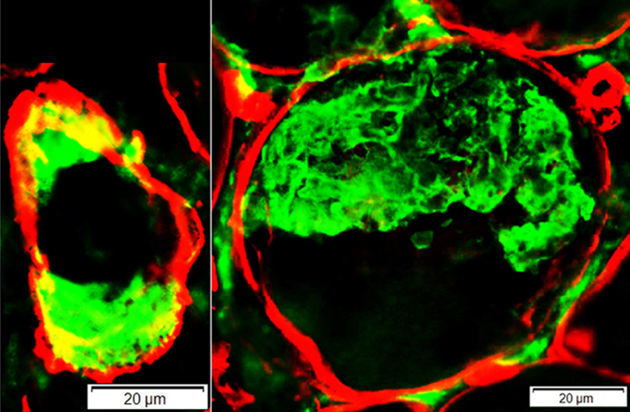 CD163 green; Collagen IV red |
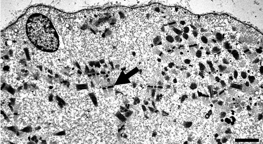
 Bar = 2 µM |
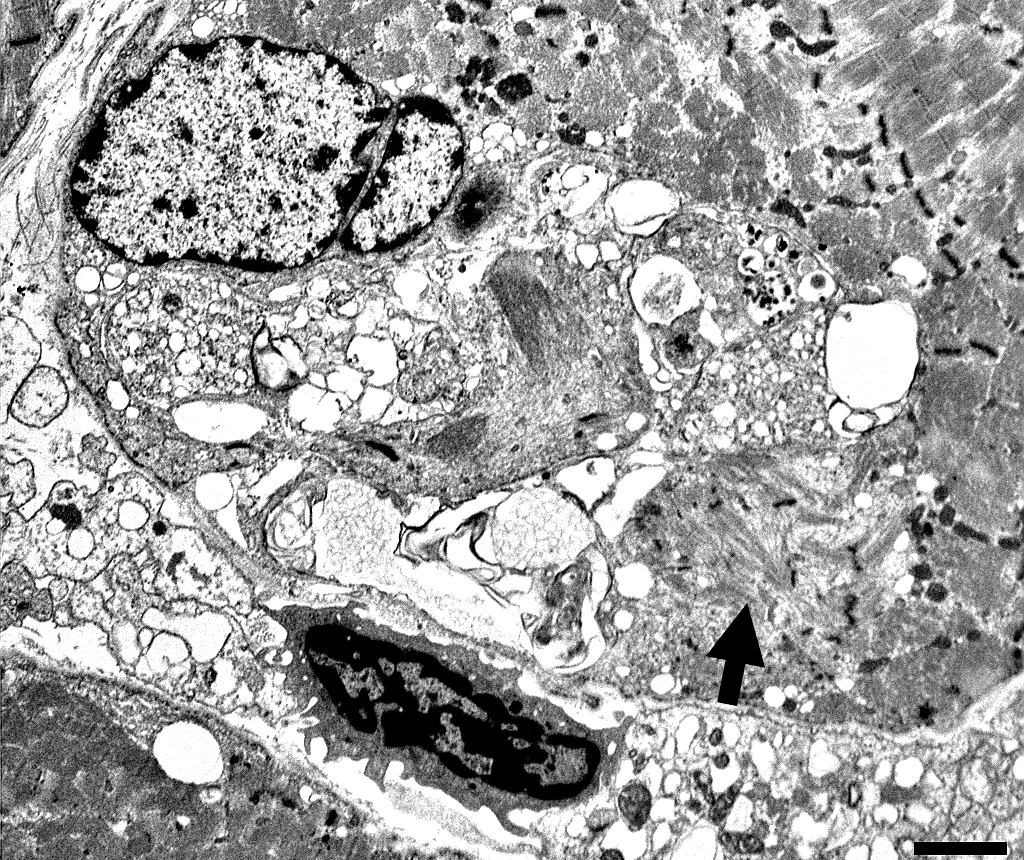
Large cells near myofibers
Surface membranes
Wavy
Pseudopodia
Thin, Irregular
Project between muscle fiber basement membrane & surface sarcolemmal membrane
Cytoplasm
Cntains abundant, variably-sized, irregularly-shaped membrane-bound phagocytic vacuoles
Vacuoles contain: Debris; Abundant smooth endoplasmic reticulum; Mitochondria
Nuclei
Irregular membranes
Markedly larger size than endomysial macrophages
Prominent nucleoli.
Some large histiocytic cells contain several nuclei
Muscle fiber: Small region of myofibrillar dissolution neighbors histiocytic cell (Arrow)
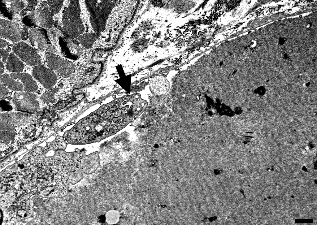
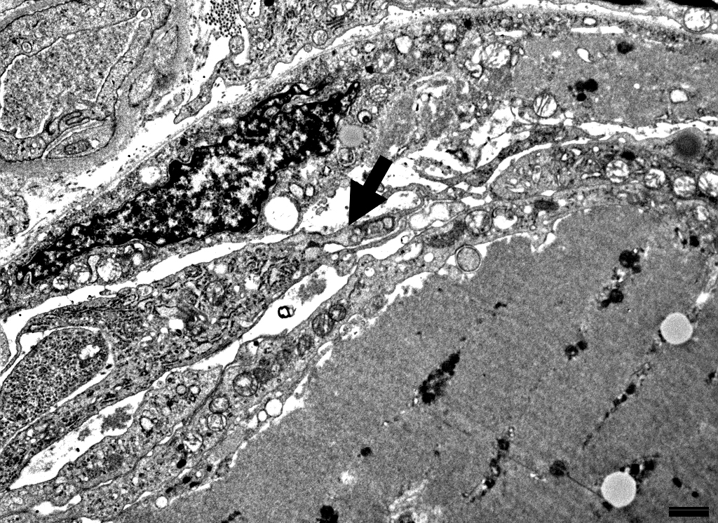 Bar = 2 µM |
Between basal lamina & Necrotic muscle fiber cytoplasm
Rich in organelles
Smooth endoplasmic reticulum
Mitochondria
Vacuoles: Clear or contain granular debris
|
Between basal lamina & Necrotic muscle fiber cytoplasm
Muscle fiber surface sarcolemma under histiocyte process
Disrupted or irregular
Return to Hemophagocytic lymphohistiocytosis
Return to Pathology index
Return to Neuromuscular Home Page
References
1. Neurology 2019;92:e1763-e1772
3/23/2021