Denervation with Reinnervation, Chronic Inactive
Muscle Fiber Type Grouping
Type groupingComparative ATPase stains
Staining patterns
Associated with, & Caused by, Axon Growth
Regeneration: Large fiber type groups
Collateral sprouting: Small fiber type groups
Denervation + Reinnervation, Chronic: Fiber Type Grouping
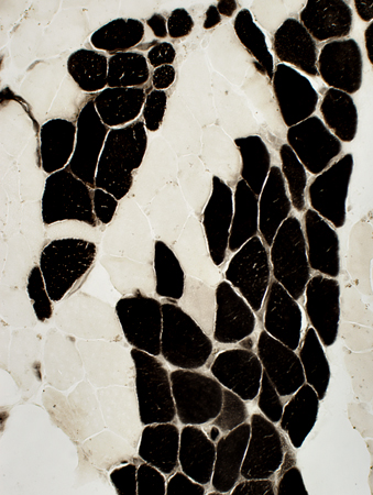
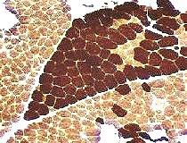  ATPase pH 9.4 stain |
 ATPase pH 9.4 stain |
|
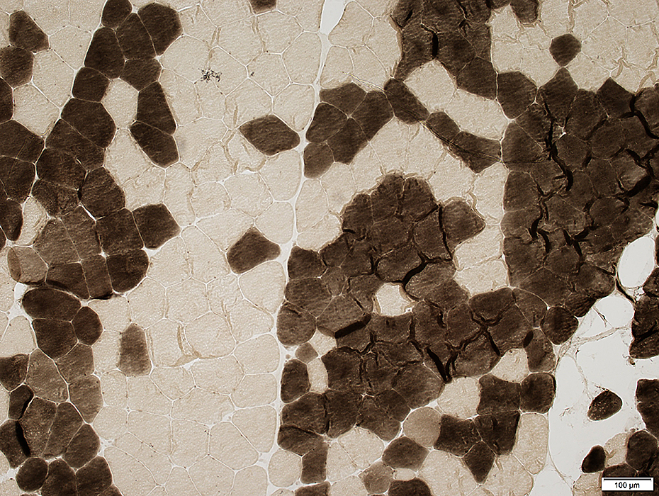
Fiber Type Grouping
Type I & Type II muscle fibers: Clustered in groups.
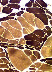
Type II fibers in this biopsy are mostly type IIB (ATPase pH 4.6 stain, below right).
 ATPase pH 4.3 stain |
 ATPase pH 4.6 stain |
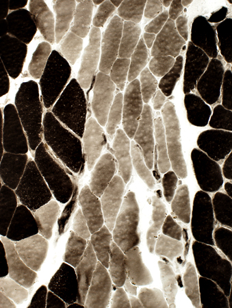
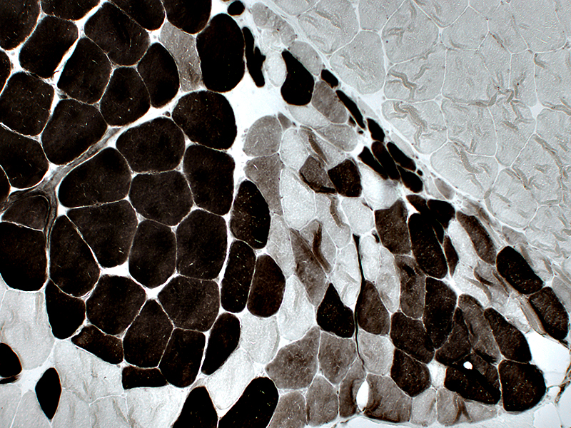
Fiber Type Grouping: Staining Patterns
 ATPase pH 9.4 stain |
Clusters of Type I & II fibers
No small muscle fibers or ongoing denervation
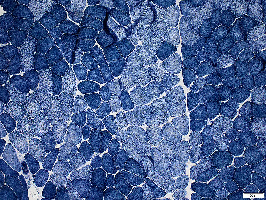 NADH stain |
 ATPase pH 4.3 stain |
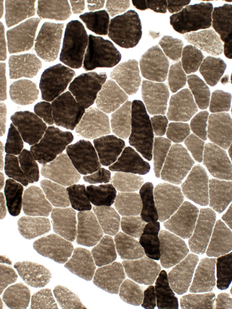
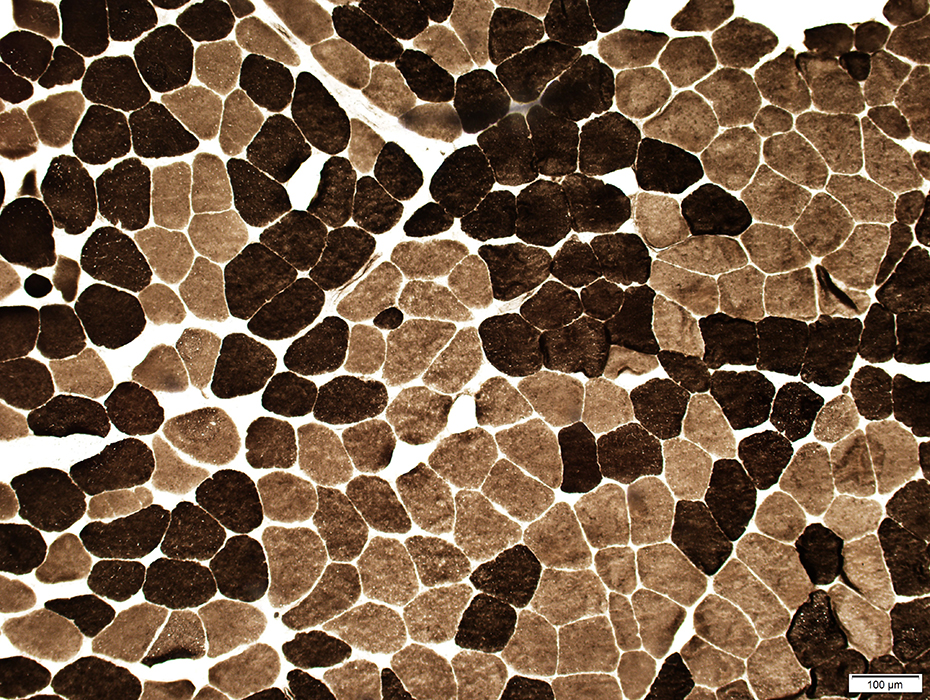
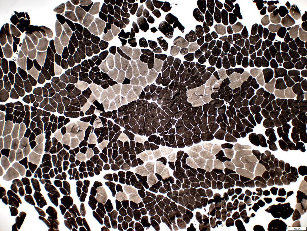
Fiber type grouping
Incomplete fiber type switching: Varied fiber types within a single group
 ATPase pH 4.3 stain |
Fiber type grouping with incomplete type switching: 2C fibers in clusters of grouped muscle fibers
 ATPase pH 4.3 stain |
Fiber Type Grouping: Comparative ATPase staining
Incomplete Fiber Type Switching within a region of Type GroupingFascicle in center of image (Dark arrows) has all type 2 fibers on ATPase pH 9.4 & 4.3, but varied subtypes at ATPase pH 4.6
 ATPase pH 9.4 stain |
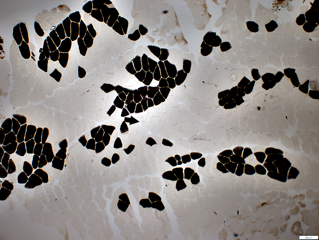 ATPase pH 4.3 stain |
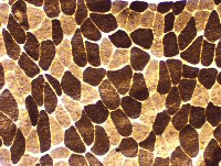
Fiber Type Switching during Grouping: Compare to ATPase pH 9.4 & 4.3
Incomplete Type Switching
Varied colors of type 2 fibers in individual groups (Black arrow)
Complete Type Switching
All fibers in a group are Type 2A (White arrow) or 2B (Red arrow)
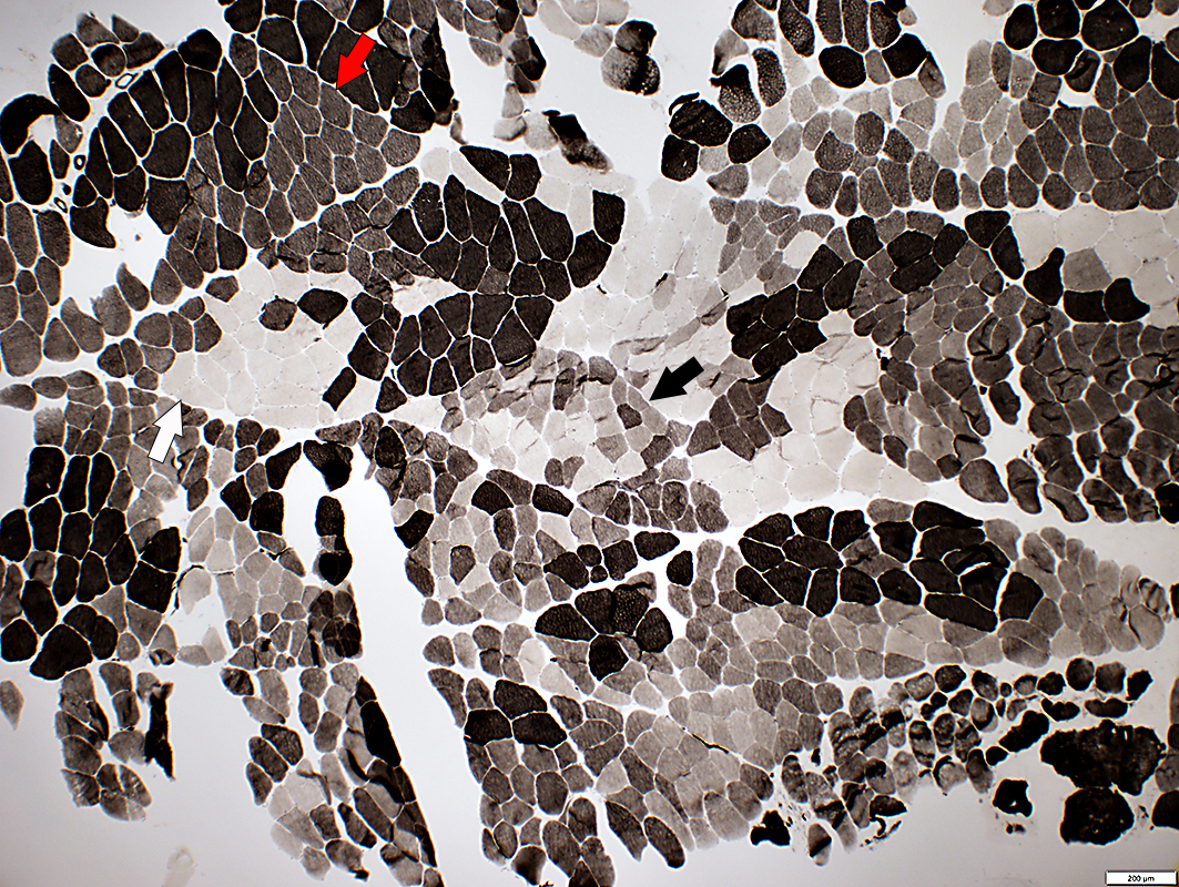 ATPase pH 4.6 stain |
Axon Loss
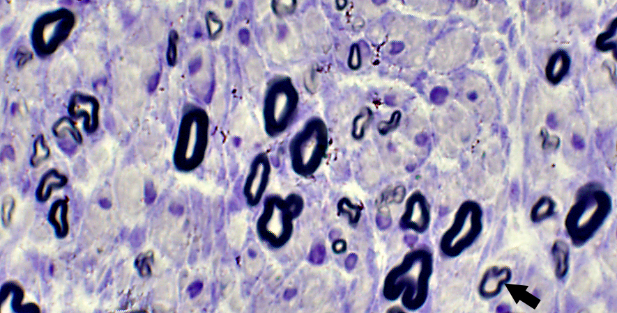
Toluidine blue stained plastic section
Nerve: Chronic Inactive Axon Loss (Denervation)
- Nerve has reduced numbers of large & small myelinated axons.
- Most large & small axons have normal myelin thickness.
- There are a few thinly myelinated (Arrow), regenerated axons.
Also see: Ongoing or Active Denervation
Return to Neuromuscular Syndromes
7/27/2023