Enterovirus-associated Inflammatory Myopathy (EVIM)
|
EVIM General pattern Cells (Inflammation) Connective tissue Muscle fibers Capillaries Also see Dermatomyopathies IMPP Dermatomyositis: Adult, IMPP Jo-1 muscle pathology |
EVIM: General Pattern
- Inflammation
- Cell types: Lymphocytes & Histiocytes
- Location: Vascular perimysium
- Connective tissue pathology
- Inflammation: Perimysium & Epimysium
- Structure: Mild damage
- Muscle fibers
- MHC I up-regulation
- COX- fibers
- Morphologic damage: Minor
- Capillaries
- Large
- Numbers: Mild loss
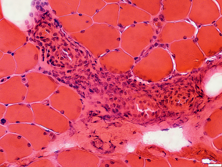
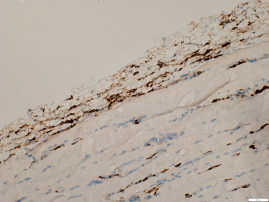
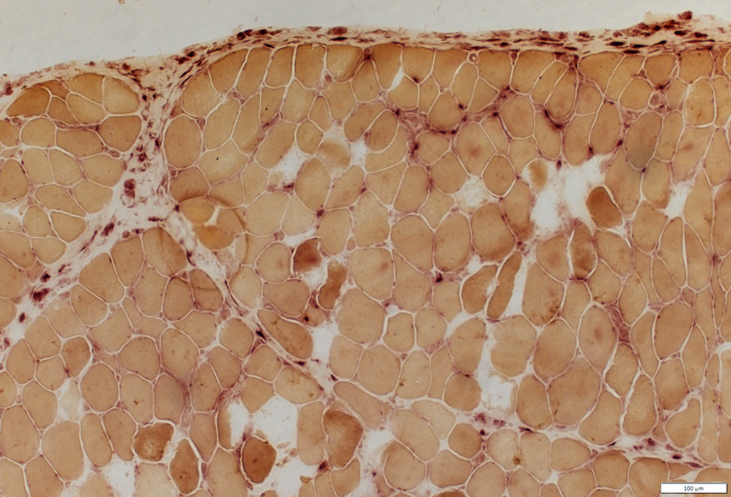
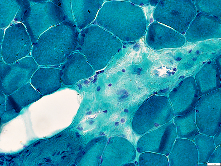
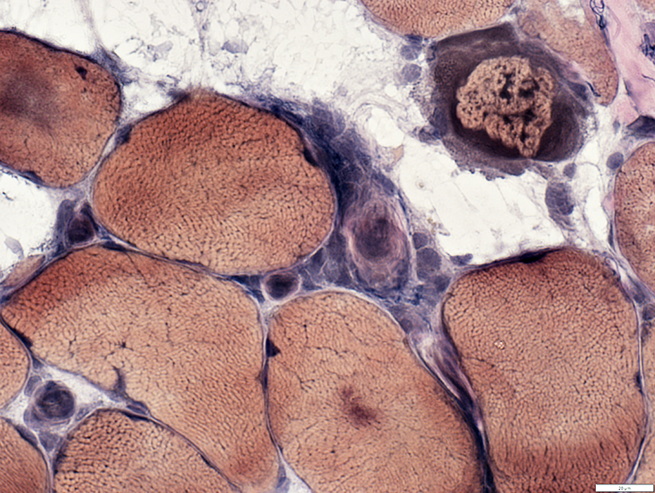
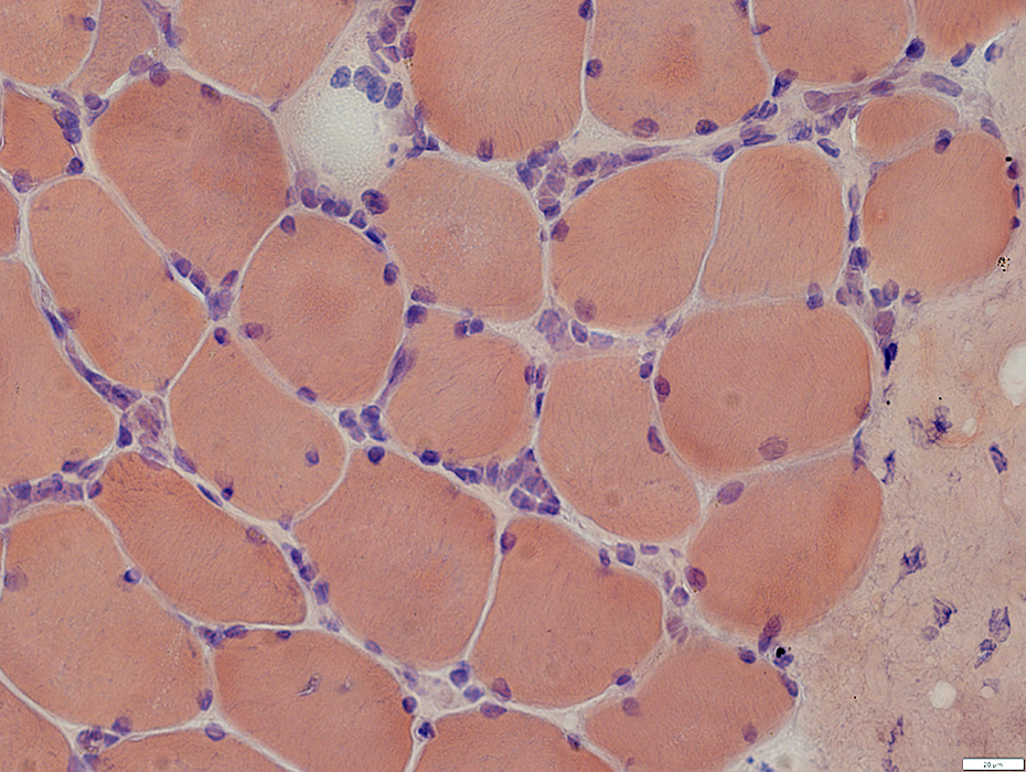
 H&E stain |
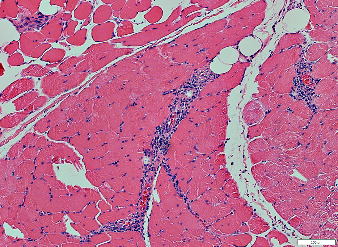
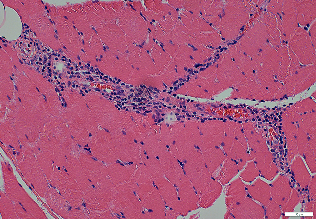
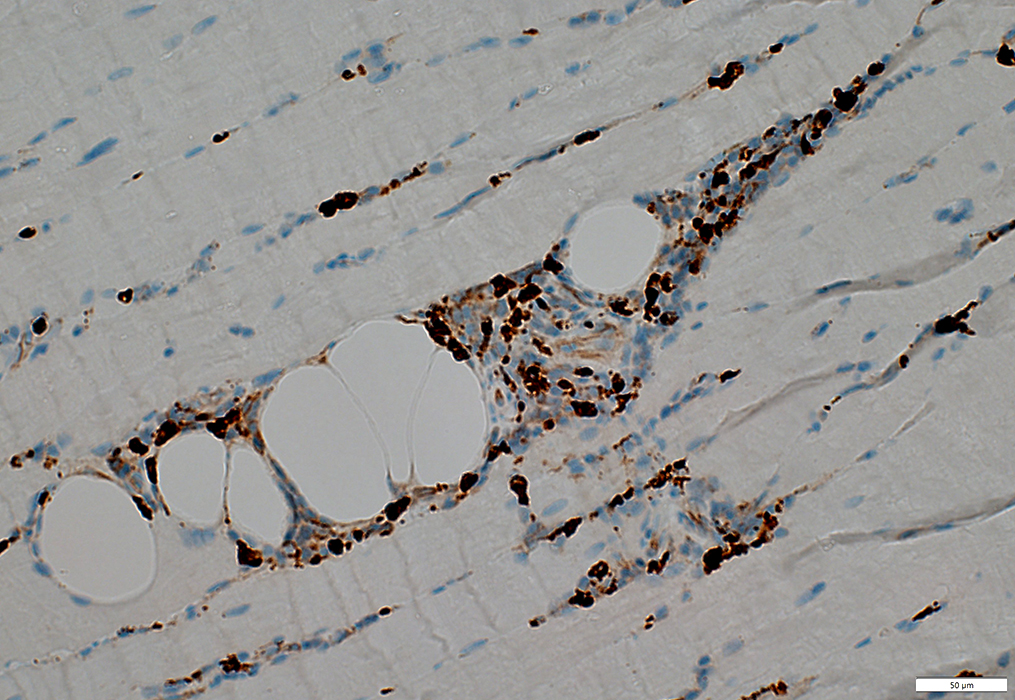
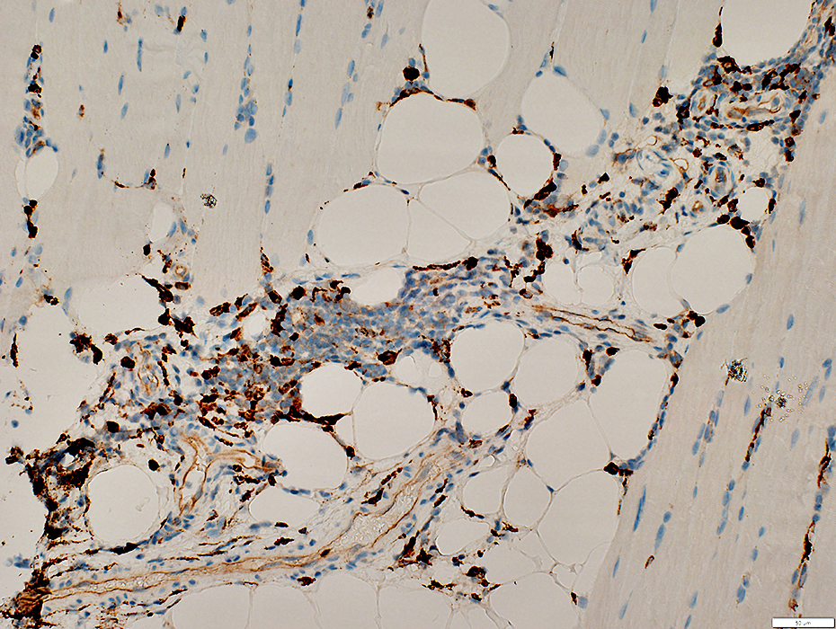
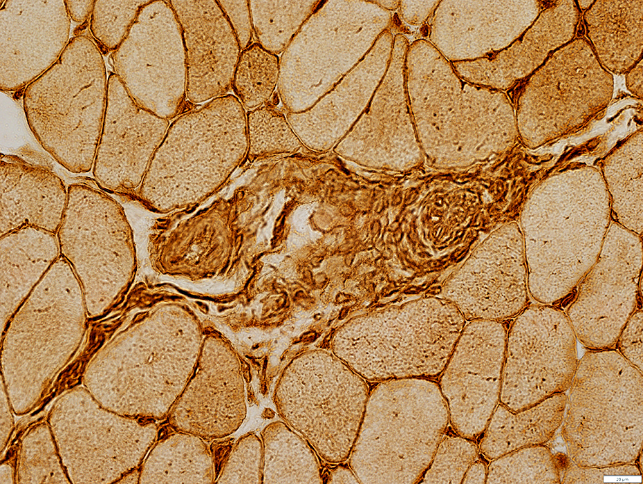
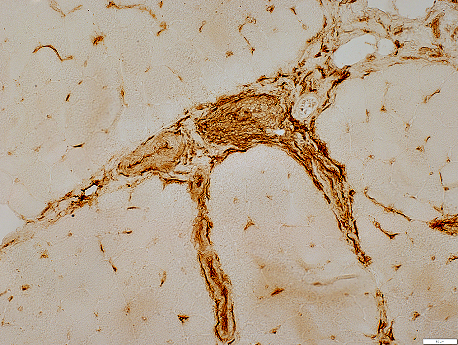
- Cell foci: Localized to vascular regions of perimysial connective tissue
- Muscle fibers: Mild, or no, involvement
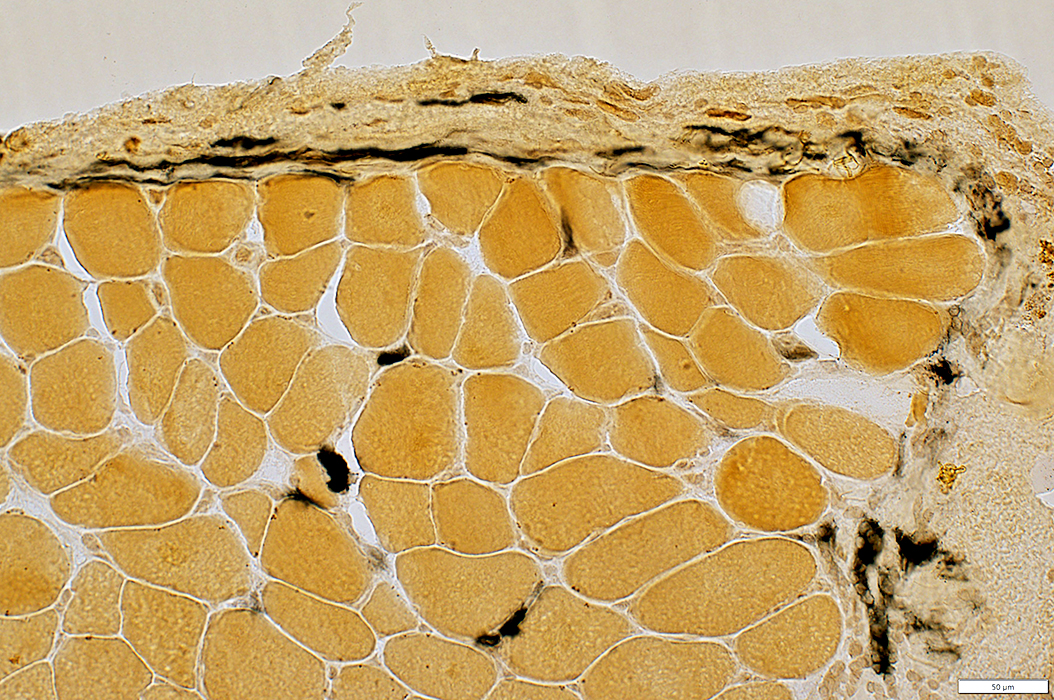
 H&E stain |
 H&E stain |
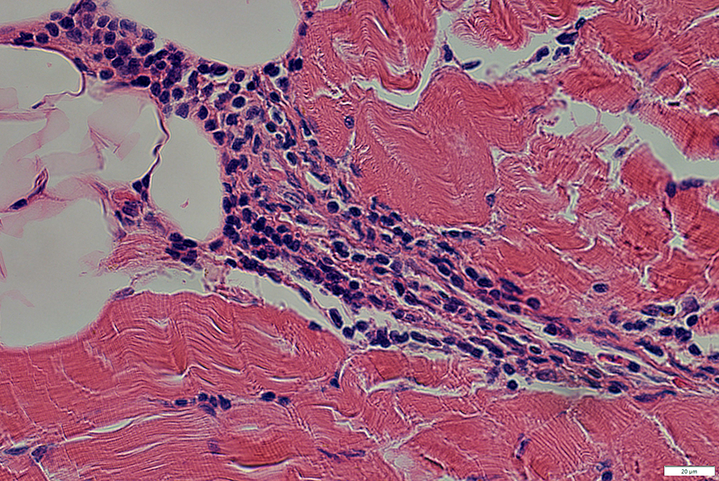
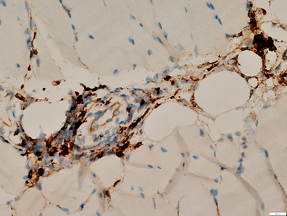
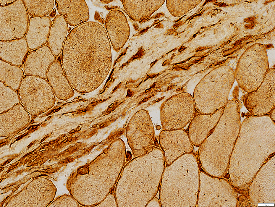
- Cells in perimysium are histiocytic (Above; Arrow). Features include
- Large nuclei
- Prominent cytoplasm
- Staining for Acid Phosphatase, Esterase & CD-68
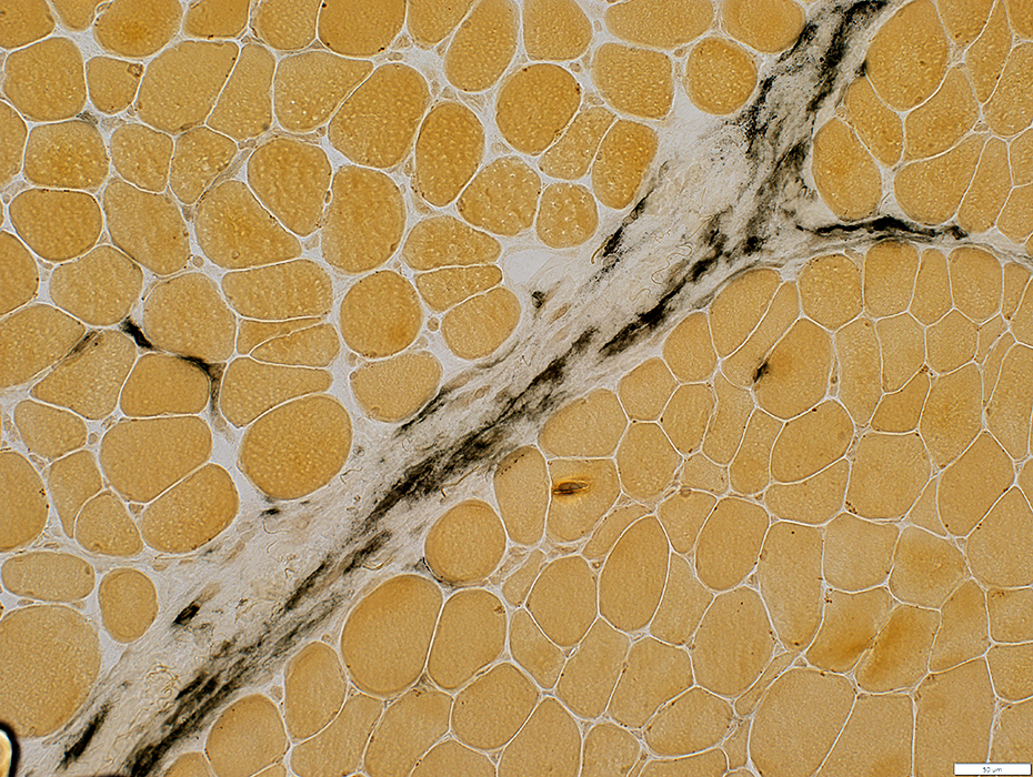
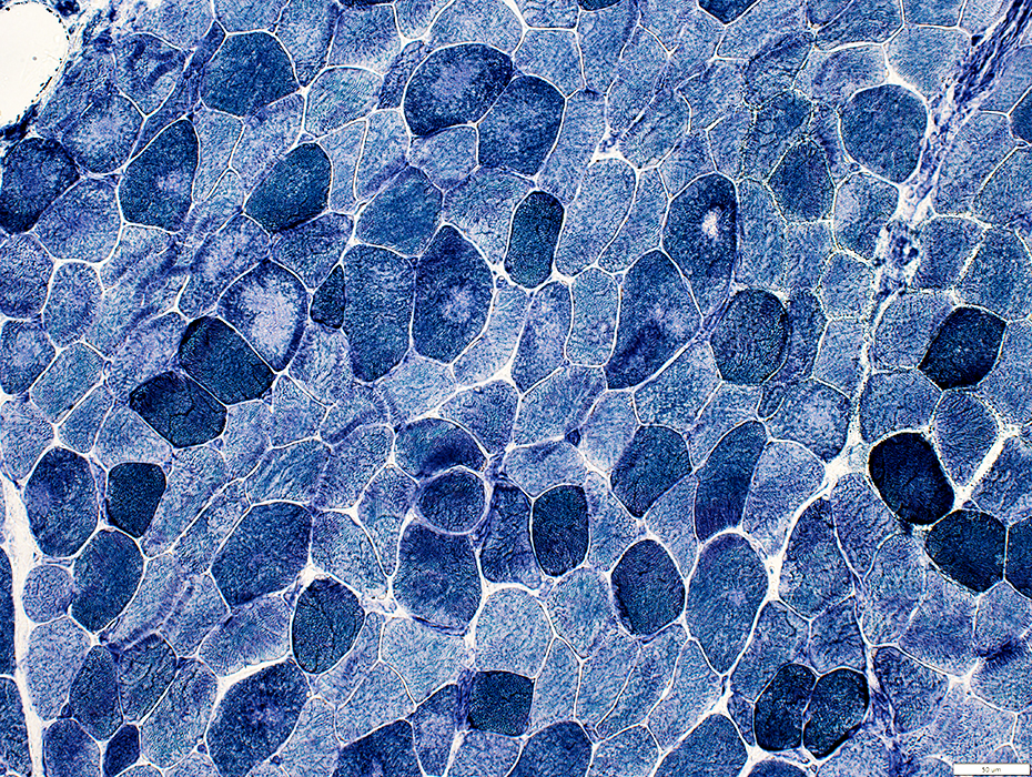
 H&E stain |
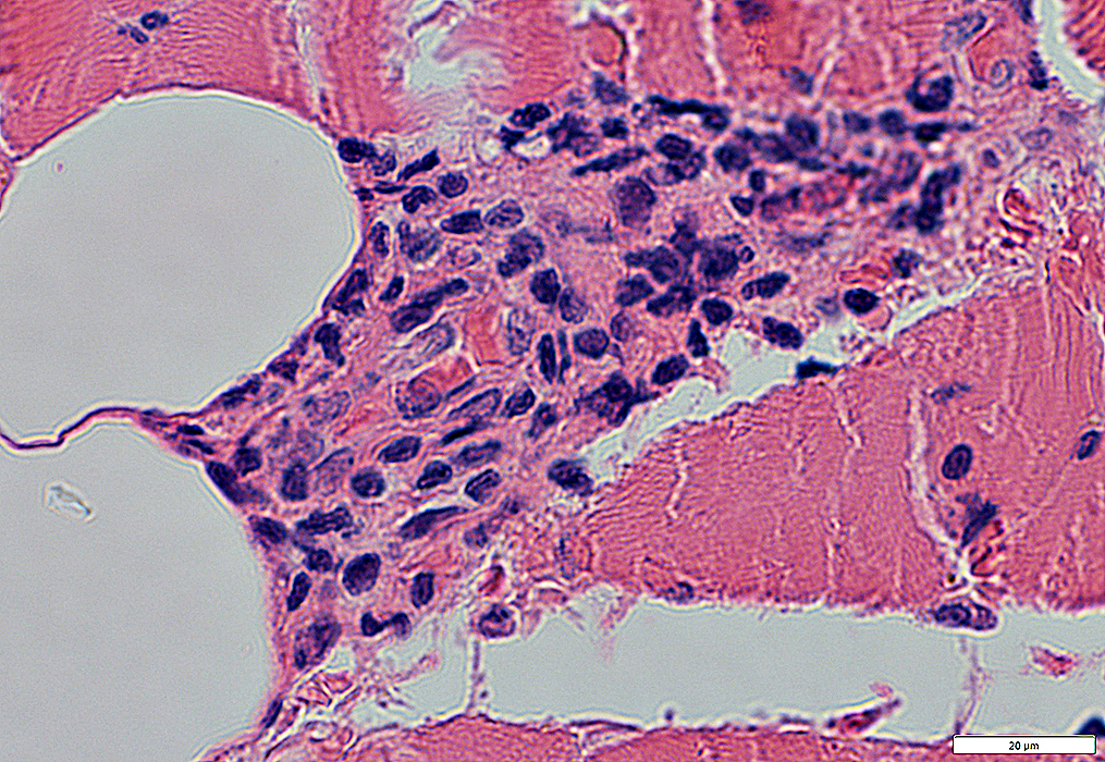
Nuclei: Moderately large
Cytoplasm: Moderate amount
 H&E stain |
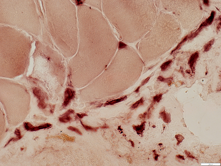
 CD3 stain |
May stain for CD3, CD4, CD68, or CD8
 CD4 stain |
 CD8 stain |
CD8 lymphocytes in perimysium & scattered in endomysium
 CD8 stain |
 Acid phosphatase stain |
Some are histocytic with Acid phosphatase & esterase staining
 Esterase stain |
 Esterase stain |
Some are histocytic with Esterase or CD68 staining
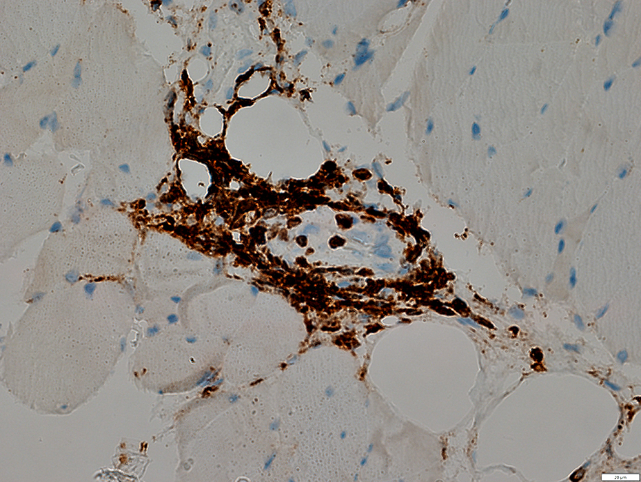
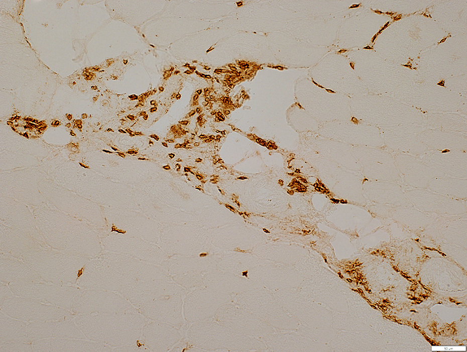
 CD68 stain |
Histiocytes stain for CD68
 CD68 stain |
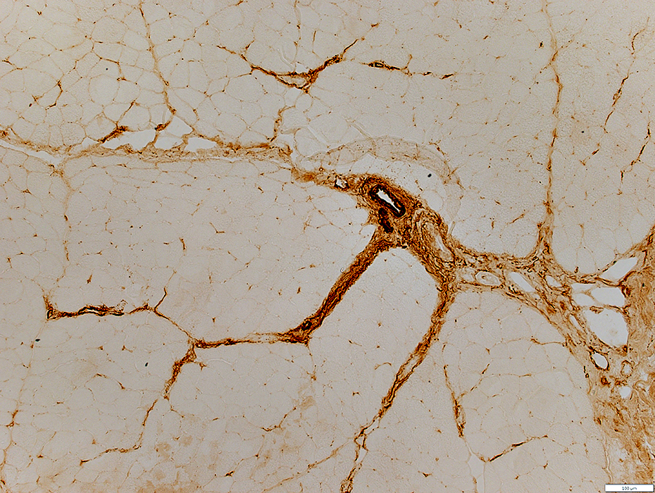
Histiocytes in perimysium stain for CD68
 CD68 stain |
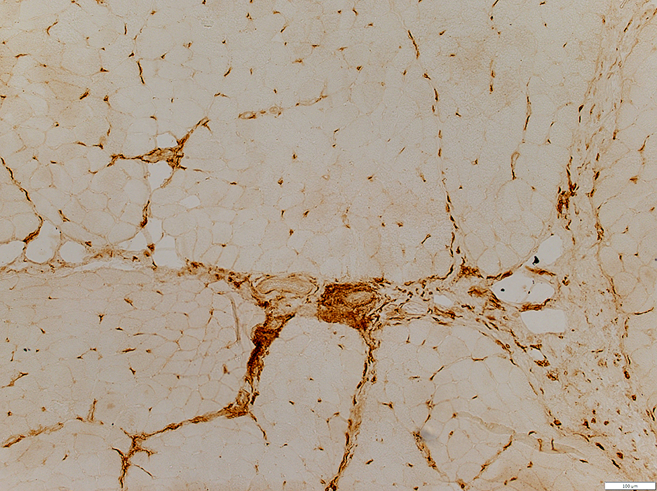
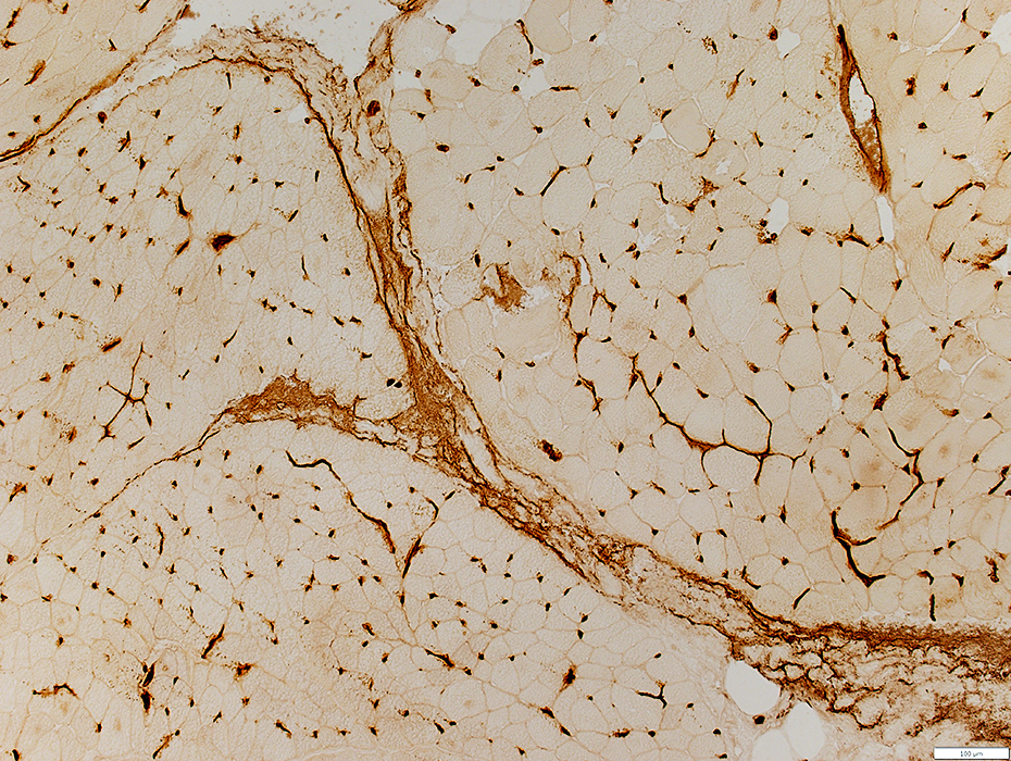
Connective tissue
 CD68 stain |
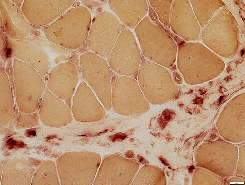
 Acid phosphatase stain |
Stains for Acid phosphatase+ cells & Alkaline phosphatase
 Alkaline phosphatase stain |
 Alkaline phosphatase stain |
Stains for alkaline phosphatase & C5b-9
 C5b-9 stain |
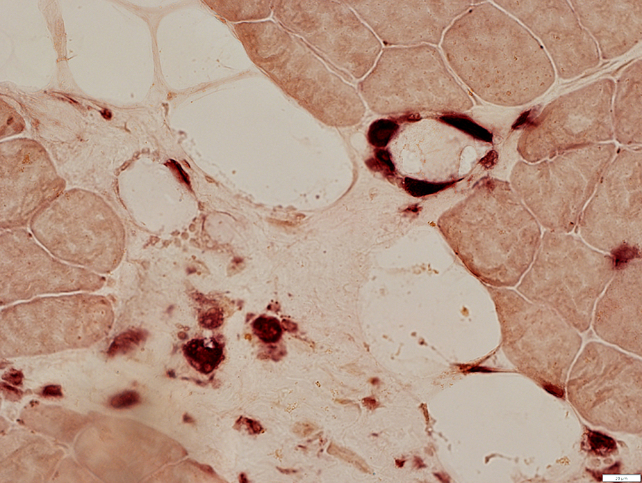
 UEA-I stain |
Unusual staining for UEA-I lectin (Usually endothelial stain)
 UEA-I stain |
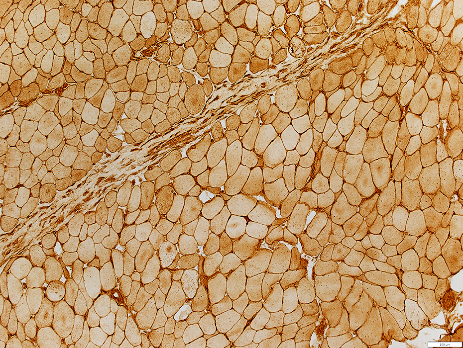
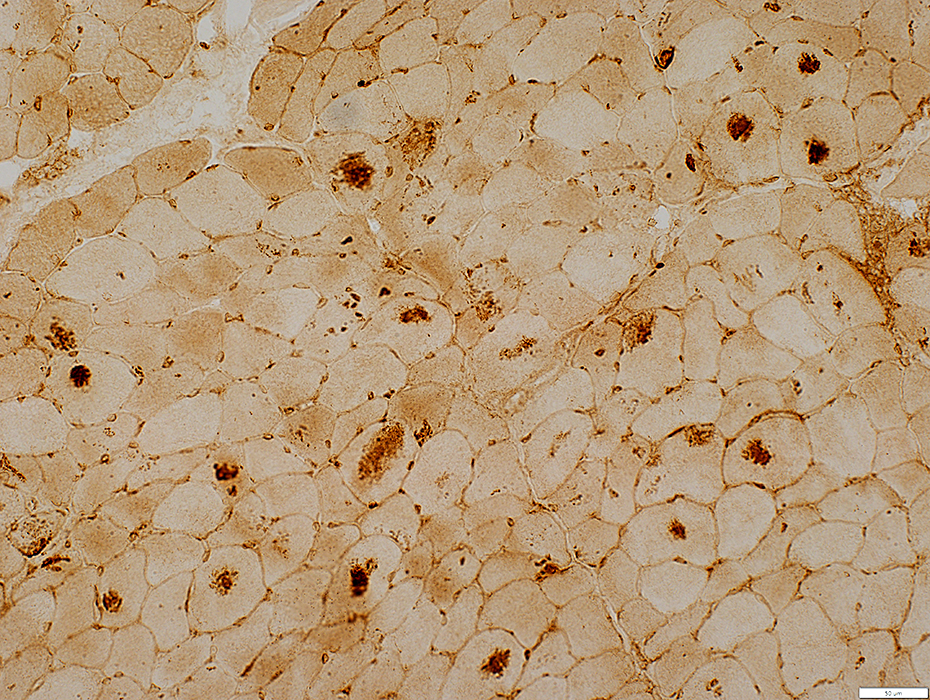
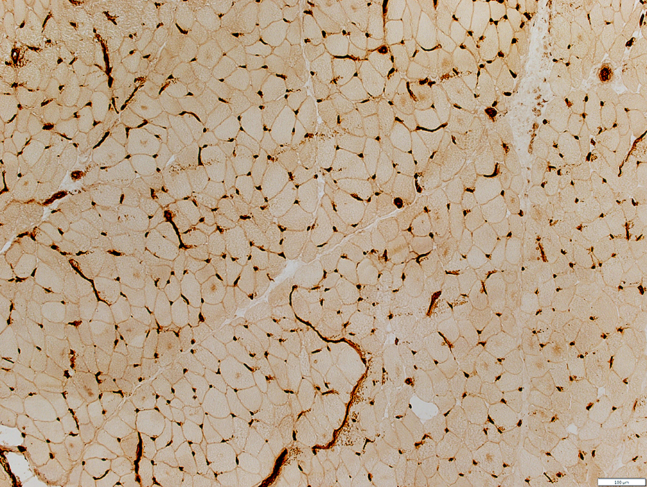
 MHC Class I stain |
Upregulated on muscle fibers
Stains cells in perimysium
 MHC Class I stain |
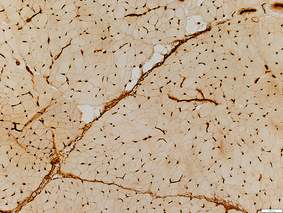
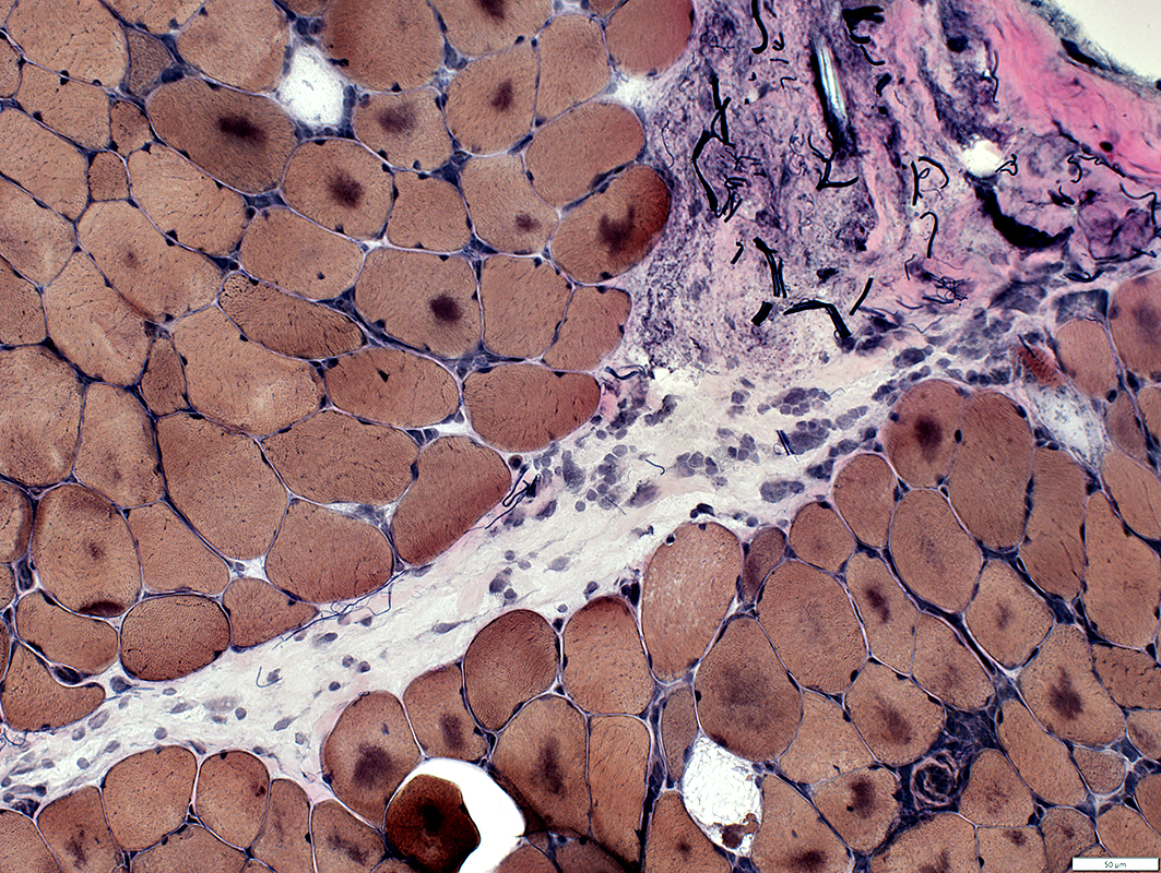
Perimysial connnective tisue structure
May be mildly damaged
 Gomori trichrome stain |
Muscle fibers: Pathology
 MHC Class I stain |
MHCI: Diffusely up-regulated on muscle fibers
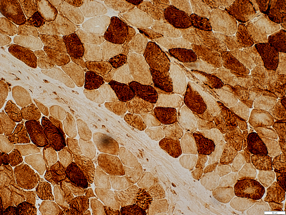
 ATPase ph 4.3 stain |
Increased numbers of immature, 2C fibers
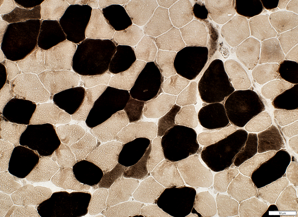
 COX stain |
Some fibers have reduced COX staining
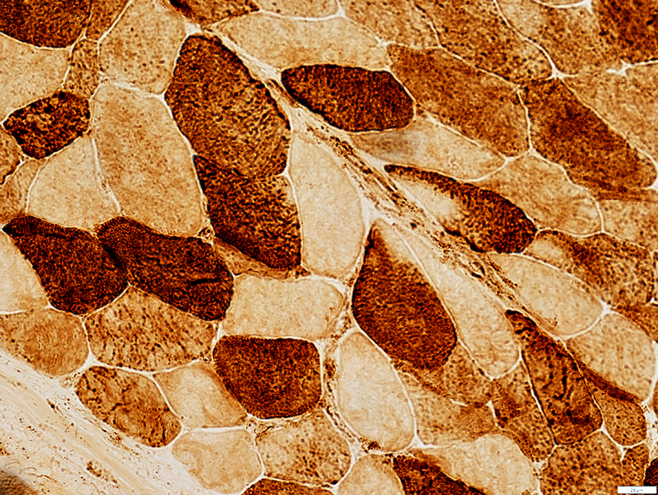 COX stain |
 VvG stain |
Stain dark on VvG & LC3
 LC3 stain |
 LC3 stain |
 VvG stain |
 NADH stain |
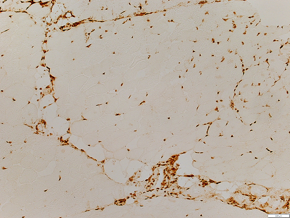
Endomysial Capillaries
 UEA-I stain |
Mildly large & dark-stained
Numbers: Mildly reduced; a few muscle fibers have no adjacent capillary
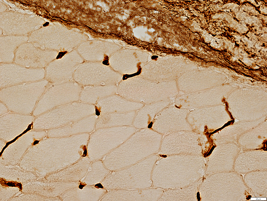 UEA-I stain |
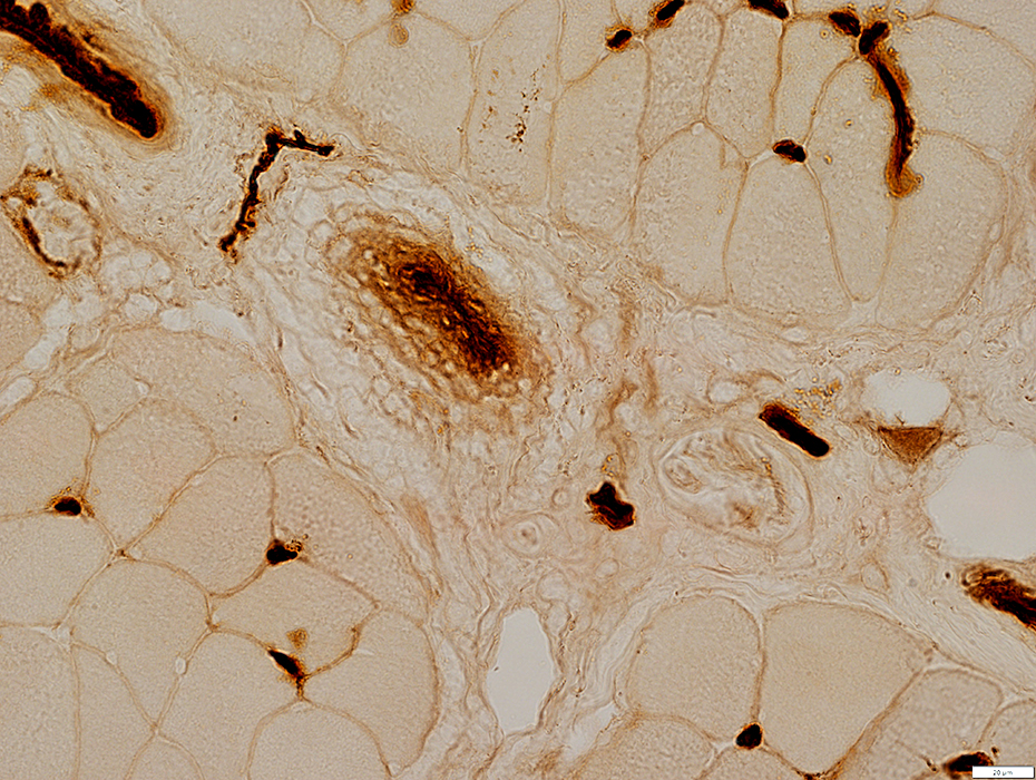 UEA-I stain |
 Esterase stain |
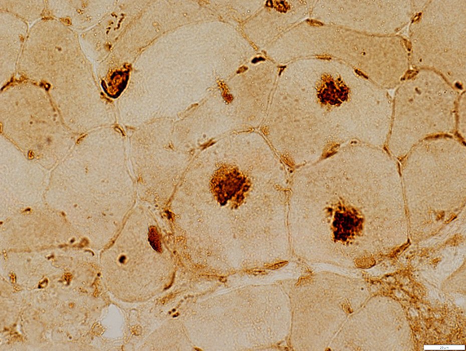
 MxA stain |
EVIM: Endomysial cells, scattered
 Congo red stain |
Return to Inflammatory myopathies
Return to IMPP
References
1. Curr Opin Rheumatol 2011;23:595-604
3/5/2025