Alcohol (Ethanol) myotoxicity: Acute Myopathy
|
Muscle pathology Necrosis Early Distribution: Scattered fibers Cytoplasm: Pale Muscle fiber properties Alkaline Phosphatase ATPase PAS Also see Necrosis: General features |
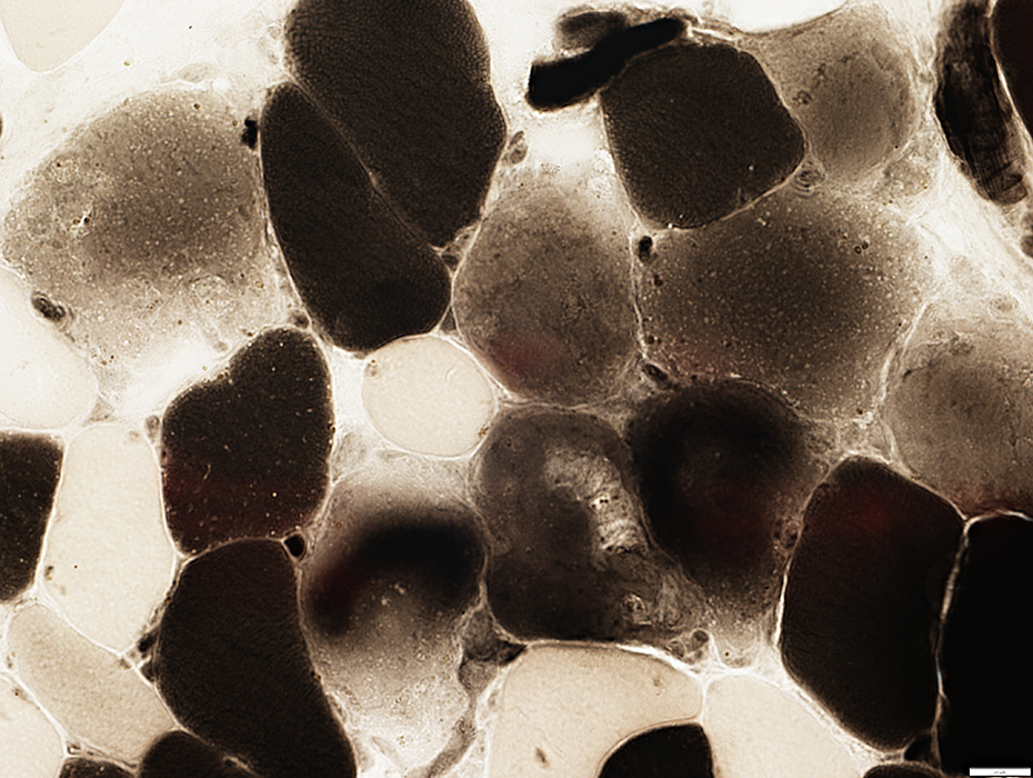
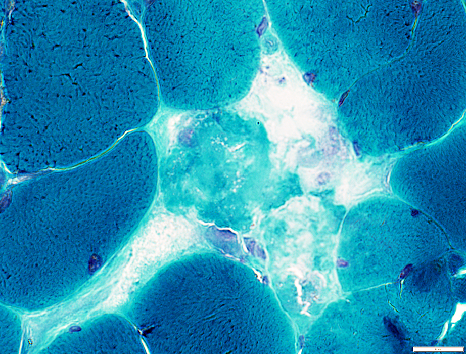
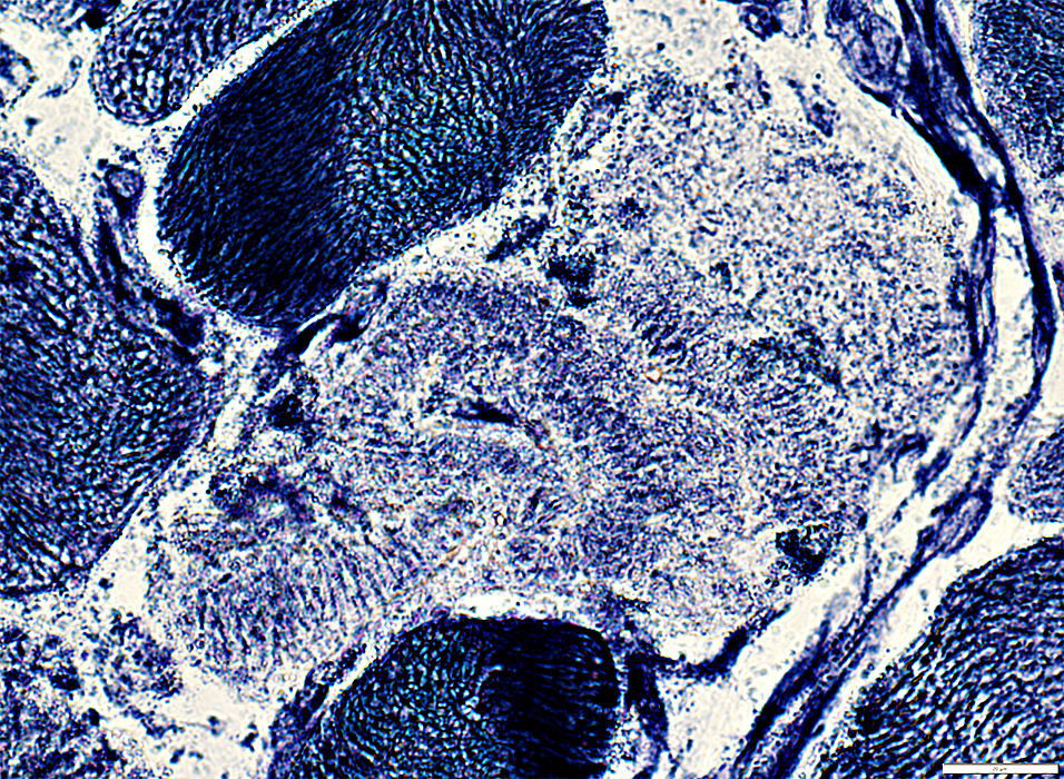
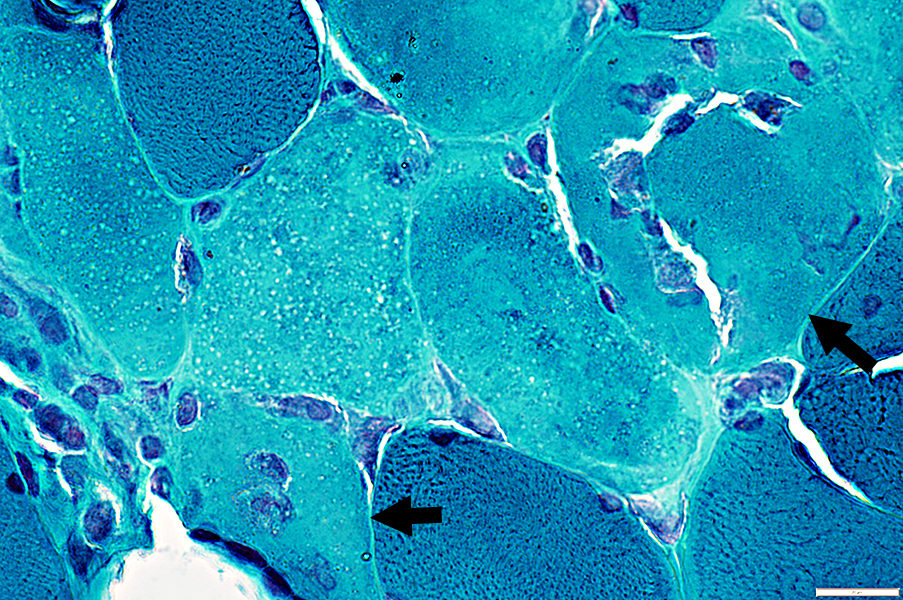 Gomori Trichrome stain Necrotic Muscle Fibers: Early Pale cytoplasm with small lipid droplets Two fibers (Arrows) invaded by phagocytic histiocytes |
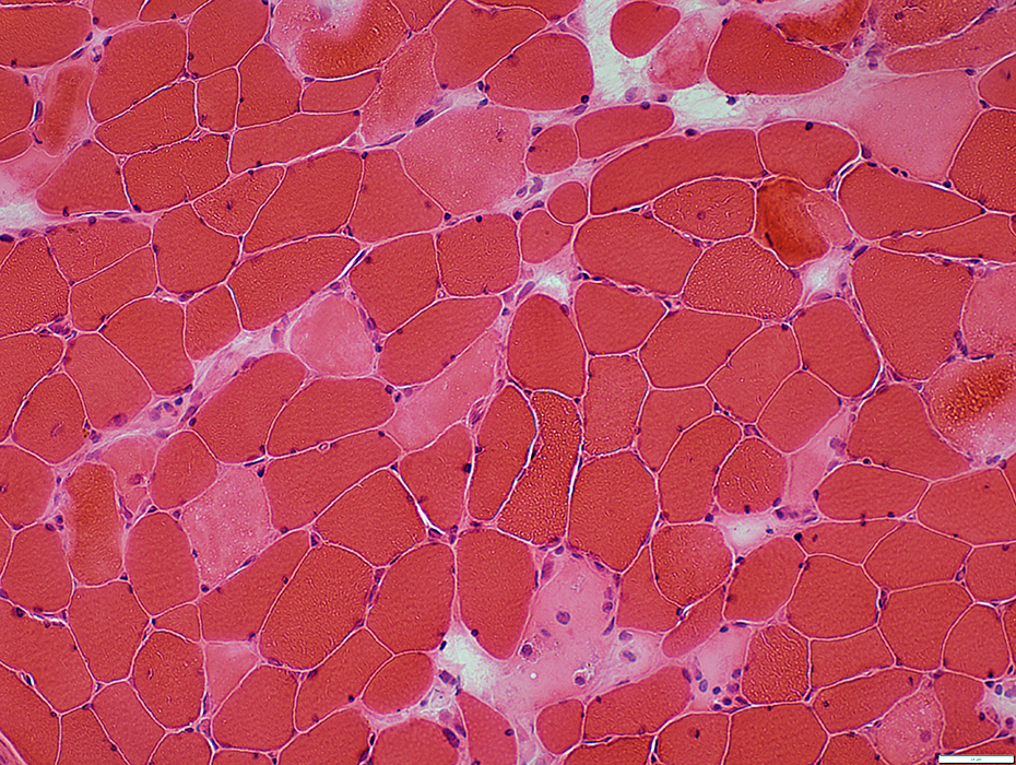
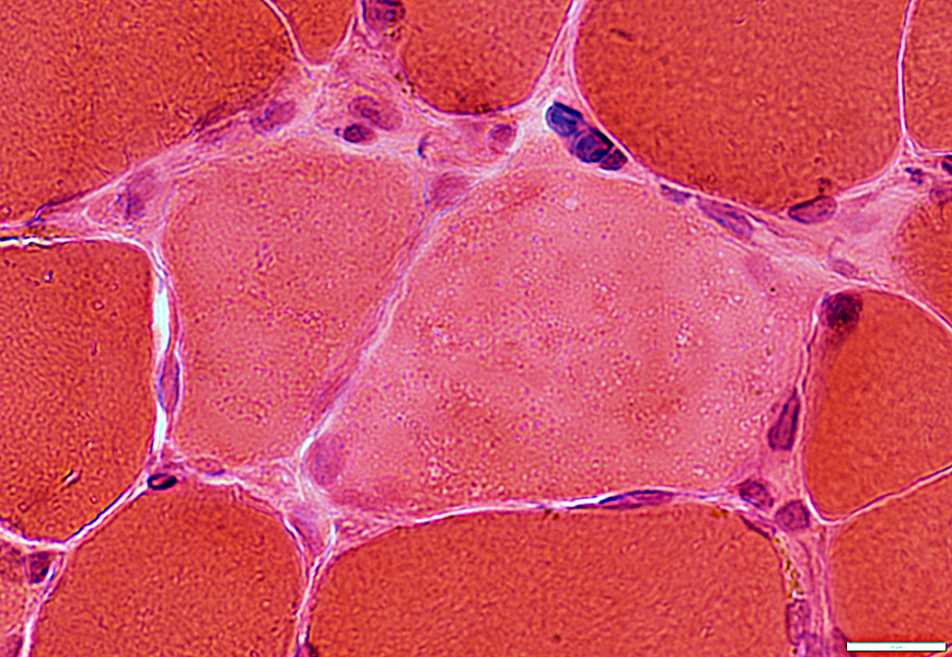
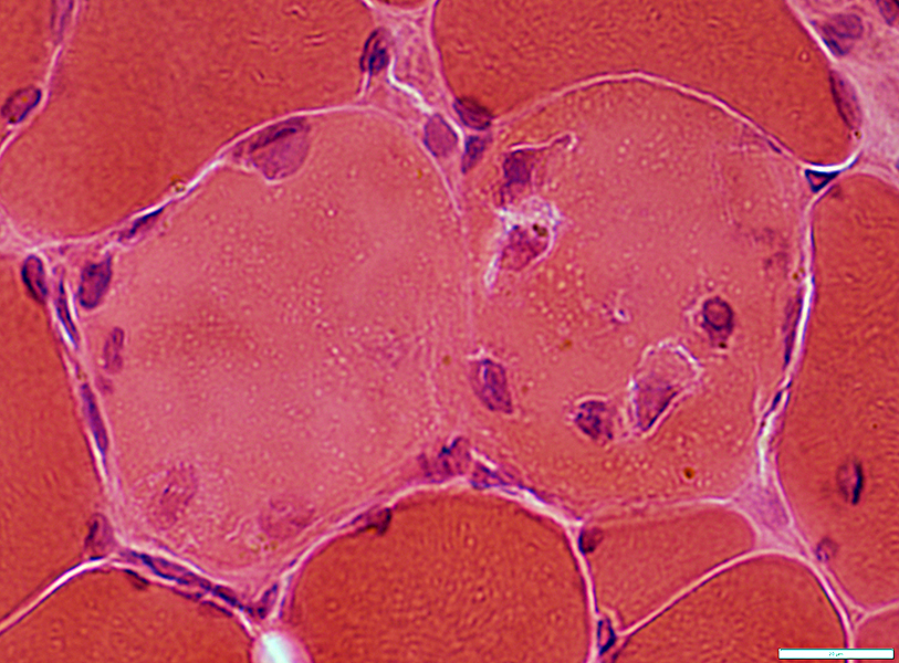
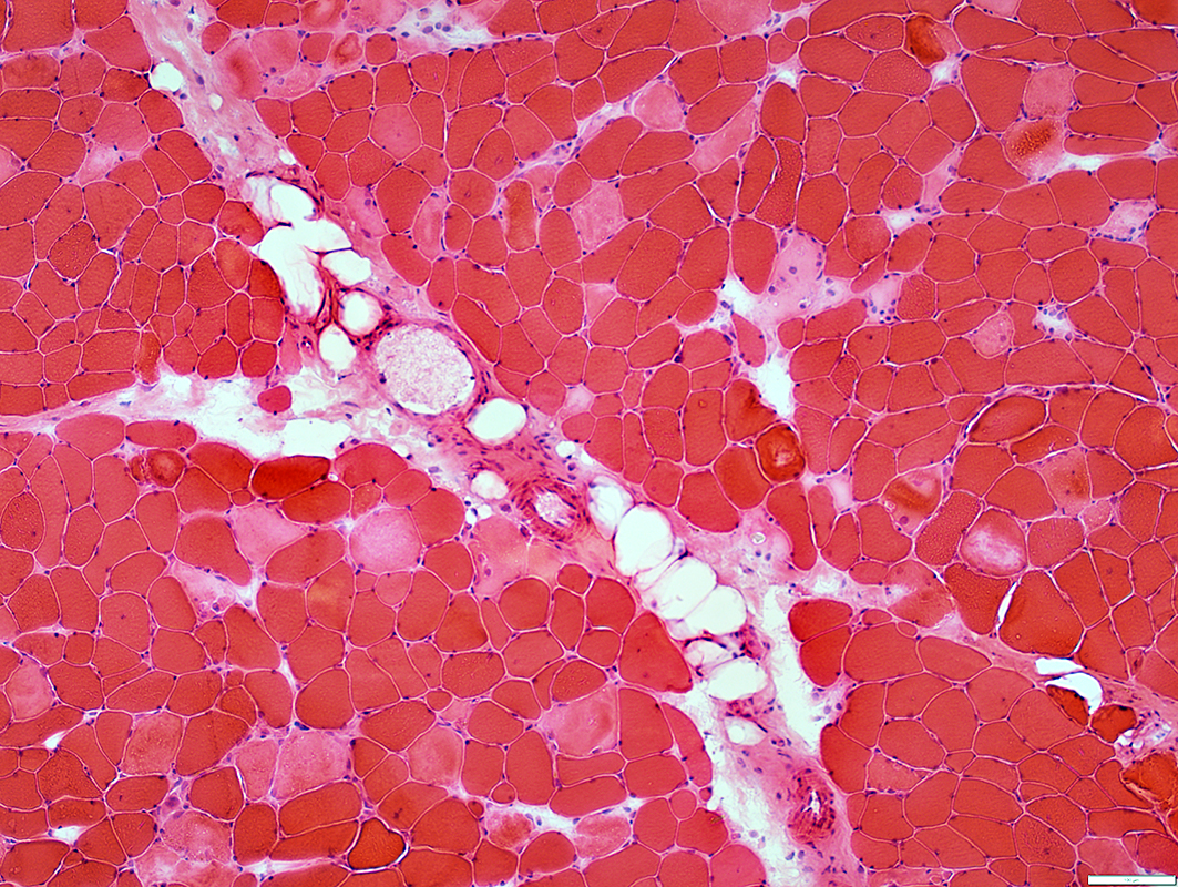 H&E stain |
Many
Distribution: Scattered
Pale staining: H&E, Gomori trichrome & NADH
Contrast to: Dark H&E stain of fibers in rhabdomyolysis with Phosphorylase deficiency
Dark staining of cytoplasm: C5b-9
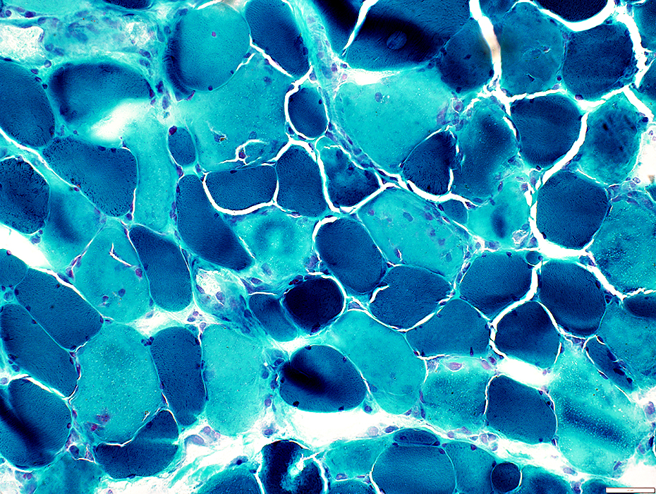
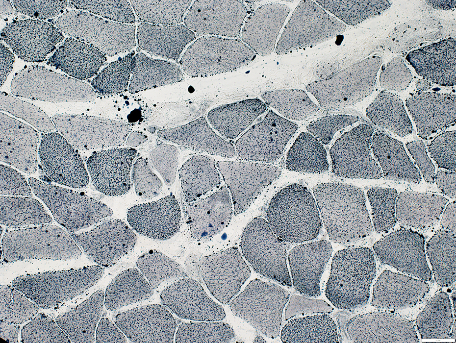
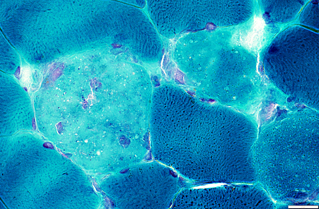
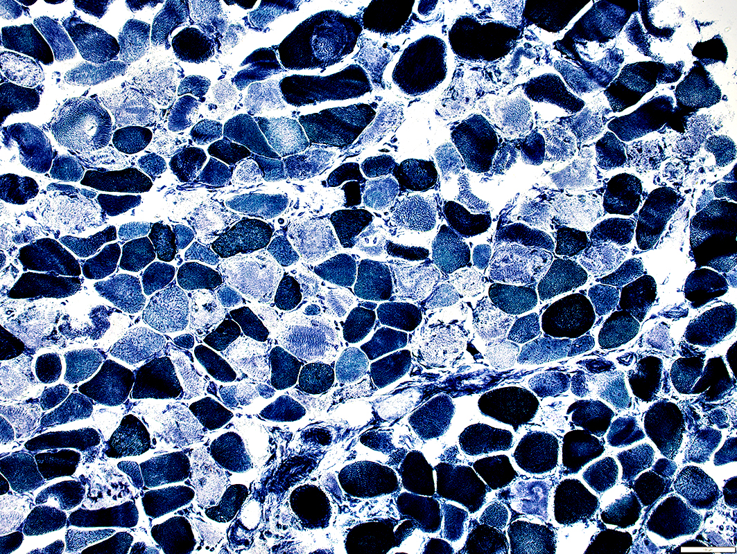 NADH stain |
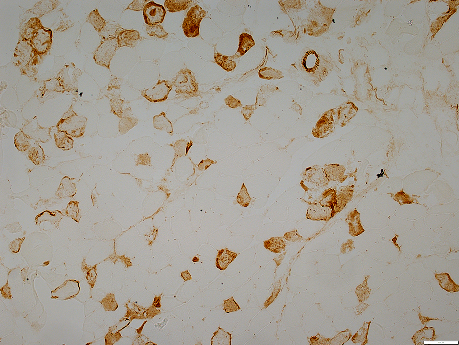 C5b-9 stain |
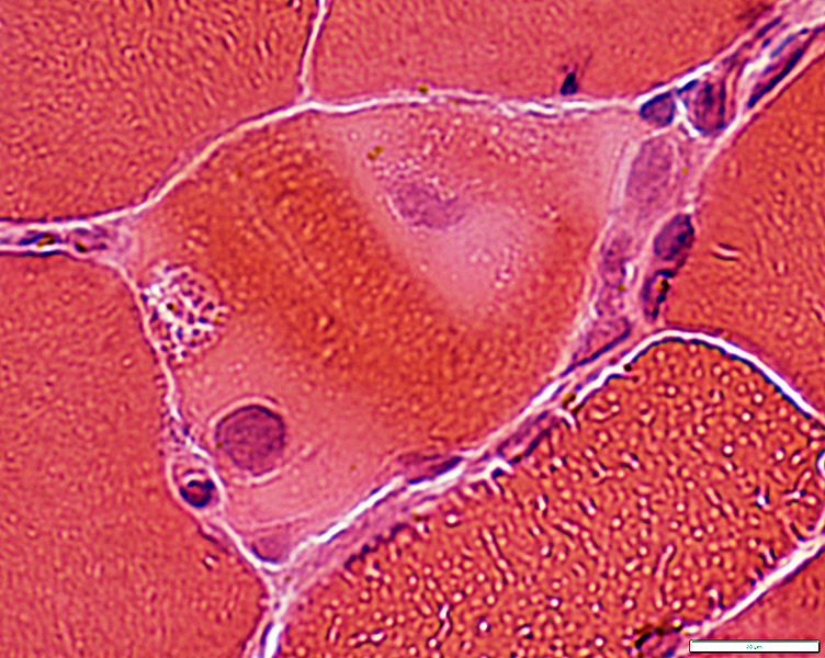
 H&E stain |
Pale: Cytoplasm staining
H&E
Gomori trichrome
NADH
ATPase pH 4.3
Dark: Cytoplasm staining
C5b9
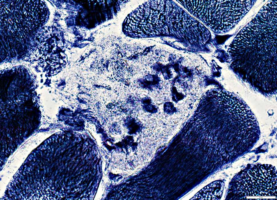
Histiocytes
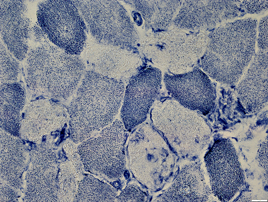
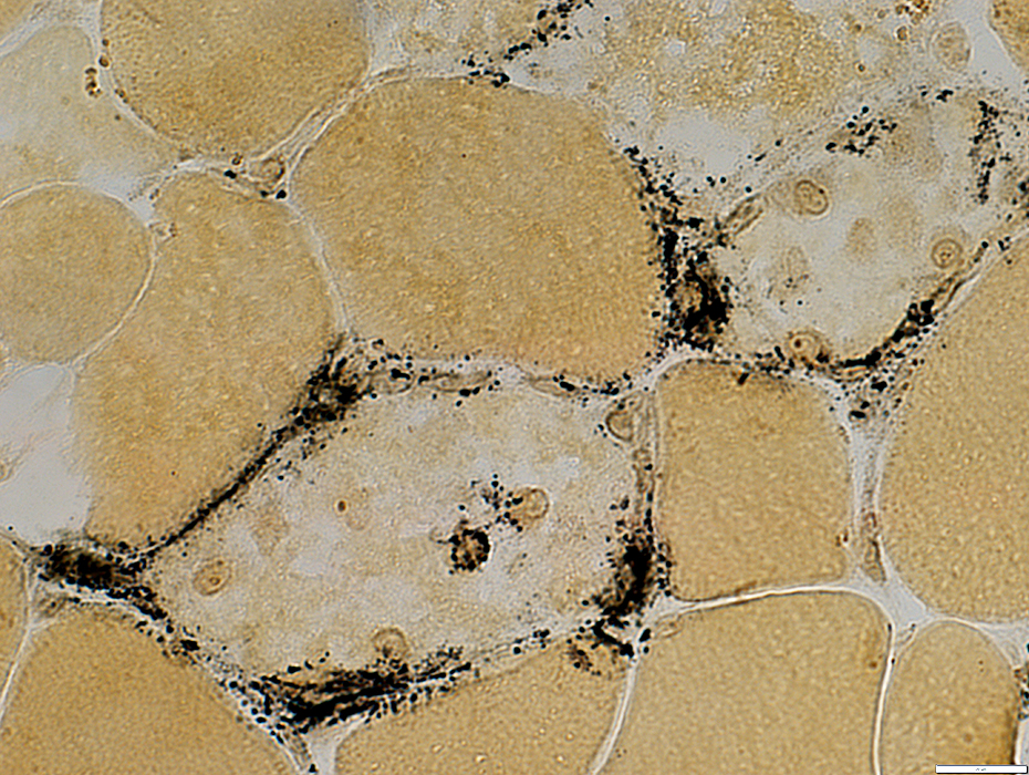
Invasion of necrotic muscle fibers: Acid phosphatase
Locations: Endomysial & Phagocytic (MHC-I stain)
 Gomori Trichrome stain |
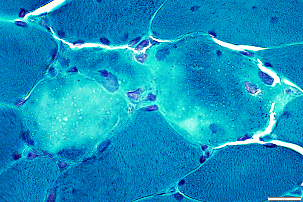
 NADH stain |
Pale stained by NADH
 Acid phosphatase stain |
 ATPase pH 4.3 stain |
Lipid droplets
Small sizes
Present in type I & II muscle fibers
 Sudan Black stain |
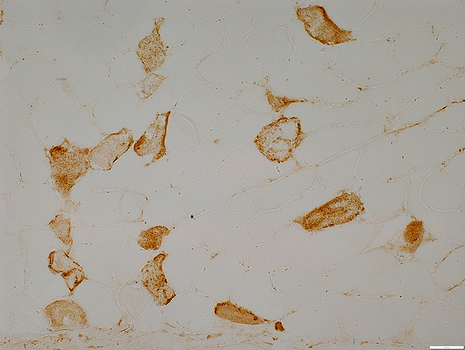 C5b-9 stain |
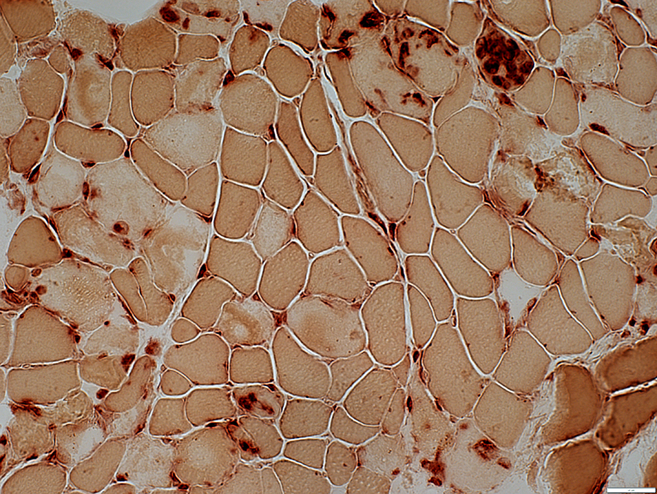
MHC Class I
Stains histiocytes scattered in endomysium & invading muscle fibers
No upregulation by muscle fibers
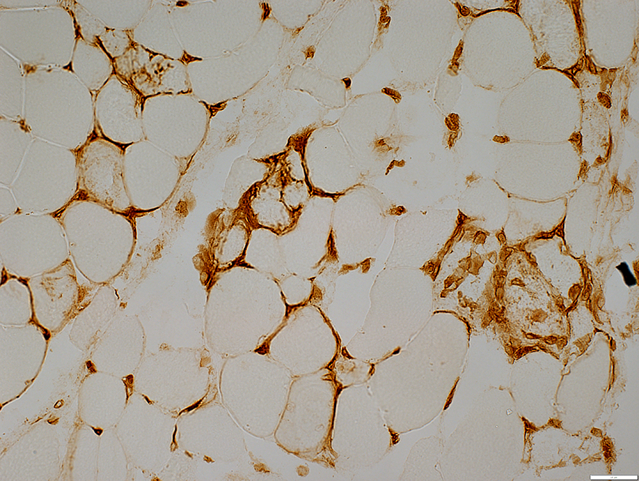 MHC Class I stain |
Acute Alcohol Myopathy: Necrotic Muscle Fibers
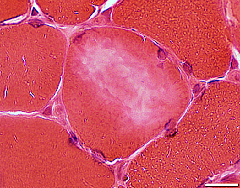
 H&E stain |
Pale: Cytoplasm staining
H&E: Pale Cytoplasm & Myonuclei
Gomori trichrome
NADH
ATPase pH 4.3
Regional Δ
Dark: Cytoplasm staining
C5b-9
Cytoplasm: Other
Lipid droplets: Small
Misoriented myofibrils
Disintegration
Histiocyte invasion
Histiocyte invasion
Invasion of necrotic muscle fibers: Acid phosphatase
Locations: Endomysial & Phagocytic (MHC-I stain)
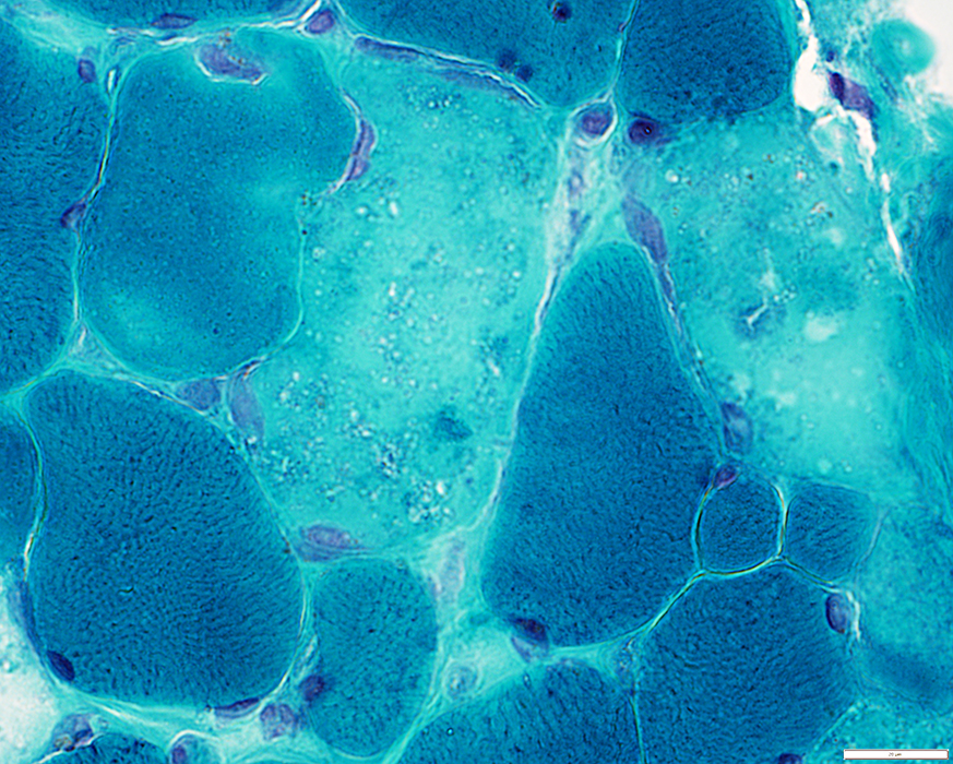 Gomori trichrome stain |
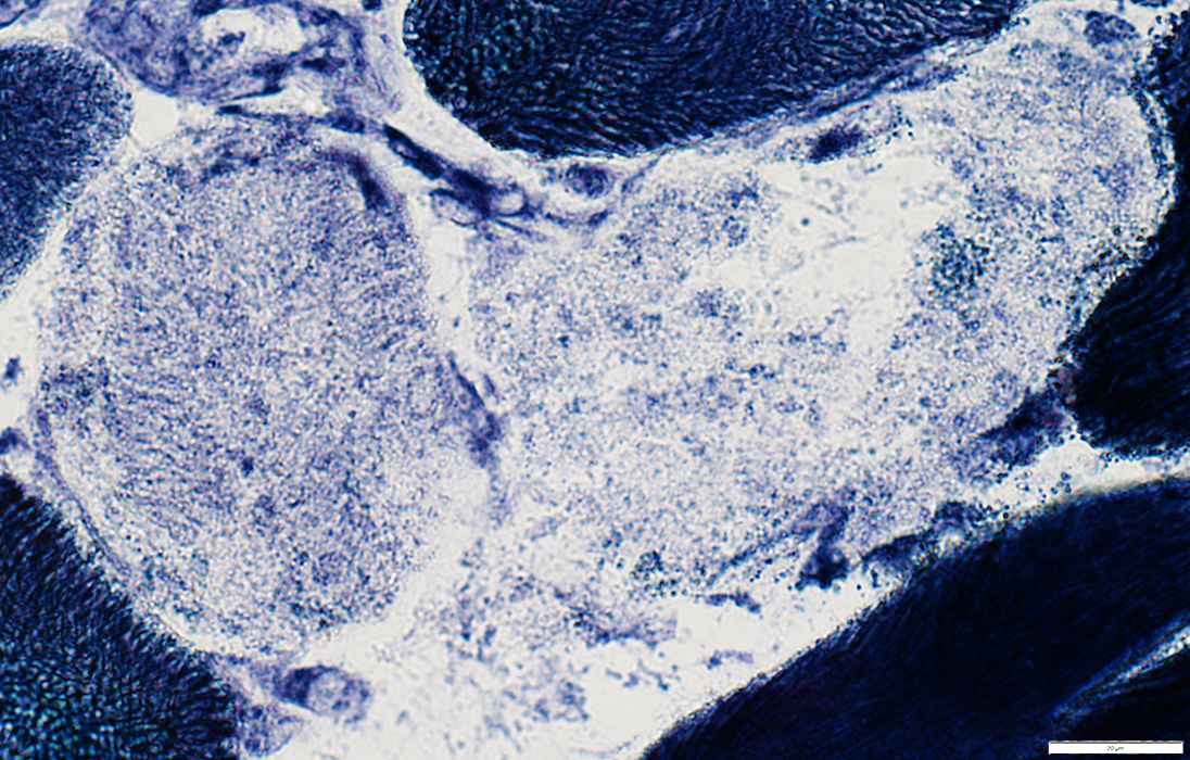 NADH stain |
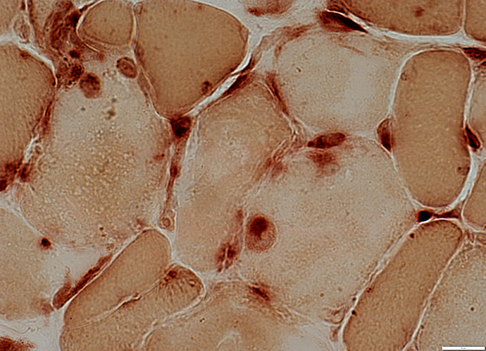 Acid phosphatase stain |
 Gomori trichrome stain |
 H&E stain |
Regional cytoplasmic palor
 H&E stain |
 Gomori trichrome stain |
 NADH stain |
 H&E stain |
 Gomori trichrome stain |
Necrotic muscle fibers: Invading histiocytes often stain for NADH
 NADH stain |
Necrotic Muslce Fibers: Surrounded by Alkaline Phosphatase staining
 Alkaline phosphatase stain |
Necrosis: Muscle fiber completely replaced by histiocytes
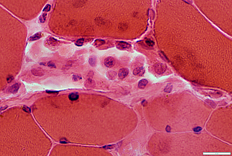 H&E stain |
Return to Necrosis
Return to Pathology & Illustrations
Return to Neuromuscular Home Page
11/6/2022