Inflammatory Myopathies with Vacuoles, Aggregates & Mitochondrial Pathology (IM-VAMP)
Myositis with mitochondrial pathology (PM-Mito) subtype
|
Inflammation Endomysial Focal invasion of muscle fibers Myopathic changes Mitochondrial disorders Other Variant syndromes IBM IM-VAMP + HIV Granulomatous myopathies |
PM-Mito: Muscle pathology
- Muscle fibers
- Inflammation
- Endomysial: CD8 & CD4 lymphocytes
- Focal invasion of muscle fibers by inflammatory cells
- Molecular: Less expression than full IBM syndrome of
- IFN-induced guanylate-binding protein (GBP6)

- Location: Macrophages
- Induced by IFN-γ
- Killer cell lectin-like receptor, subfamily G, Member 1 (KLRG1)
- CD8+ T-cell function–related
- T-cell exhaustion marker
- IFN-induced guanylate-binding protein (GBP6)
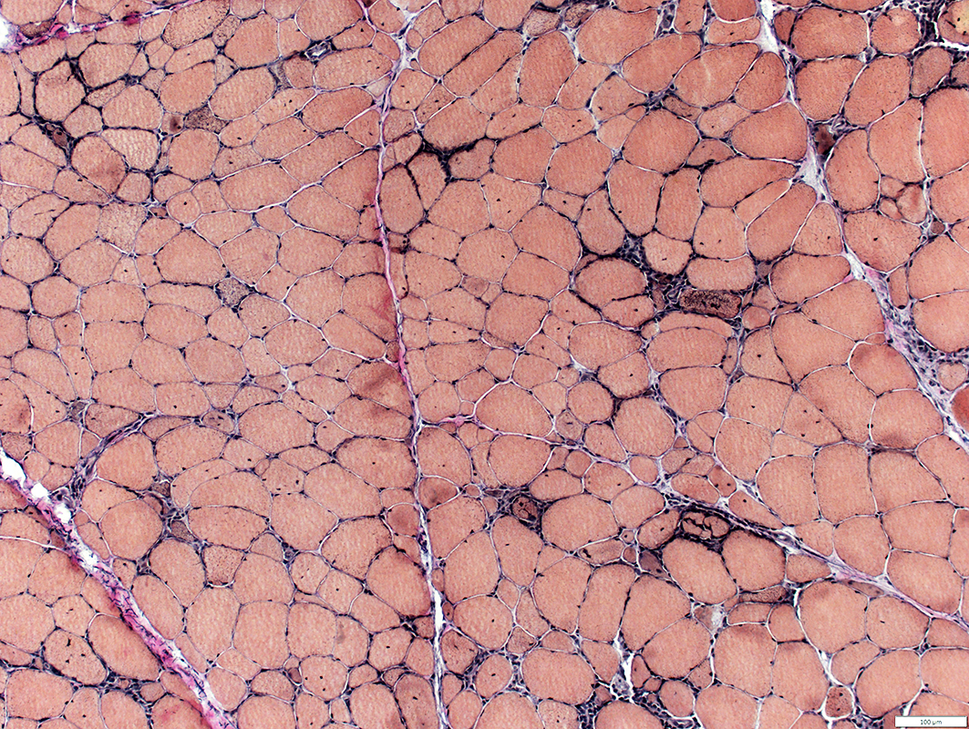
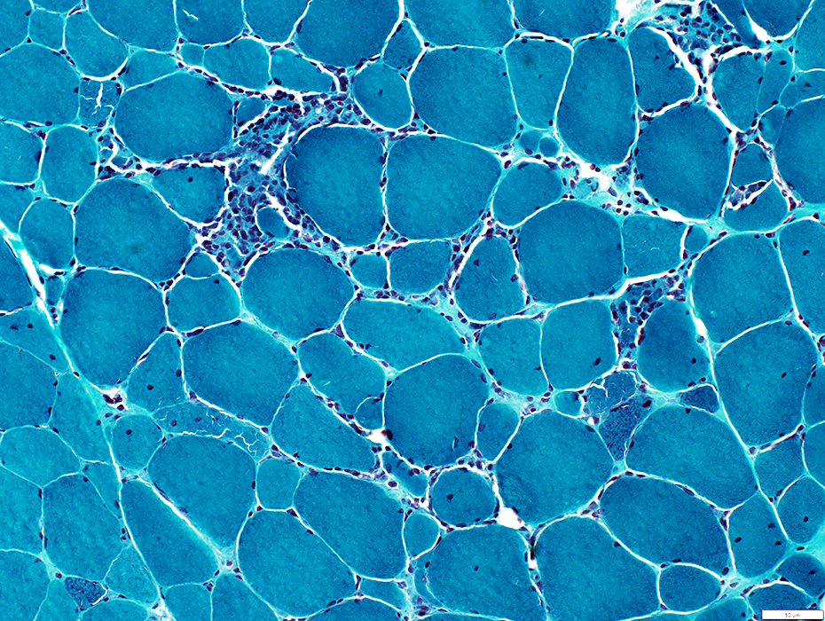
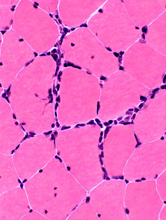
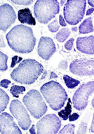
PM-Mito: Myopathy, Inflammatory
 VvG stain |
Sizes: Varied; Largest fibers hypertrophied
Internal nuclei
Immature
Small, Darker stained cytoplasm, Large nuclei
Distribution: Scattered
Connective tissue, endomysial
Increased, mild to moderate
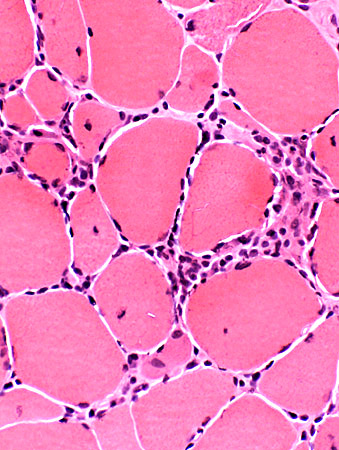
Inflammation
Endomysial lymphocytes
Some focal invasion of muscle fibers
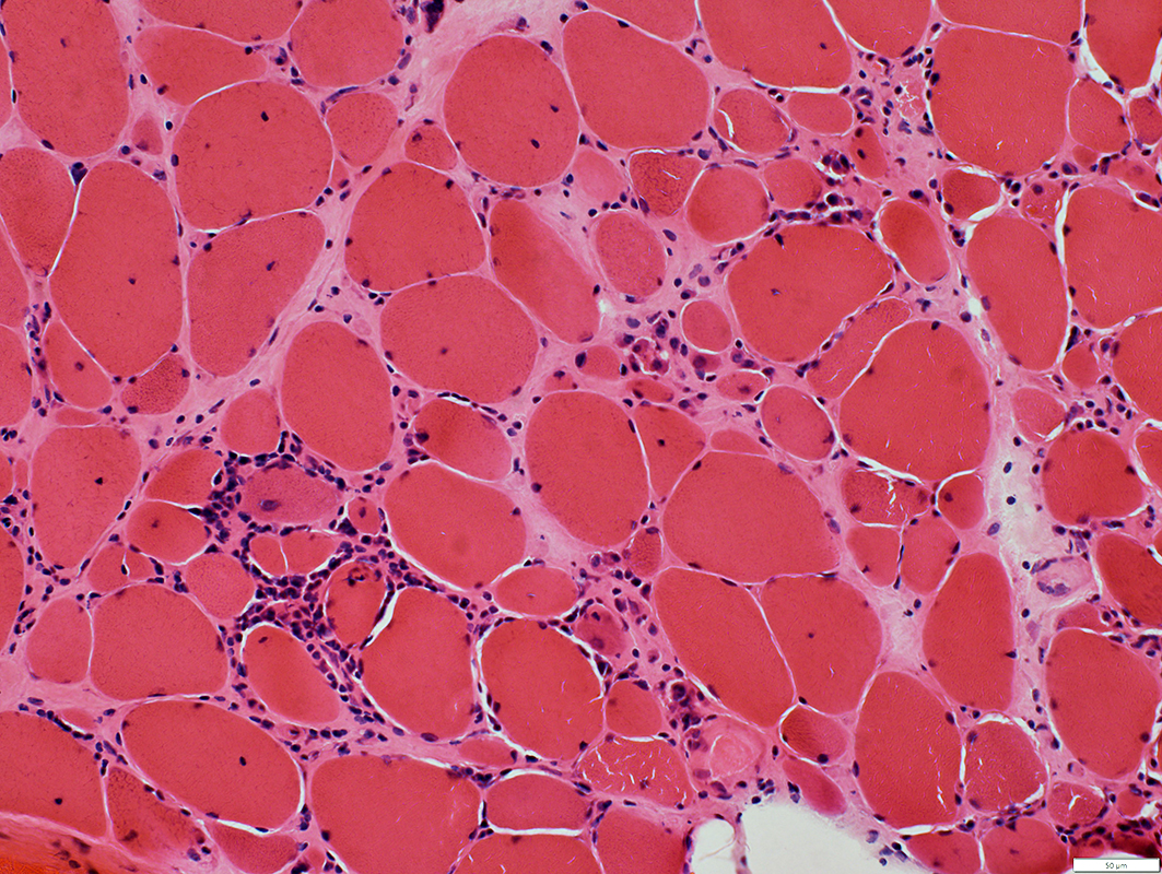 H&E stain |
 Gomori trichrome stain |
 H&E stain |
 Gomori trichrome stain |
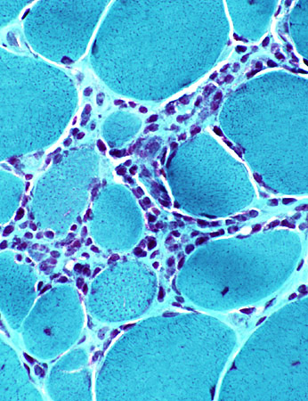
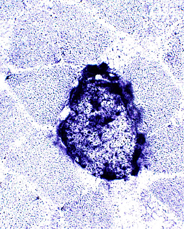
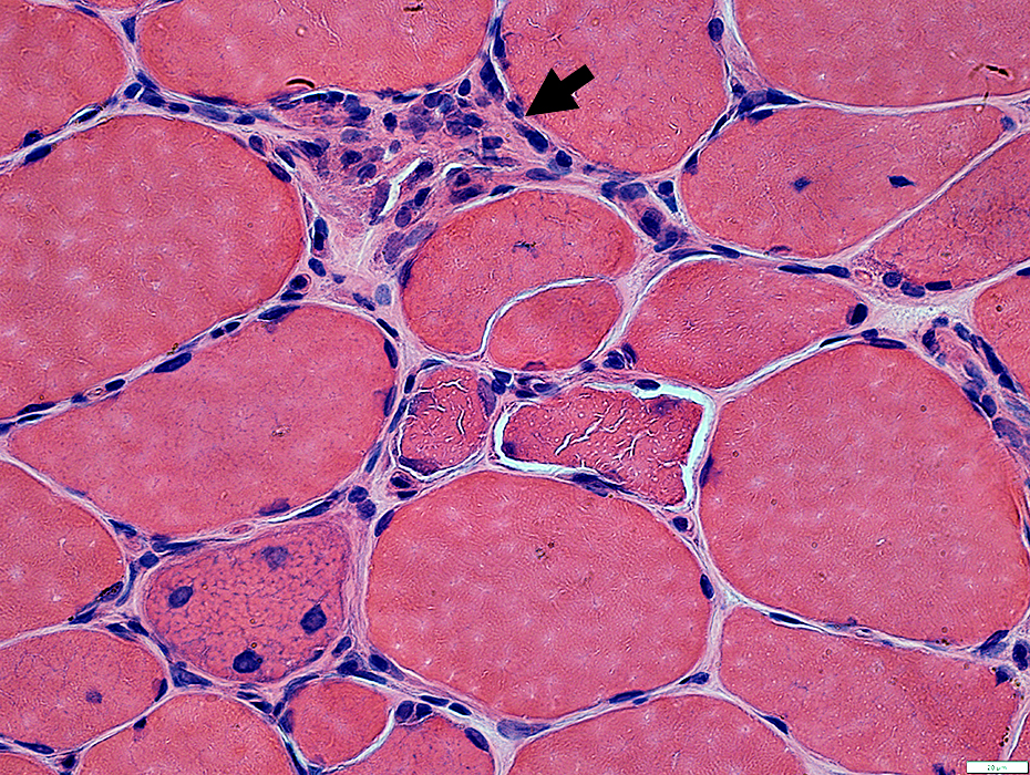
PM-Mito: INFLAMMATION
 H&E stain |
Focal invasion of muscle fibers by immune cells
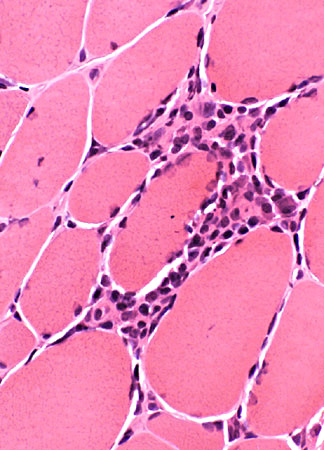
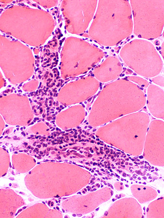
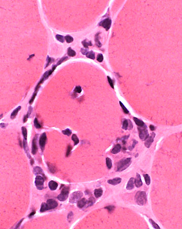 H&E stain |
Cell foci contain: Lymphocytes & Histiocytes (Acid phosphatase positive)
 Acid phosphatase stain |
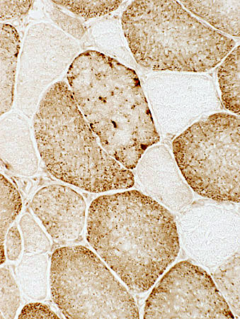
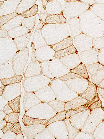
IM-VAMP: Mitochondrial Pathology
 Cytochrome oxidase stain |
COX- muscle fibers: Scattered in muscle
Normal fibers have: Dark (Type 1) or Intermediate (Type2) stain
 Cytochrome oxidase stain |
COX staining absent or severely reduced
Normal fibers have: Dark (Type 1) or Intermediate (Type2) stain
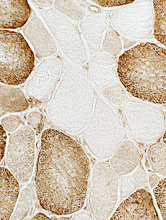 Cytochrome oxidase stain |
Mitochondrial Proliferation: Succinate Dehydrogenase (SDH) positive Muscle fibers
 SDH stain |
Scattered large & small muscle fibers are abnormally dark
Normal fibers have: Intermediate (Type 1) or Moderately pale (Type2) stain
 SDH stain |
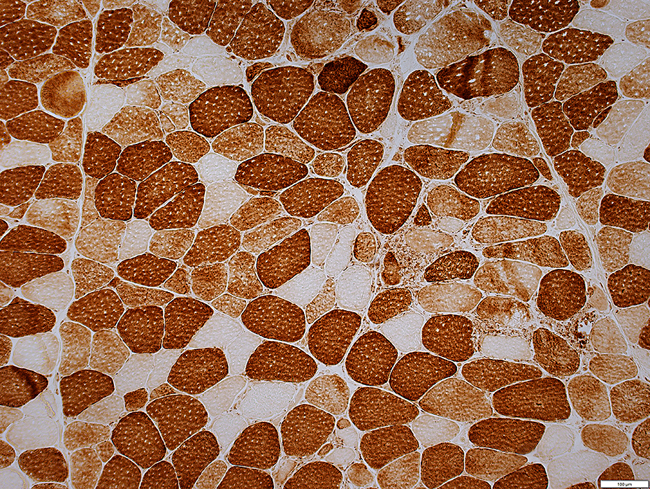
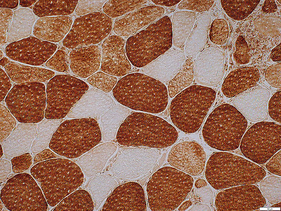
PM-Mito: MHC 1 upregulation
|
LC-3 aggregates in muscle fibers
Varied shapes: Diffuse, punctate, or irregular, dark aggregates
|
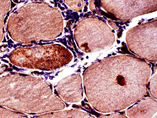
PM-Mito: Capillary & Vessel pathology
Capillary sizes: Some are largeEndothelial cells: May stain for acid phosphatase (Dark arrow)
Neighboring histiocytic cells (White arrow)
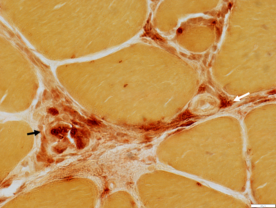 Acid phosphatase stain |
 H&E stain |
Return to Inflammatory myopathies
Return to Polymyositis with mitochondrial disorders
12/12/2024