Hereditary Neuropathy with Liability to Pressure Palsies (HNPP)
|
Myelin patterns Tomaculae: Teased axons Ultrastructure: 1; 2 |
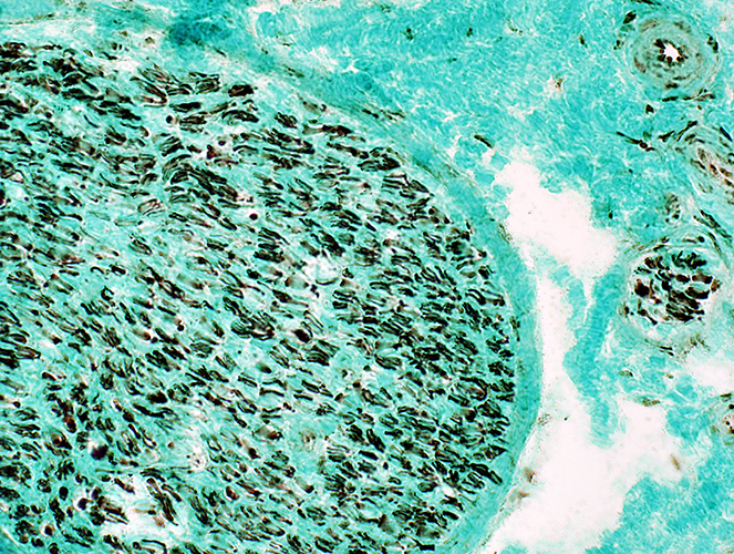
HNPP Nerve: Frozen sections
Axon Loss: Reduced numbers of myelinated axons
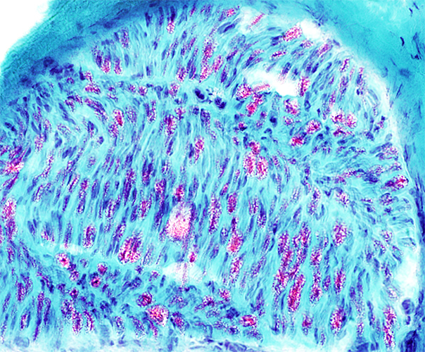 Gomori trichrome stain |
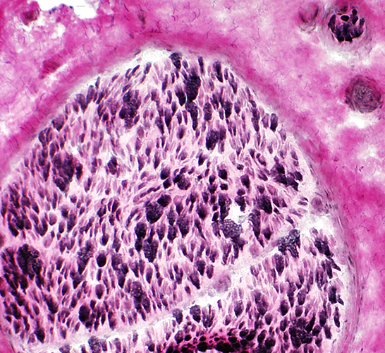 VvG stain |
HNPP Nerve: Fixed |
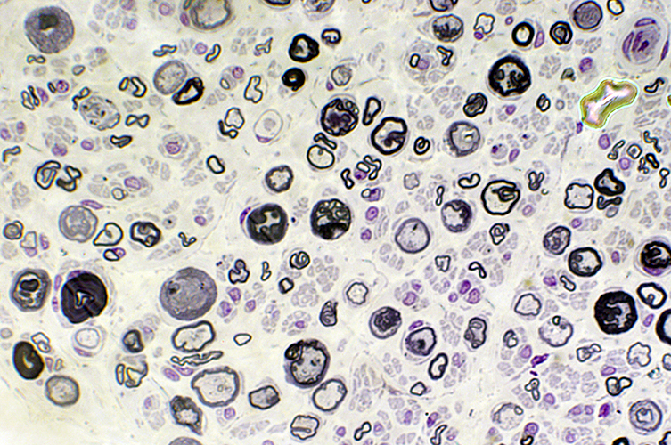 Toluidine blue stain |
|
Reduced numbers of large axons Abnormal myelination of axons includes Hypomyelination Abnormally folded (Tomaculae) or shaped myelin sheaths Small onion bulbs are also present (Below; Arrow) 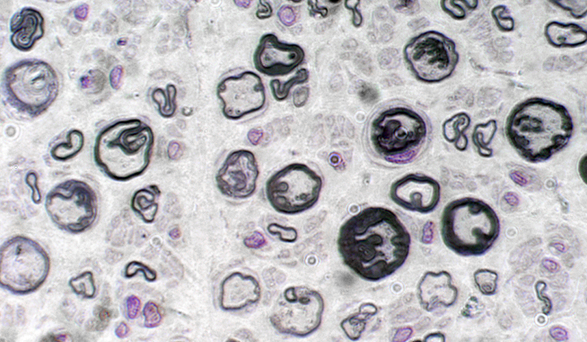 Toluidine blue stain 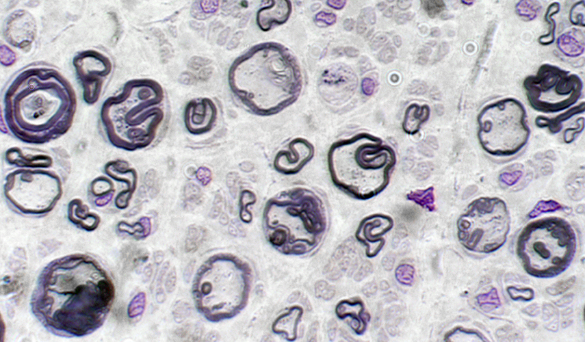 Toluidine blue stain 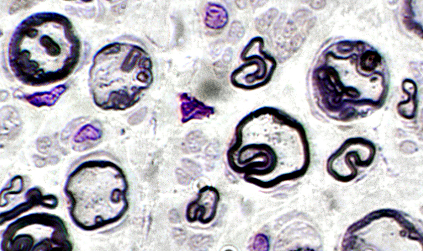
|
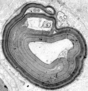
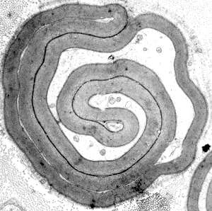
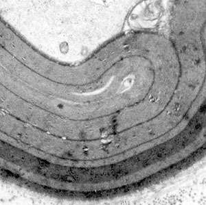 Thickened myelin sheath due to multiple layers of folded myelin |
|
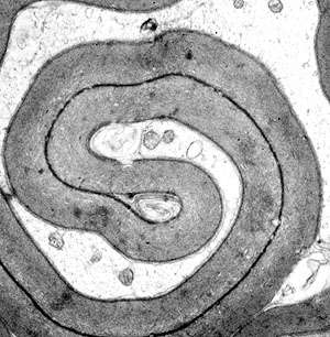 Folding of myelin sheath Outside axon or invaginating into axon |
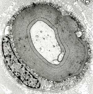
Myelin sheath: Thick & Folded Schwann cell nucleus: Peripheral |
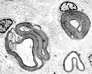
Two axons with tomaculae One thinly myelinated large axon |
HNPP: Axons & Myelin components
Most myelinated axons are smallCompare to Normal nerve
Myelinated axons are large & small
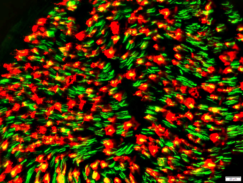 Neurofilament stain (Green) P0 stain (Red) |
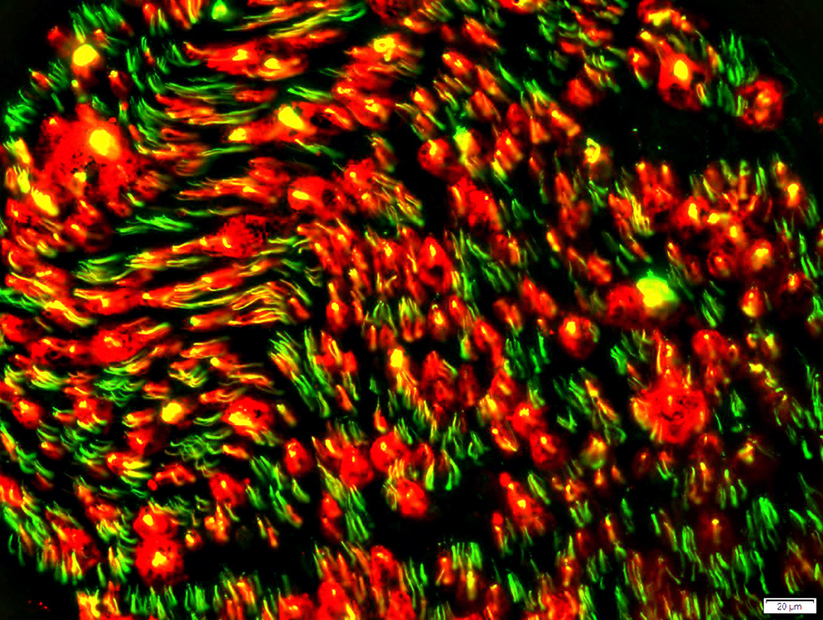 Neurofilament stain (Green) Myelin basic protein stain (Red) |
Compare to Normal nerve
MBP is only in sheaths around large axons
HNPP: MBP is in most myelin sheaths
Compare to Normal nerve
MBP is only in sheaths around large axons
P0 is in myelin sheaths around small & large axons
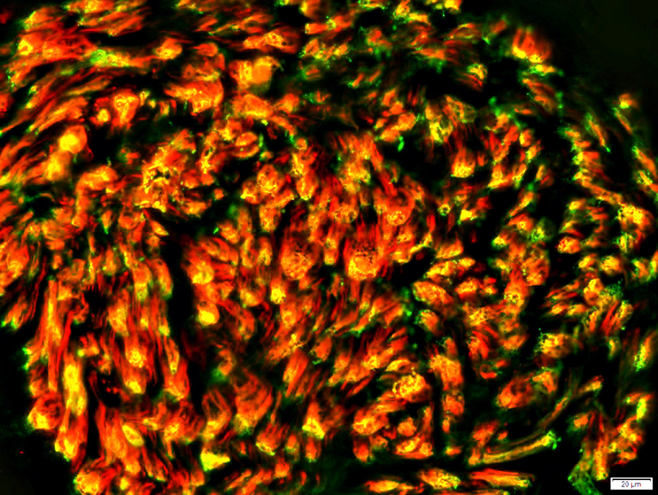 P0 stain (Green) Myelin basic protein stain (Red) |
 Neurofilament (SMI31) stain Axon Loss Reduced numbers of large axons Relative preservation of numbers of small axon |
HNPP: Muscle |
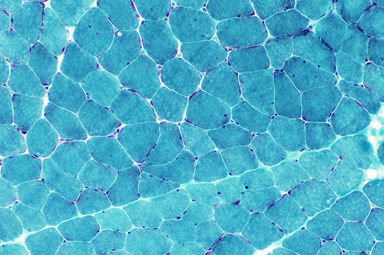 Gomori trichrome stain Mild atrophy |
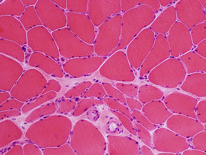 H& E stain Occasional: Grouped atrophy & Pyknotic nuclear clumps |
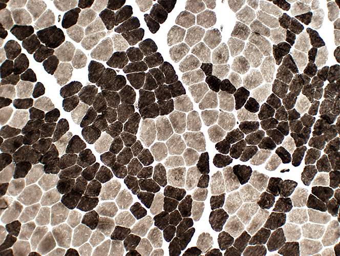 ATPase pH 9.$ stain Fiber type grouping |
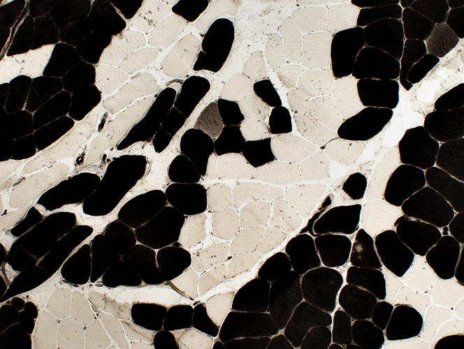 ATPase pH 4.3 stain Fiber type grouping; Rare Type 2C muscle fiber |
Return to HNPP
Return to Demyelination: Chronic
Return to Nerve biopsy: Normal
Return to Biopsy: Pathology index
Return to Neuromuscular Home Page
Return to Nerve biopsy
Tomaculae
Tomaculae: Ultrastructure
Return to Demyelinating neuropathies
Return to Active demyelination
4/24/2024