AMYLOID: PATHOLOGY
|
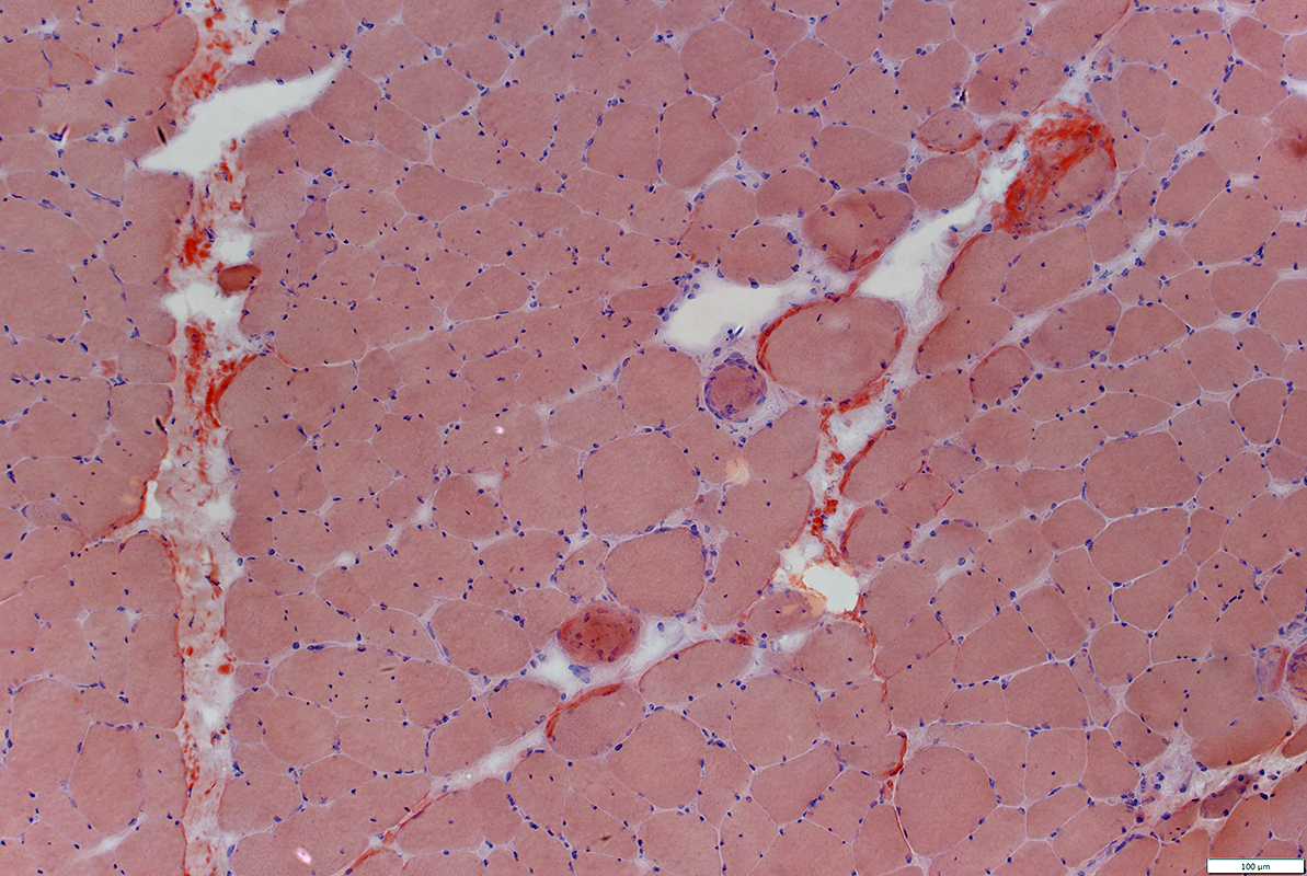
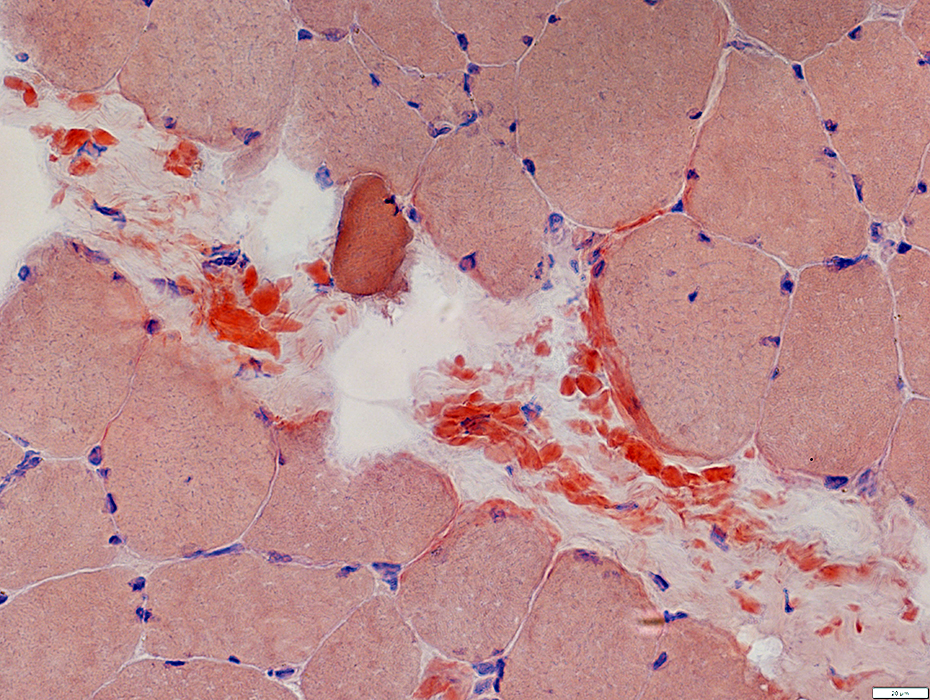
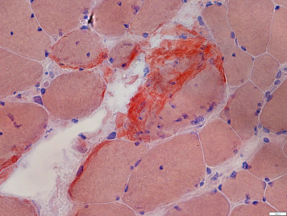
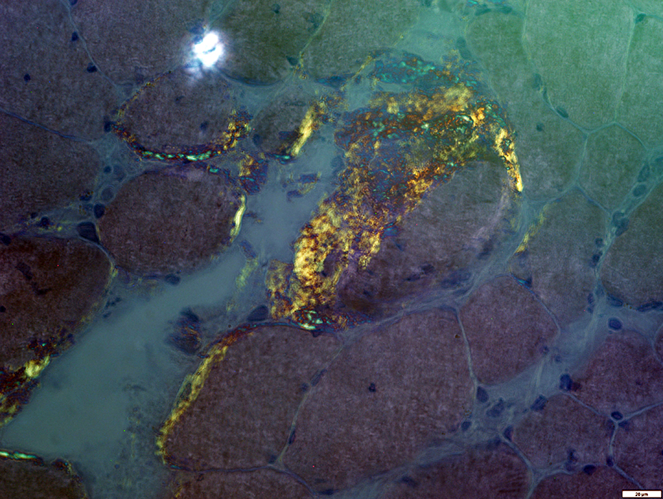
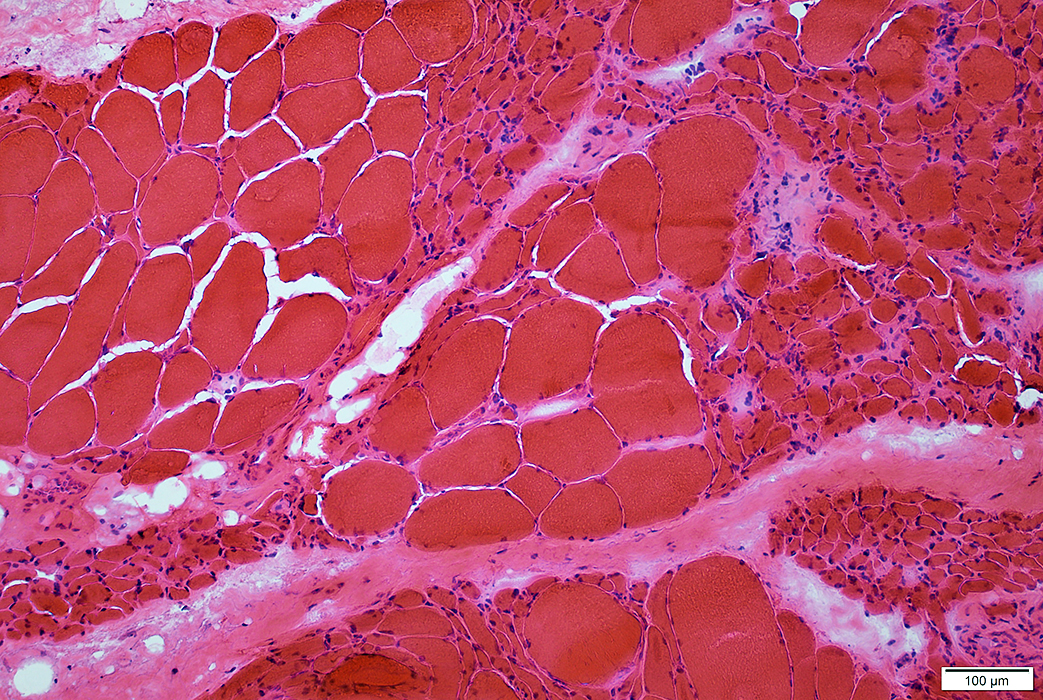
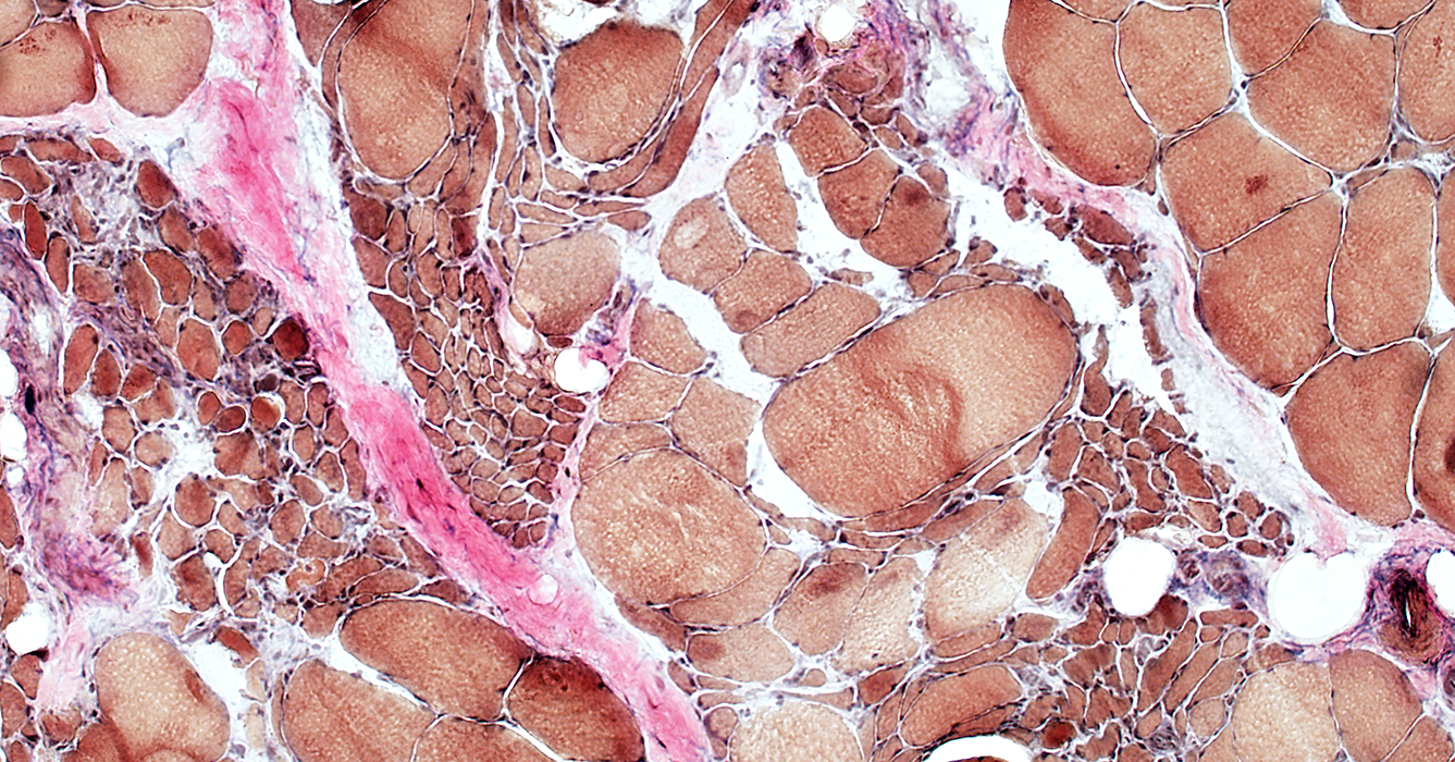
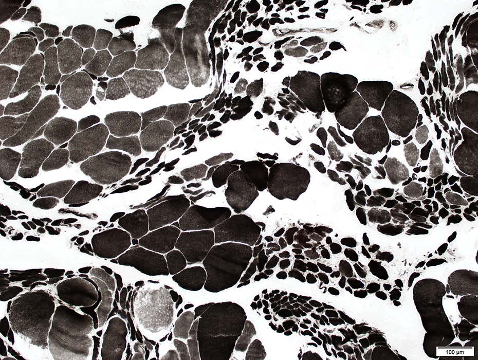
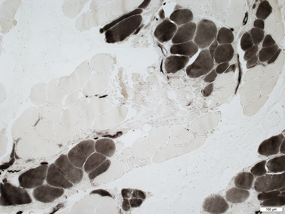
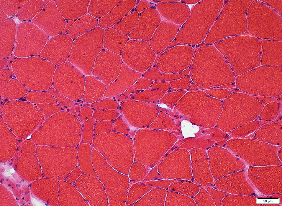
MUSCLE
Amyloid myopathy
|
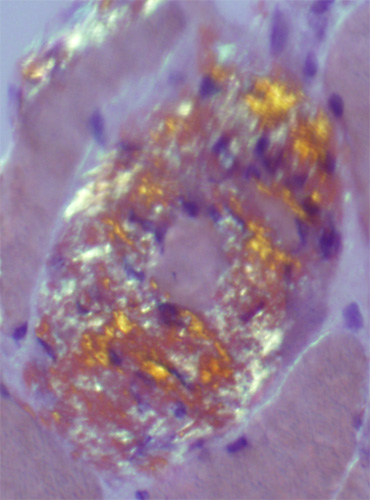
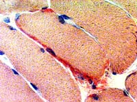
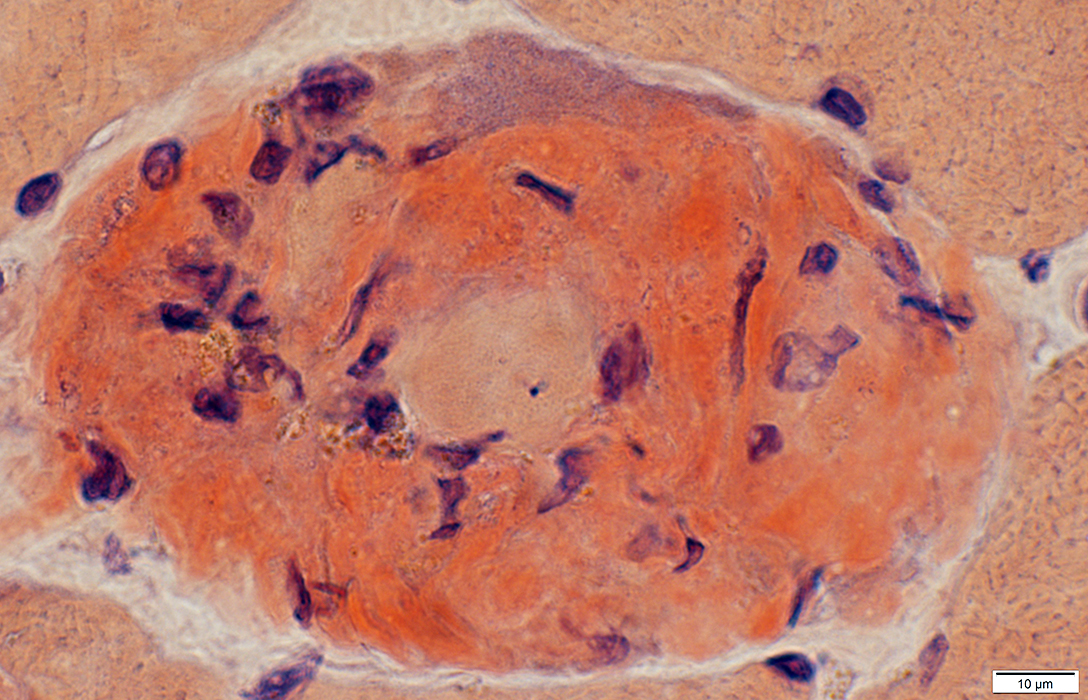
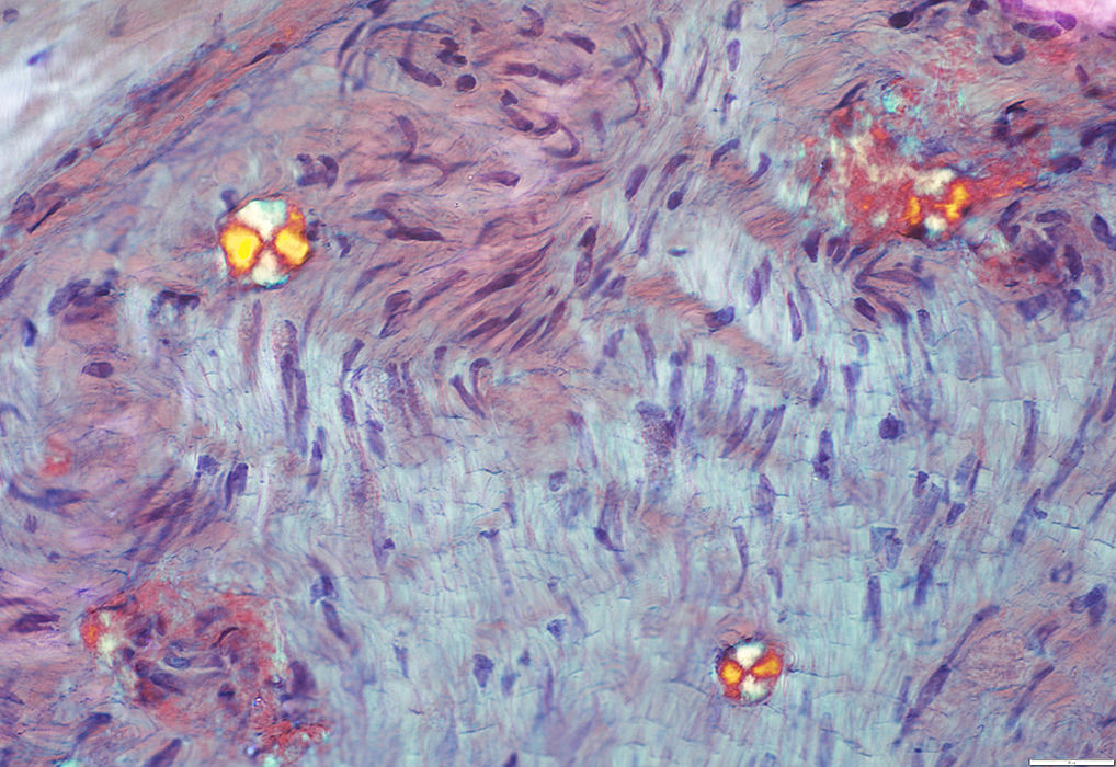
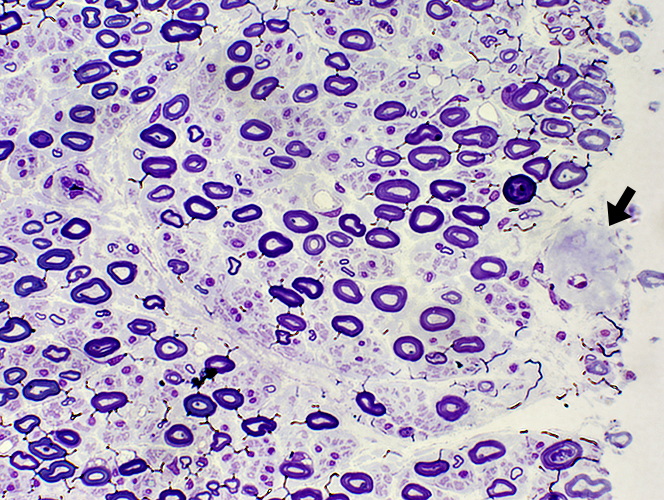
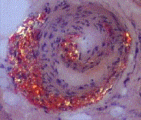
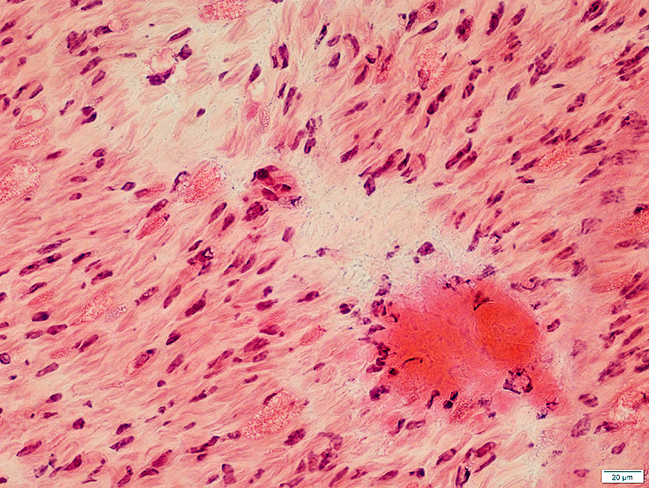
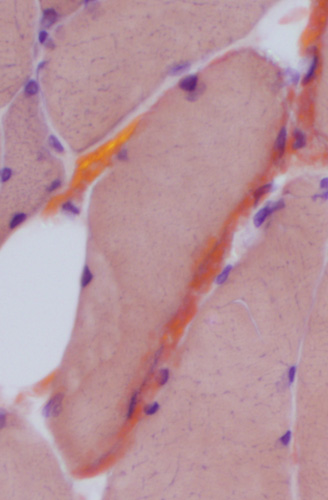 Congo red |
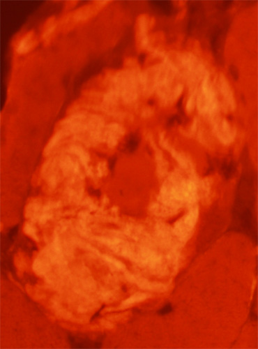
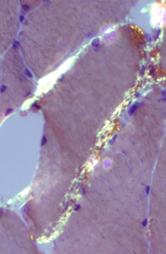 Congo red (Polarized light) |
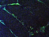
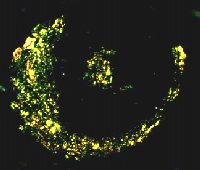
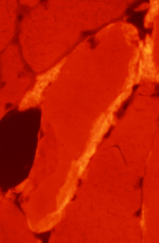 Congo red (Fluorescence) |
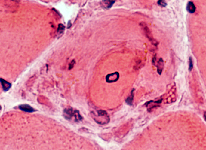
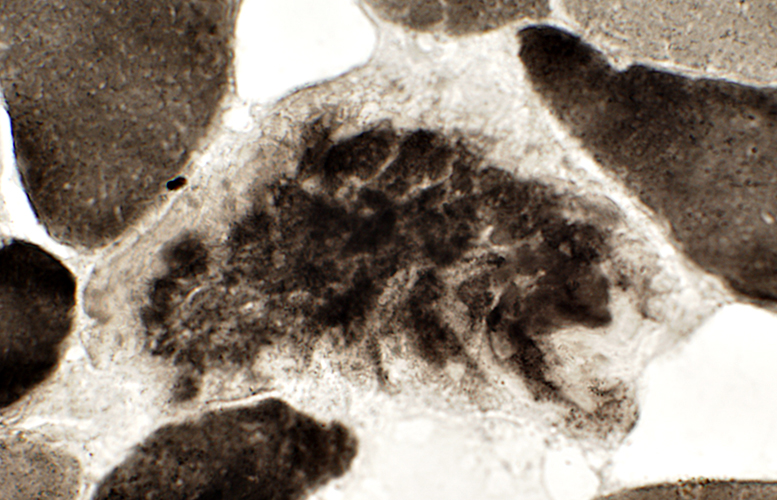
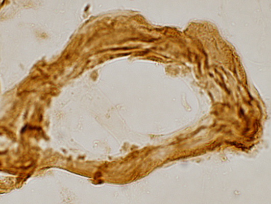
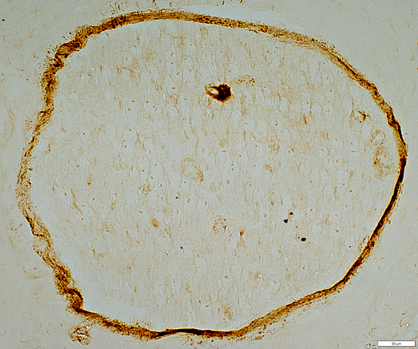
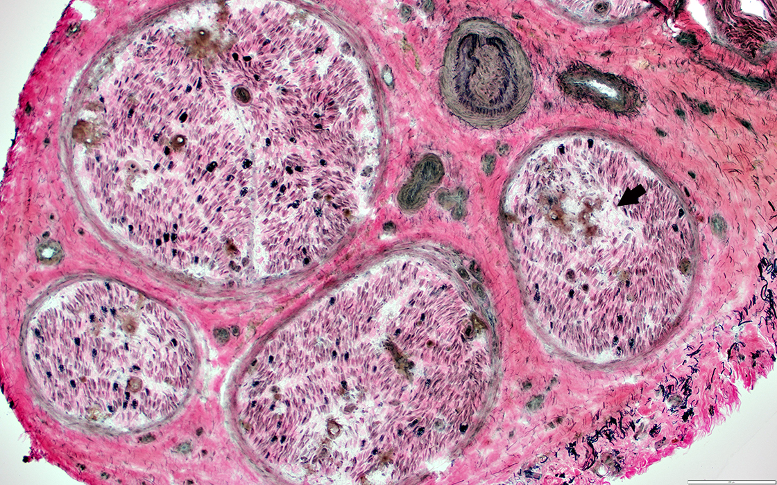
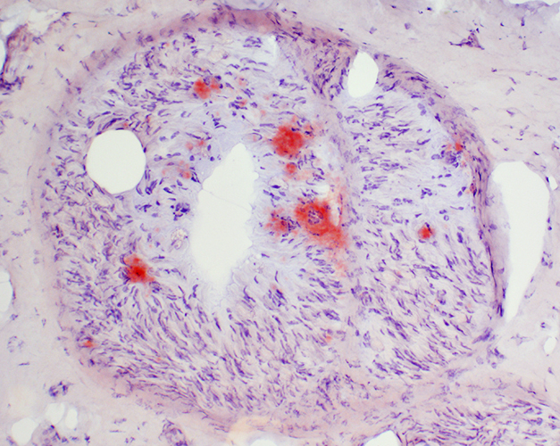
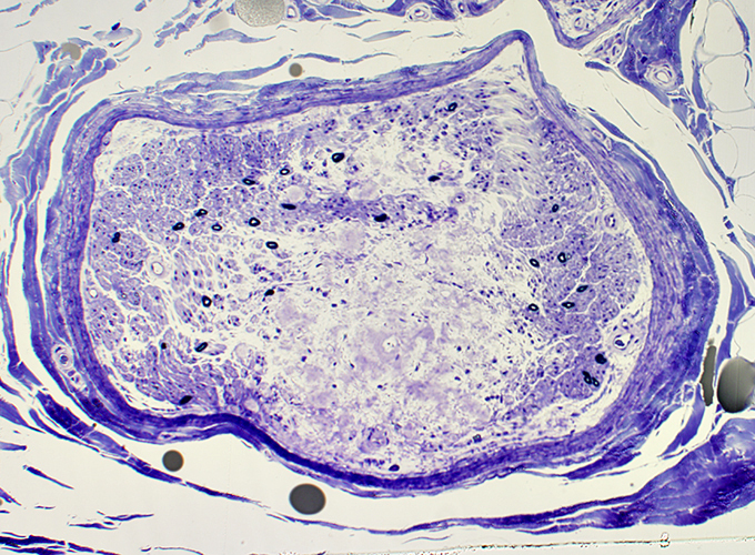
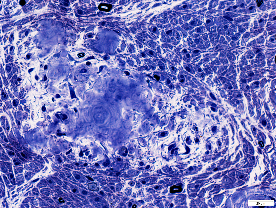
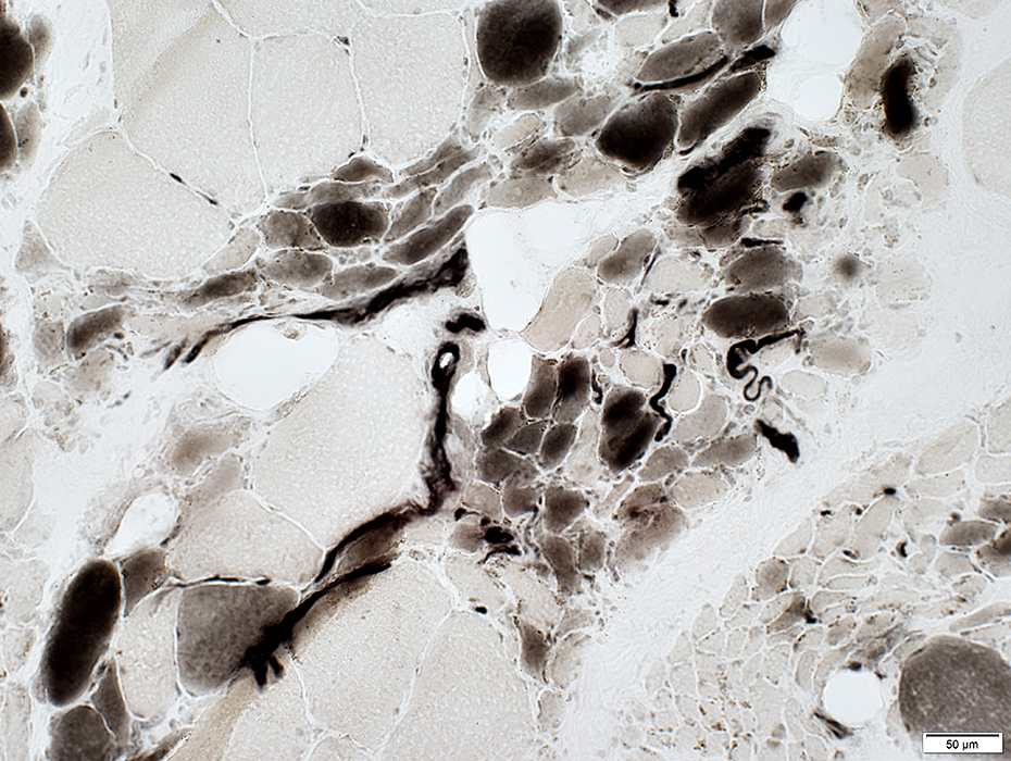
Amyloid Muscle Fibers
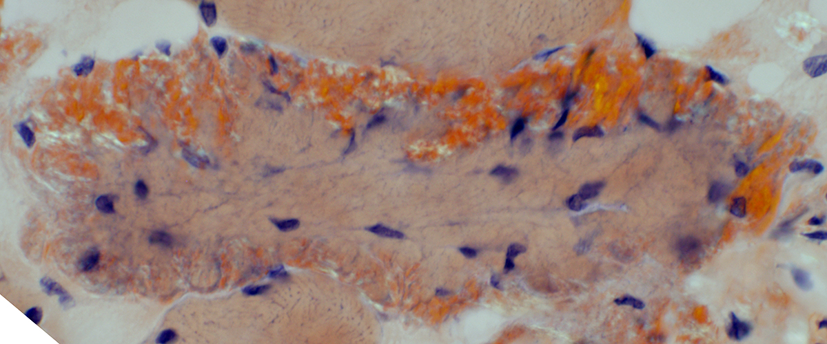
Amyloid surrounding, and on, muscle fibers- Mild pathology (Above): Thin layer of amyloid around normal sized muscle fiber
- More severe changes (Below)
- Thick deposit of amyloid around, and on, small muscle fibers
- Nuclei and NADH-positive membranes are present in regions of amyloid deposits
- Myofibrillar contractile apparatus (ATPase staining) is absent from areas of amyloid deposition but present in the center of fibers
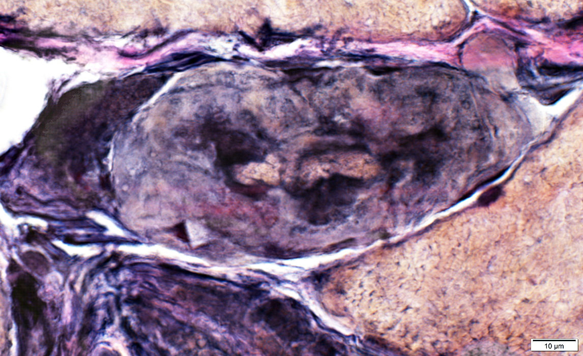
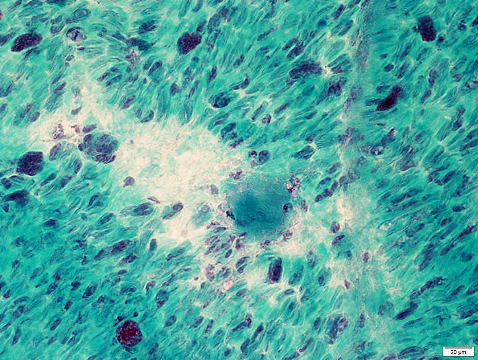
 Congo red |
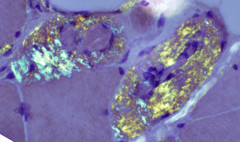
 Congo red (Polarized light) |
 Congo red (Fluorescence) |
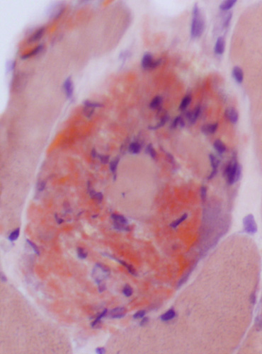
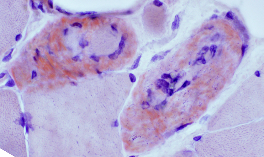
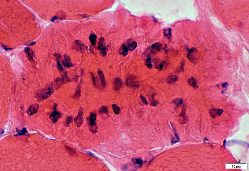
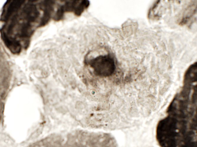
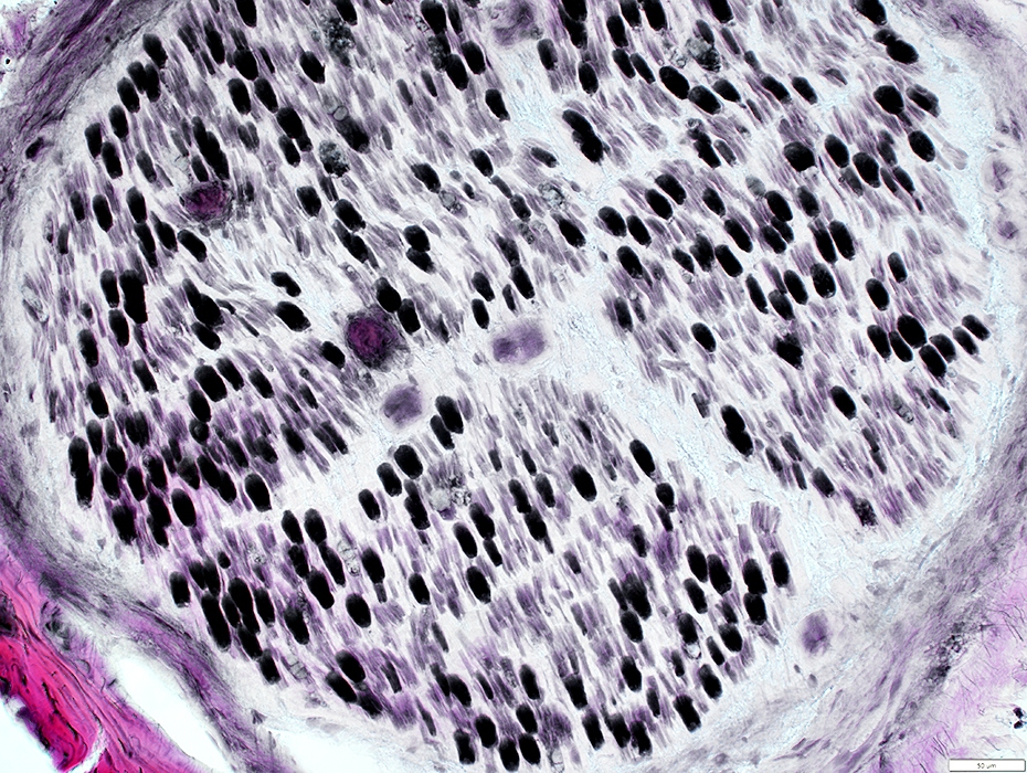
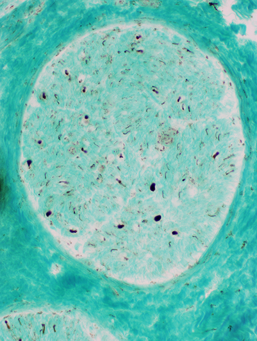
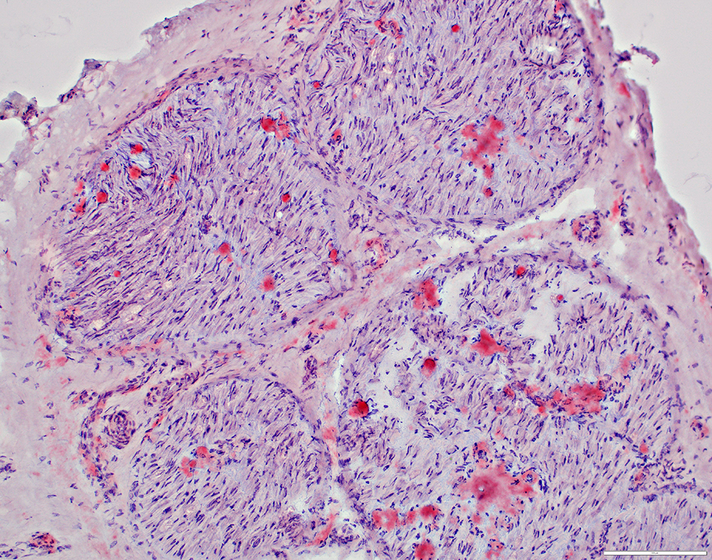
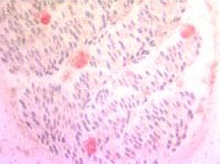
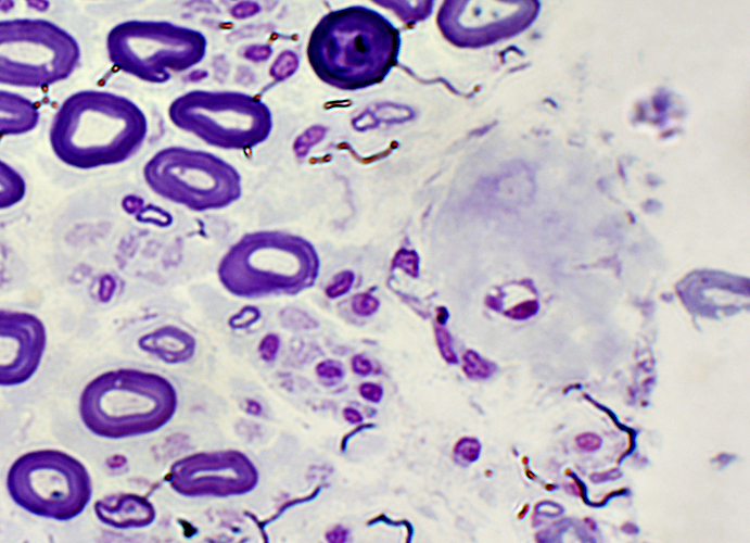
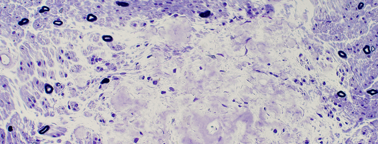
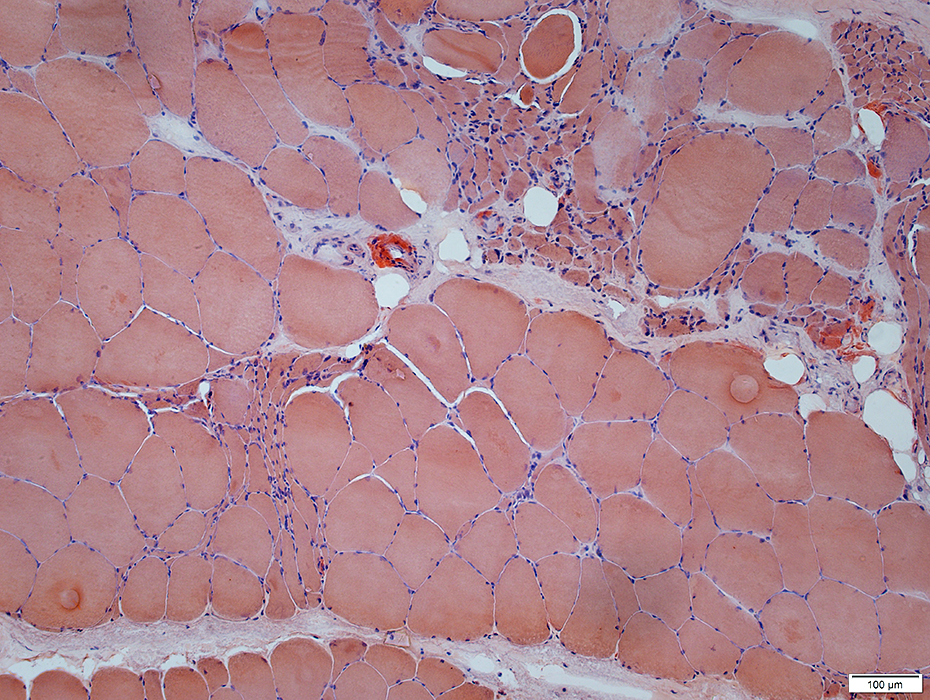
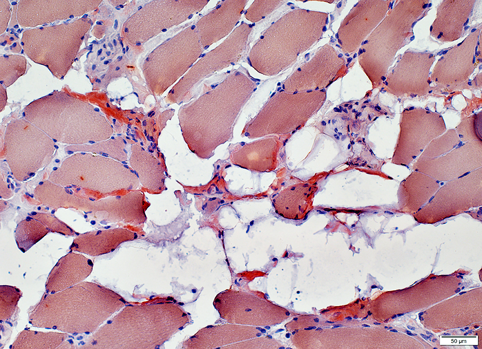
Muscle fiber pathology in amyloid myopathy
- Abnormal nuclei: In ring within muscle fibers
- Irregular deposits of amyloid: On muscle fibers outside nuclear ring
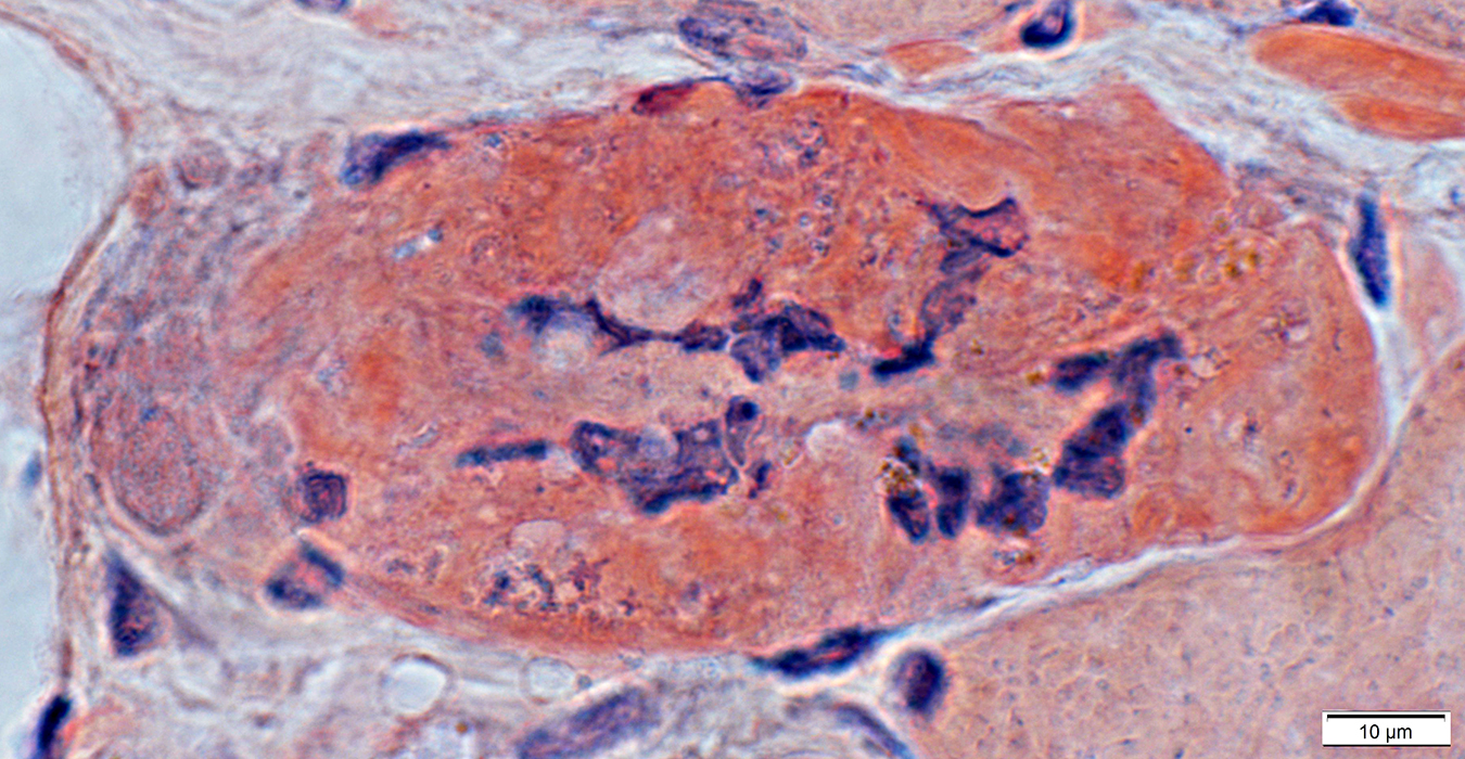
 Congo red |
 Congo red stain |
 Congo red stain |
|
 Congo red stain (Polarized light) |
|
 Congo red, polarized light |
 Congo red |
|
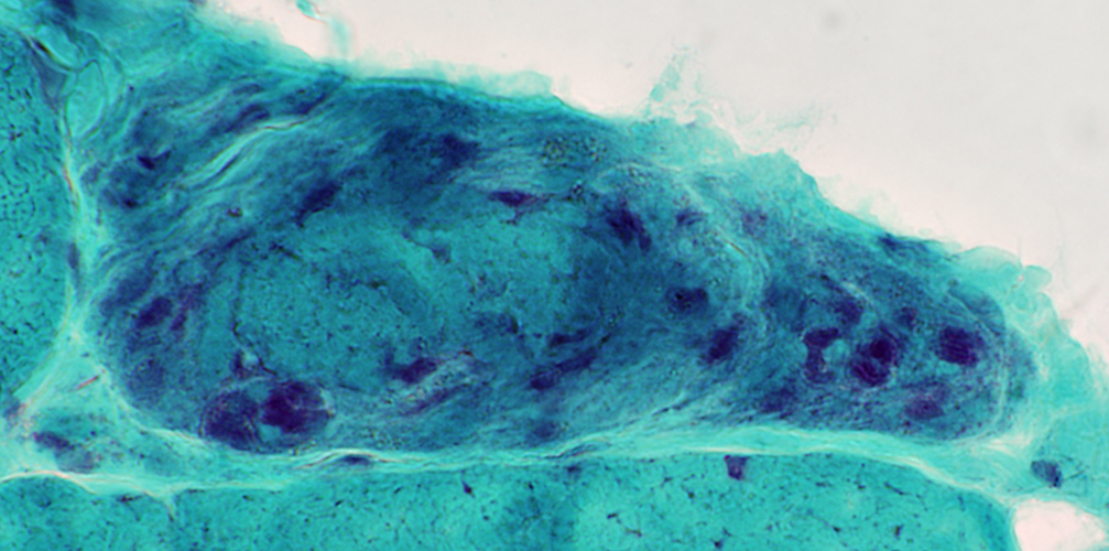
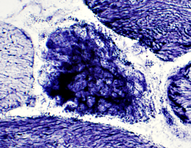
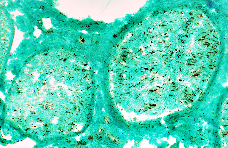
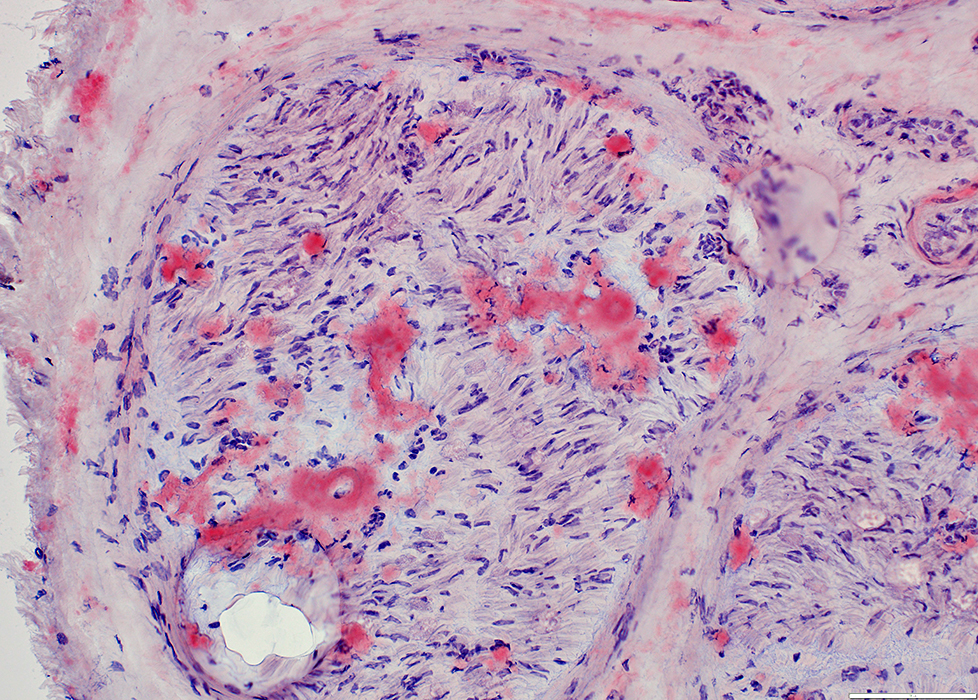
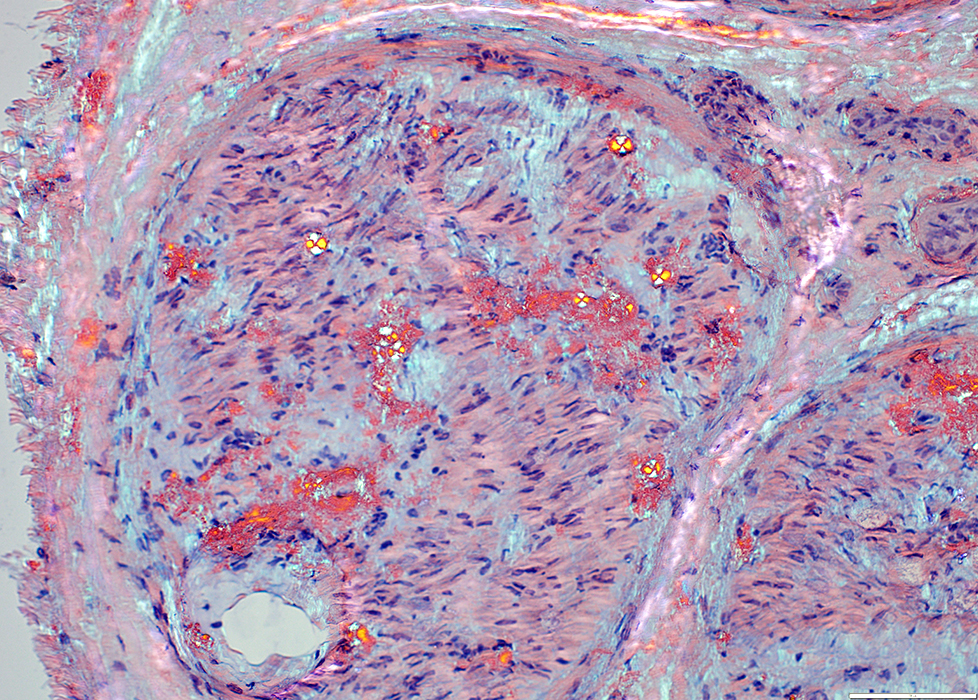
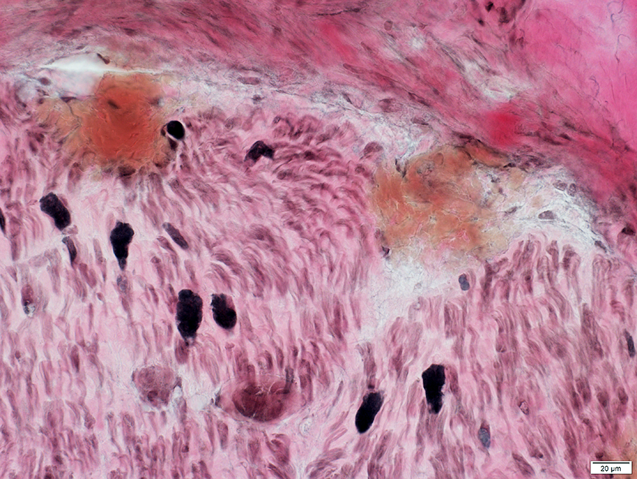
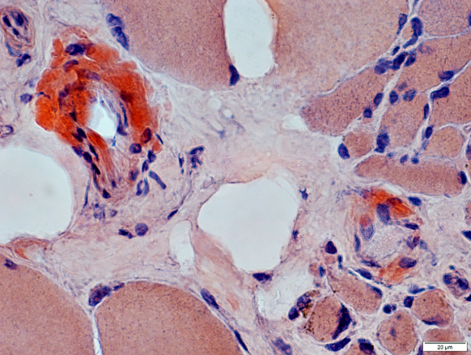
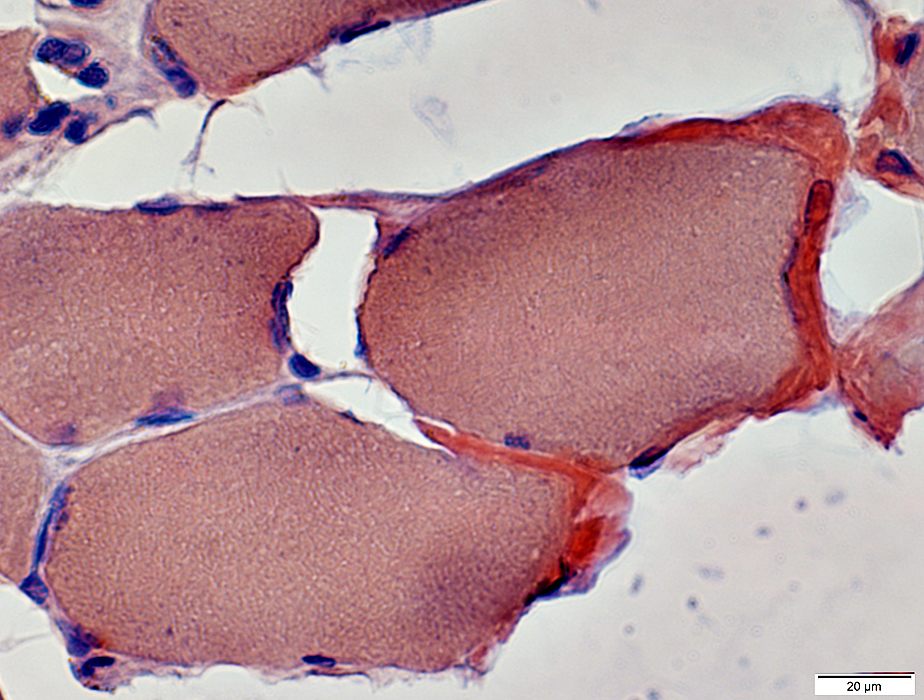
Amyloid Fibers Muscle fiber pathology in amyloidosis
|
 H&E stain |
 H&E stain |
 Gomori trichrome |
 VvG |
 Congo red |
Amyloid Fibers
 VvG |
 Congo red |
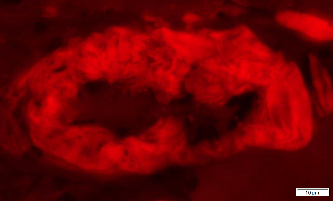 Congo red: Fluorescence |
Amyloid Fibers
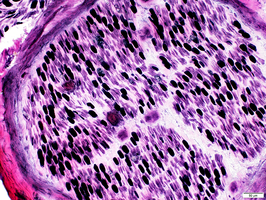
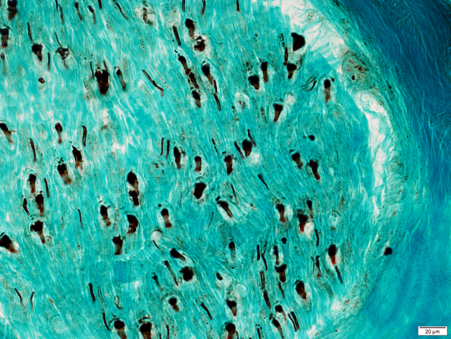
 ATPase pH 9.4 |
 ATPase pH 9.4 |
 NADH |
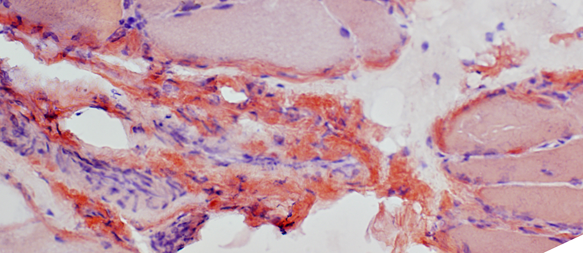
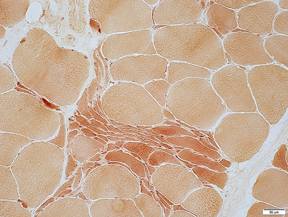
 Dystrophin |
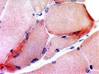
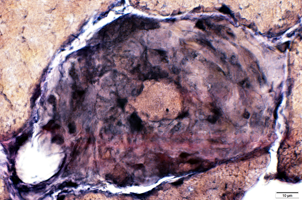
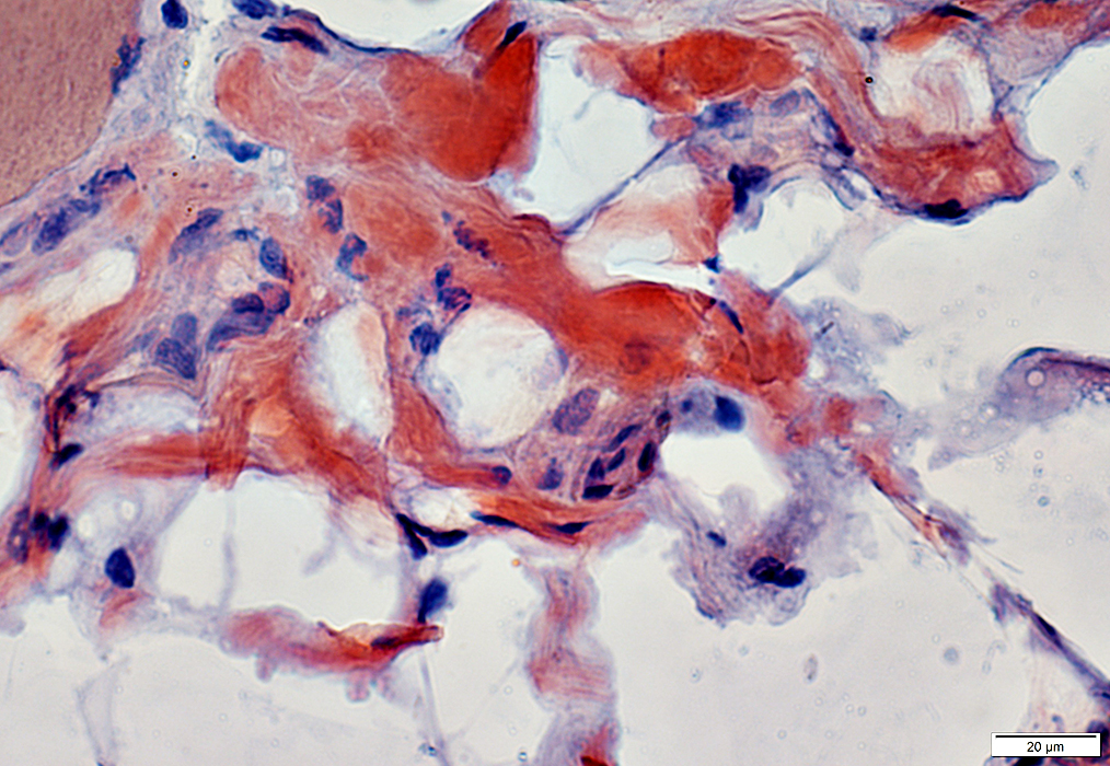
| Surface membrane (Above) & Basal lamina (Below): Thickened & reduplicated around muscle fibers |
 α2-Laminin |
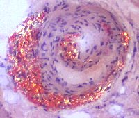
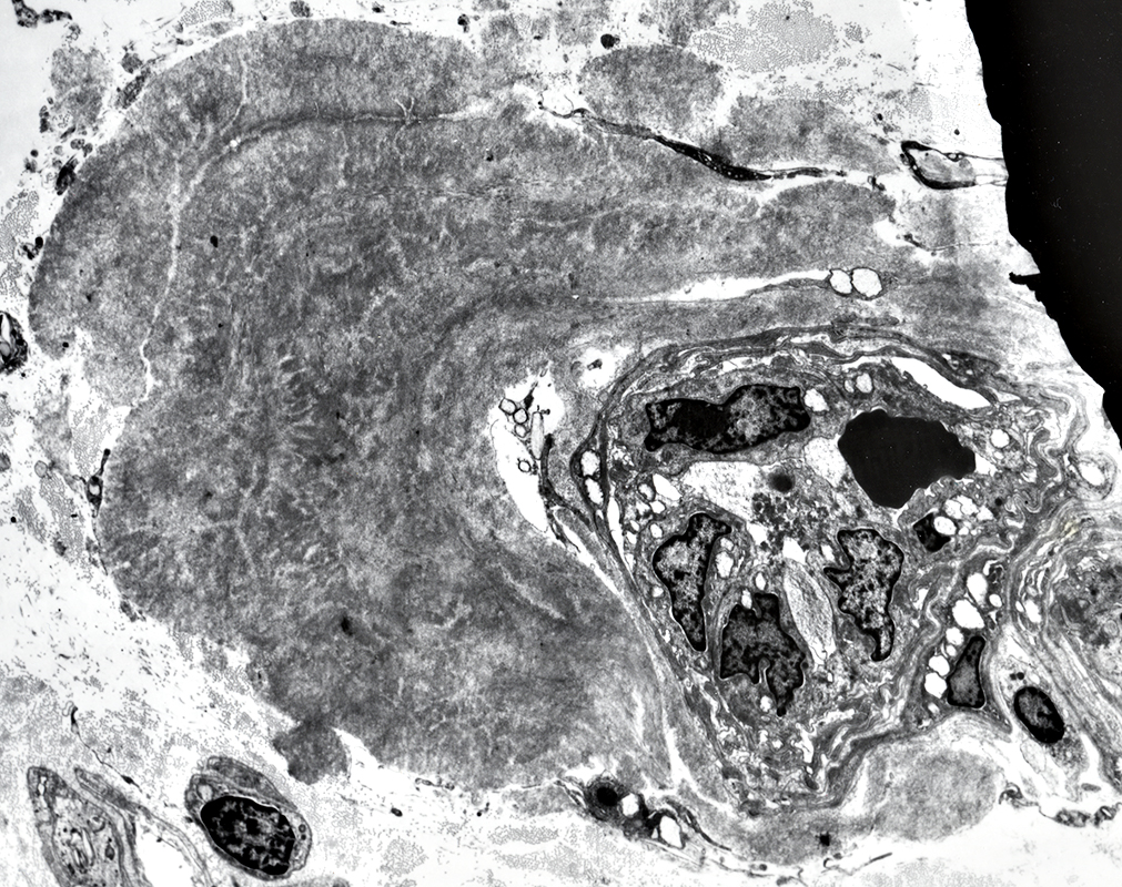
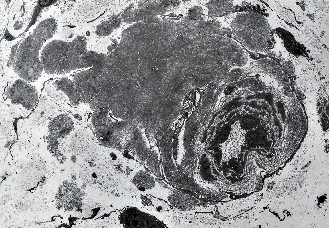
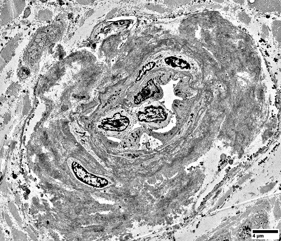
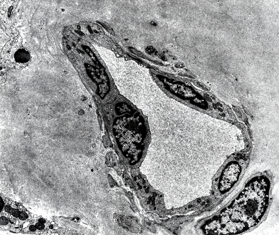
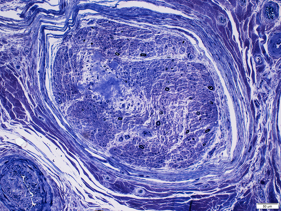
IgAλ M-protein: Amyloid in Nerve
 Congo red |
Congo red stained amyloid on endoneurial microvessels
 Congo red |
Red-Green birefringent Congo red stained amyloid on endoneurial microvessels
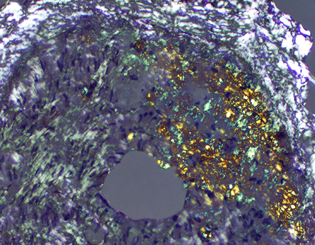
 Congo red, Polarized |
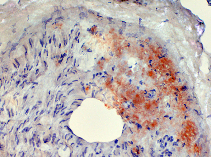
C5b-9 stains focus of endoneurial amyloid
 C5b-9 |
Amyloid
Amorphous material in wall of endoneurial vessel
Moderate & ongoing loss of myelinated axons
 Toluidine blue stain |
Amyloid
Moderate & ongoing loss of myelinated axons
 VvG stain |
Amyloid
Axon loss: Small > Large
 |
Amyloid
Non-myelinating Schwann cells: Diffusely distributed in endoneurium
 |
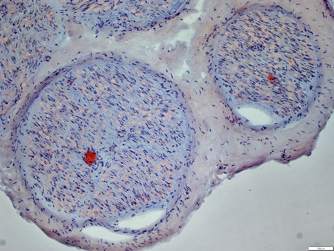
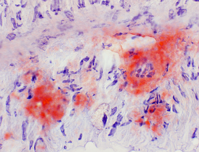
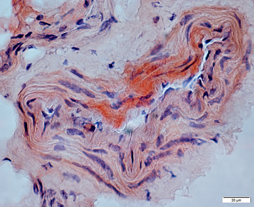
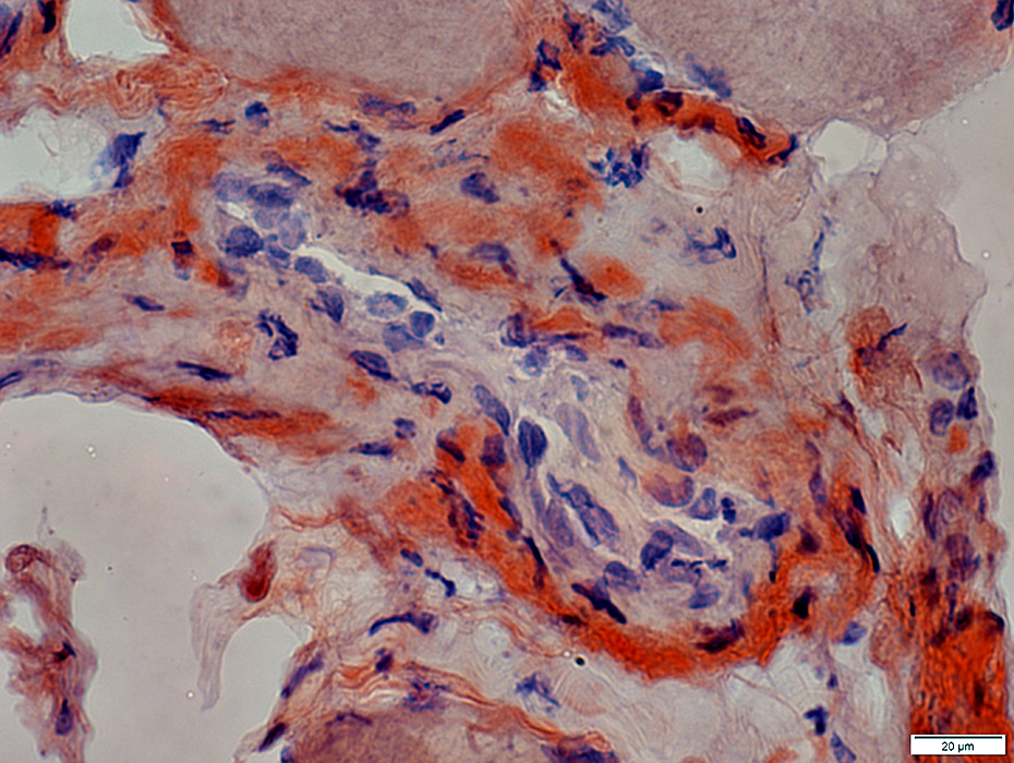
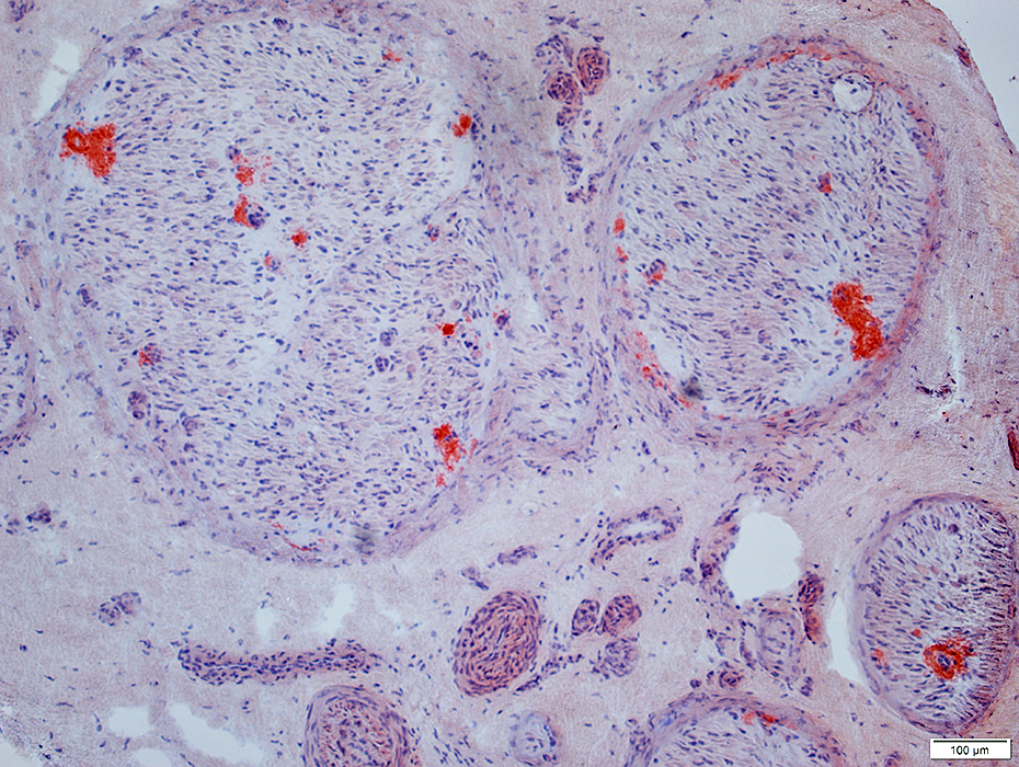
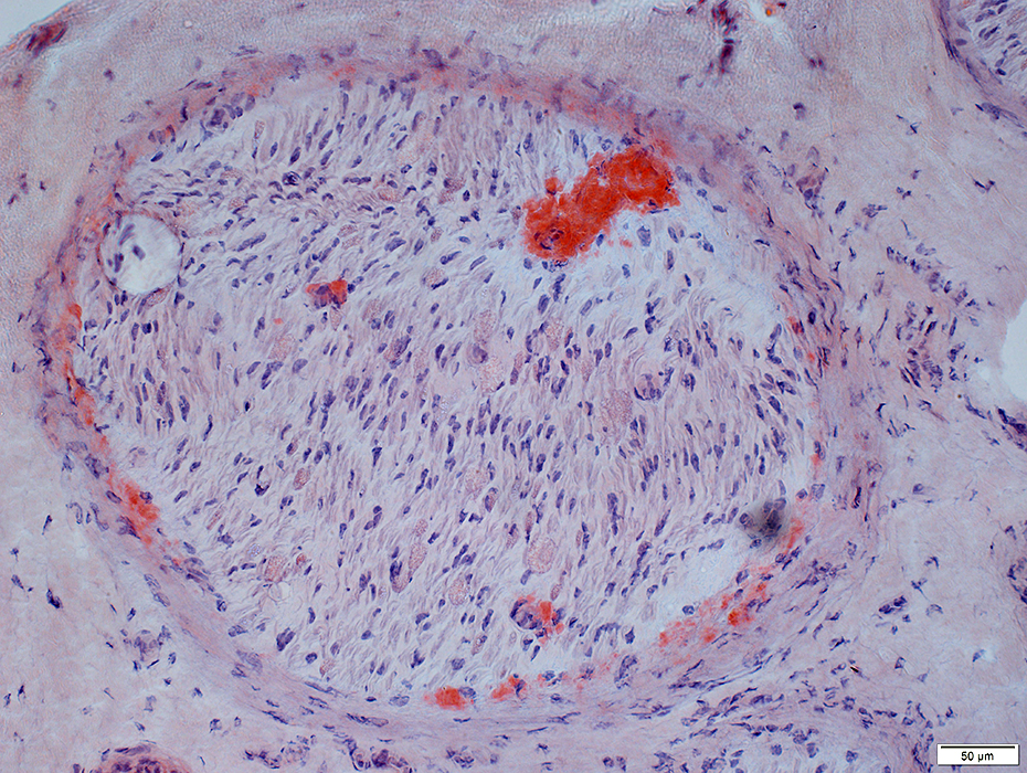
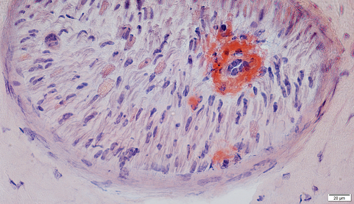
IgAκ M-protein: Amyloid in Muscle
 Congo red |
| Amyloid deposits in perimysium and along the perimysial surface of muscle fibers |
 Congo red |
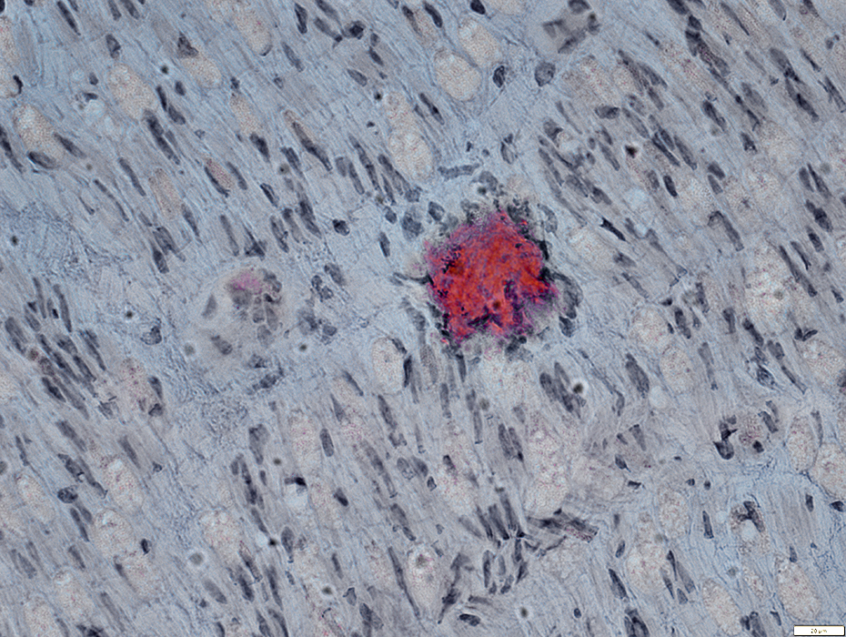
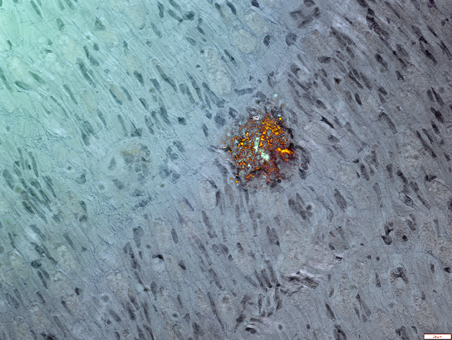
IgGλ M-protein: Amyloid
 Congo red |
 Congo red |
 Congo red |
 Congo red |
NERVE: Amyloid
Patterns of axon loss- Size
- Moderately severe: More loss of unmyelinated & small myelinated axons than large myelinated axons
- Severe: Loss of Large & Small axons
- Endoneurial
- Subperineurial
- Epineurial
Amyloid: Distribution
- Organs: Many
- Vessels: Common
- More abundant in large nerves
|
Severe Axon Loss Marked axon loss Small axons are even more involved than large axons See: Control nerve 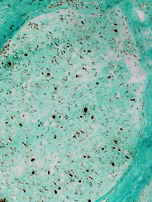 Neurofilament stain |
 Neurofilament stain |
Moderate axon loss
Differential fascicular loss: More axon loss in some areas than others
 Neurofilament stain |
Moderate axon loss
 VvG stain |
 Neurofilament stain |
Small axons: Severe loss
 Neurofilament stain |
 NCAM stain |
Axon loss: Normal or Increased Numbers
Amyloid deposition: Patchy loss of Schwann cells & axons
 NCAM stain |
 VvG From: R Bucelli |
Focal endoneurial regions have loss of cells (Arrows)
NCAM(r)ar.jpg) NCAM (Red); Neurofilament (Green & Yellow) |
Patchy loss of Schwann cells & Axons (Arrows)
Some remaining myelin is abnormal with NCAM staining (Below; Yellow)
P0(r)ar.jpg) NCAM (Green); P0 (Red) |
 Congo Red From: R Bucelli |
Locations
Endoneurial connective tissue
Around endoneurial microvessels
Perineurium
Epineurial connective tissue
Epineurial vessel walls
 Congo Red From: R Bucelli |
 Congo Red From: R Bucelli |
Maltese cross pattern in some areas
 Congo Red From: R Bucelli |
 From: R Schmidt |
 From: R Schmidt |
 From: R Schmidt |
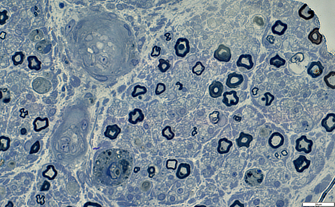
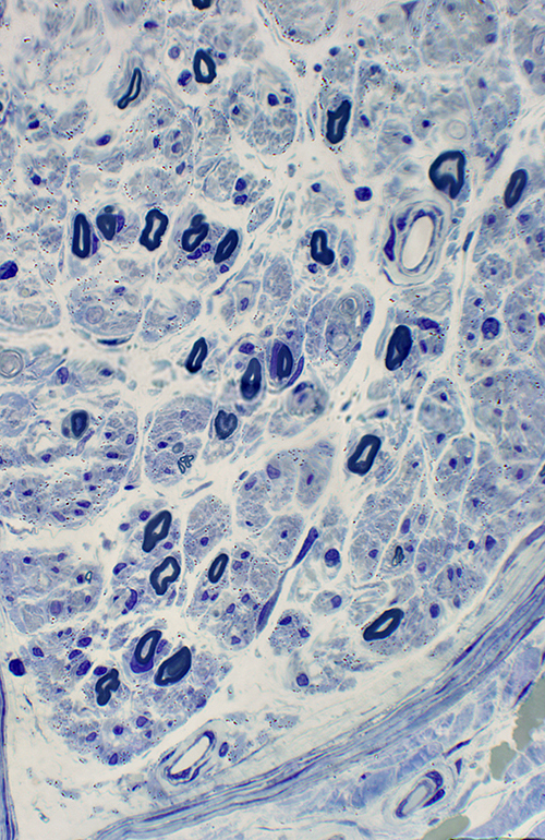
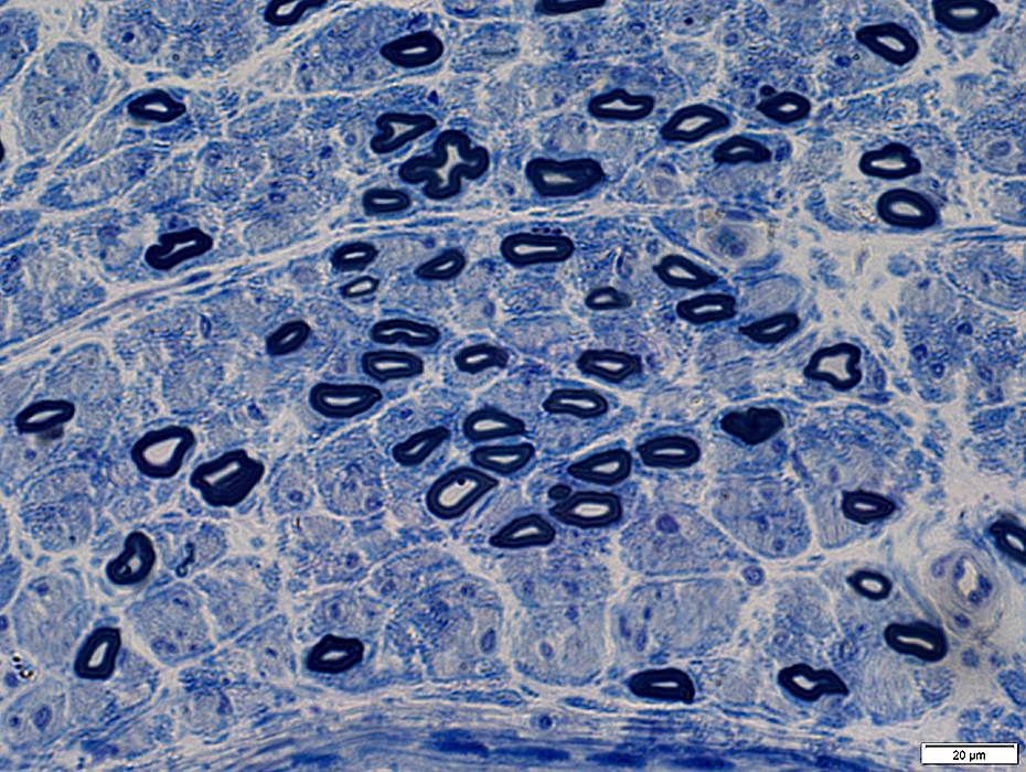
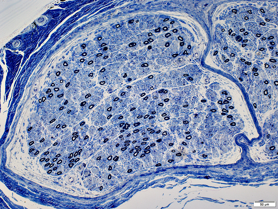 Toluidine blue stain |
|
Moderate axon loss More loss of small, than large, myelinated axons Subperineurial edema in some fascicles 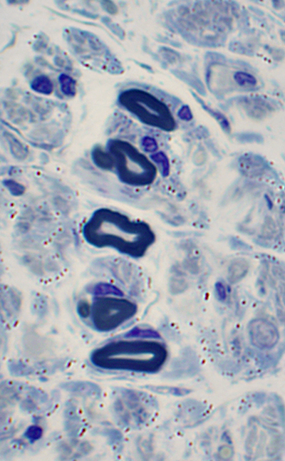 Toluidine blue stain |
 Toluidine blue stain |
 Toluidine blue stain |
Relative preservation of larger myelinated axons
Loss of Small axons with collagen pockets
 From: R Schmidt |
Amyloid Deposition
Capillary walls: Thick
Sub-Perineurial edema
 H&E stain |
 Congo red stain |
Contain red-stained amyloid
Amyloid is birefringent
 Congo red stain |
Endoneurial amyloid, Multifocal, often near endoneurial vessels (Arrow)
|
 Congo red stain 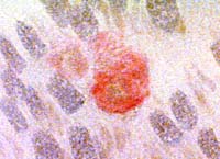 Congo red stain |
 Congo red stain |
 Toluidine blue stain |
 Toluidine blue stain |
Endoneurial amyloid, Diffuse
 Congo red stain |
 Congo red stain |
 Toluidine blue stain |
 Toluidine blue stain |
|
SUBPERINEURIAL AMYLOID (Arrow) 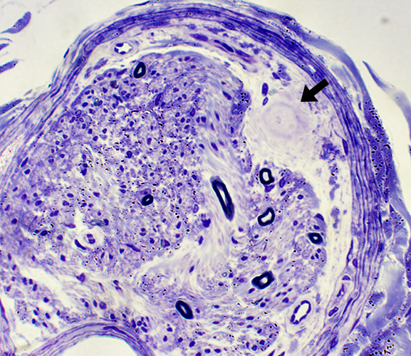 Congo red stain |
Moderate sized vessels: Amyloid
Vessels in Perimysium & Epineurium: May have amyloid deposits
 Congo red stain |
 Congo red stain (± Polarized light) |
 Congo red stain (Polarized light) |
|
Amyloid in vessel walls Red-Stained |
Birefrengence ± Polarization |
Amyloid in vessel wall Apple green with polarized light. |
Amyloid: Artery
 Congo red stain |
 Congo red stain |
 From: R Schmidt |
Around wall of endoneurial microvessel Asymmetric deposits
 From: R Schmidt |
 From: R Schmidt |
 From: R Schmidt |
Replaces basal lamina of endoneurial microvessel
Surrounds endoneurial microvessel
 From: J Karamchandani |
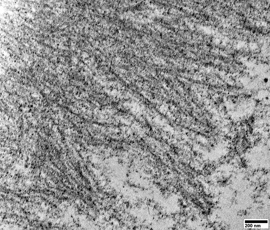
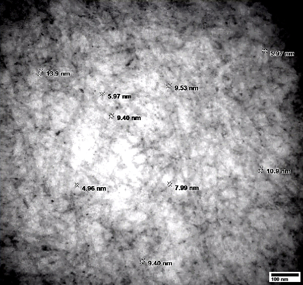
Typical widths
TTR: 6 to 8 nm
Imunoglobulin light chain: 10 to 20 nm
 From: R Schmidt |
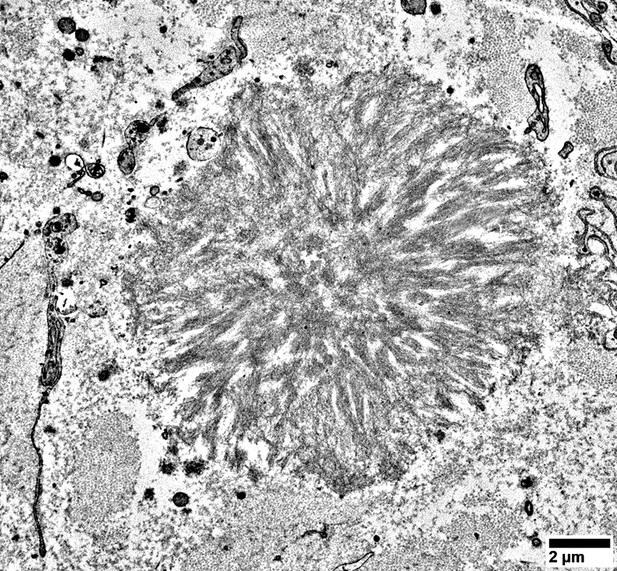
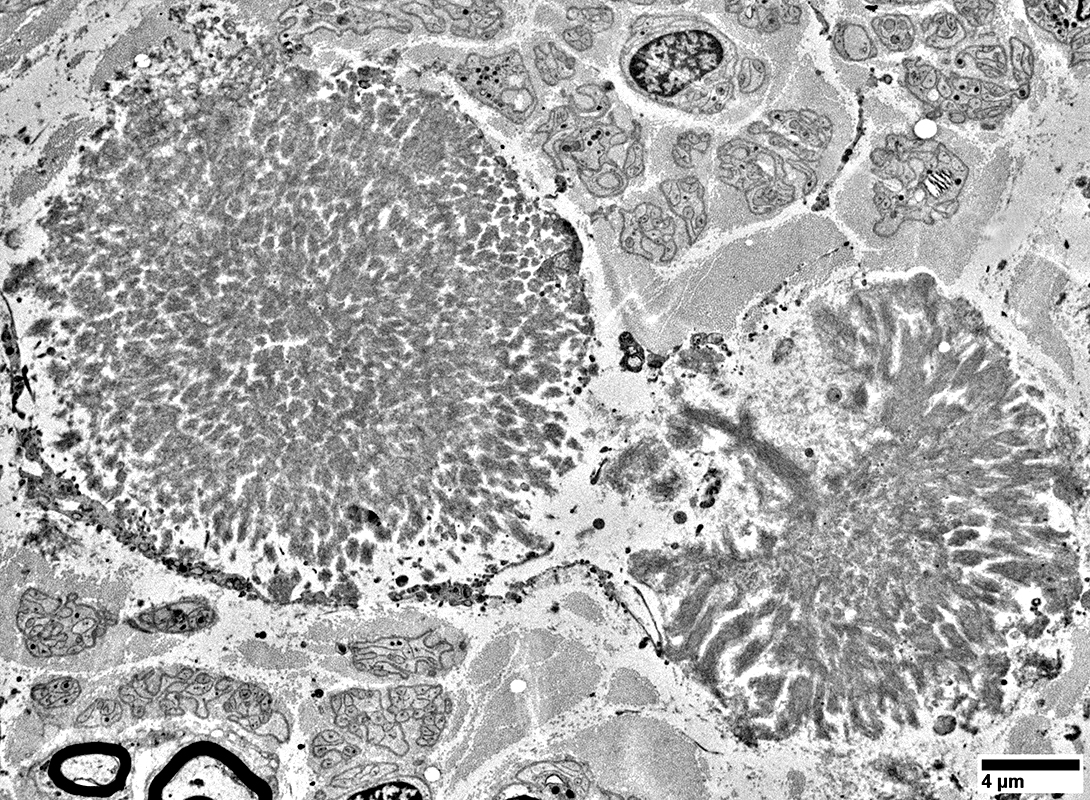
Amyloid: TTR Val30Met mutation
Nerve
 H&E stain |
 H&E stain |
 H&E stain |
 Gomori trichrome stain |
 VvG stain |
 Toluidine blue stain |
Amorphous, rounded clusters in endoneurium
Amyloid surrounded by pale cell-free regions
 Toluidine blue stain |
 Congo red stain |
Amorphous, rounded clusters in endoneurium
Perineurial deposition, scattered
 Congo red stain |
Amyloid
Surrounds endoneurial microvessel
 Congo red stain |
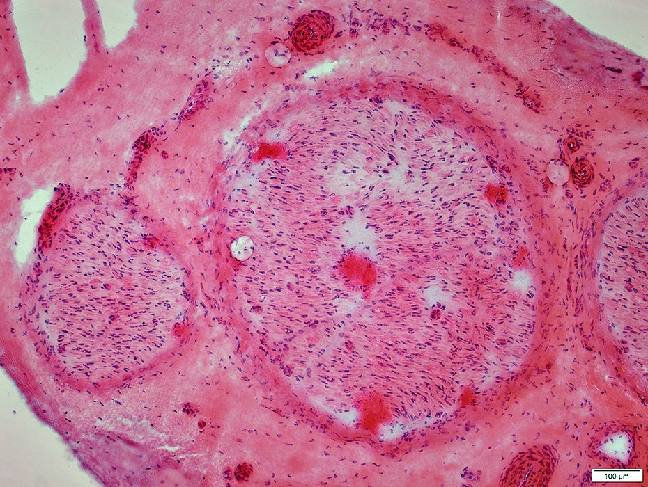
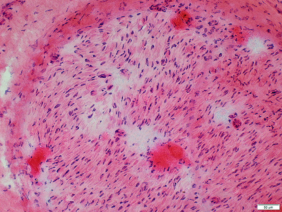
Amyloid (TTR Val30Met): Muscle
 H& E stain |
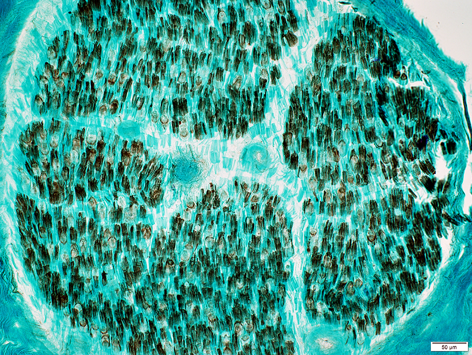
Grouped atrophy
Larger muscle fibers are hypertrophied
 VvG stain |
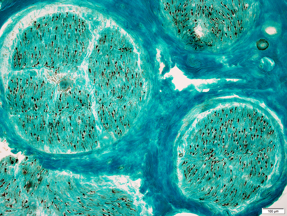
 ATPase pH 9.4 stain |
Fiber type grouping
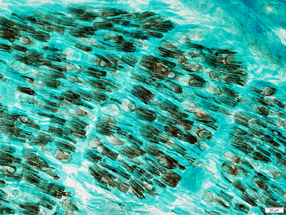
 ATPase pH 4.3 stain |
 ATPase pH 4.3 stain |
Muscle fibers in region of grouped atrophy
Varied degrees of immaturity (Type 2C)
Esterase positive cytoplasm
 Esterase stain |
 Congo red stain |
Most prominent in smaller perimysial vessels
 Congo red stain |
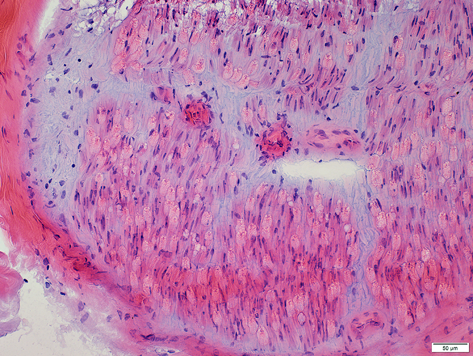
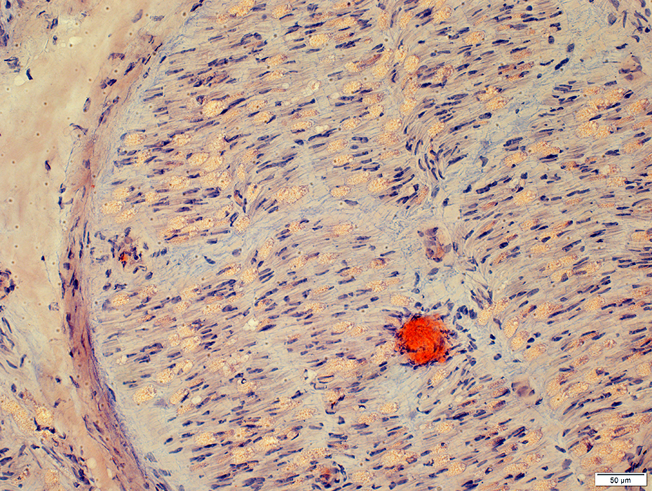
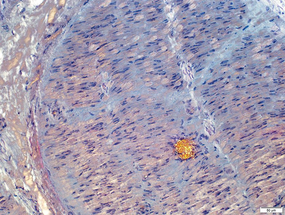
Amyloid (TTR Thr60Ala): Muscle
 H&E stain |
 Congo red stain |
Most prominent in connective tisue & around muscle fibers
 Congo red stain |
 Congo red stain |
Return to Neuromuscular Home Page
Return to Amyloidosis
8/30/2025