TNFα Inhibitor Myopathy
|
General pattern Endomysium Inflammation Cell types Muscle fibers Incomplete fusion Necrosis Perimysium |
General pattern
Necrotic muscle fibers: Scattered
Perimysial connective tissue: Damage & Cellularity
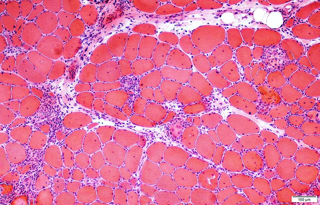 H&E stain |
ENDOMYSIAL CELL PATHOLOGY
Muscle fibers
Necrotic & Regenerating: Scattered; Varyied stages
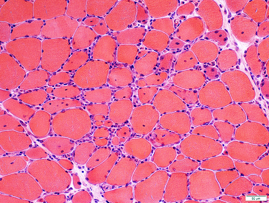 H&E stain |
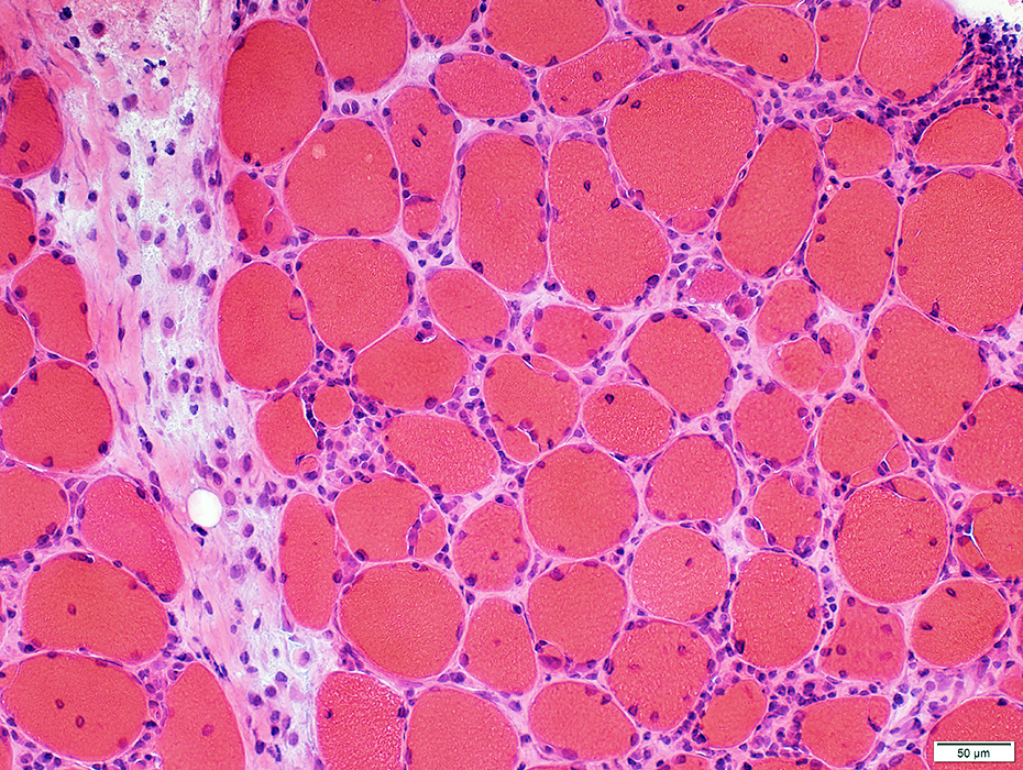 H&E stain |
Endomysial: Lymphocytes
Perimysium: Histiocytes
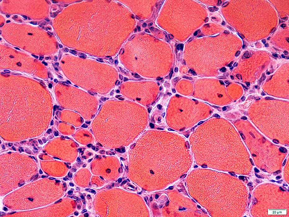 H&E stain |
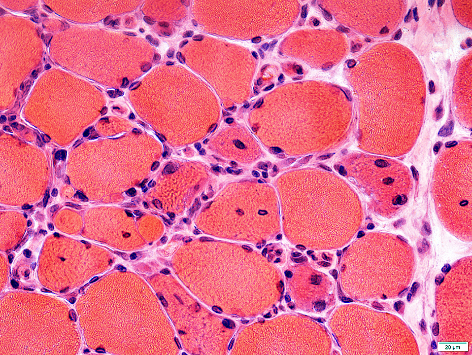 H&E stain |
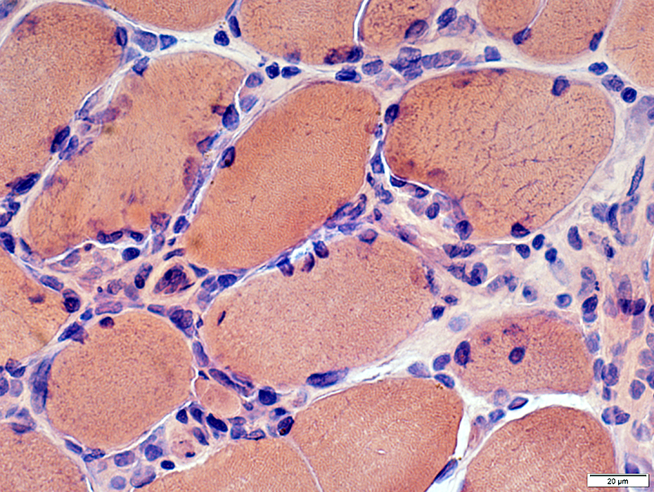
Congo red stain |
Lymphocytes: Endomysial
Histiocytes: Replacing necrotic muscle fibers
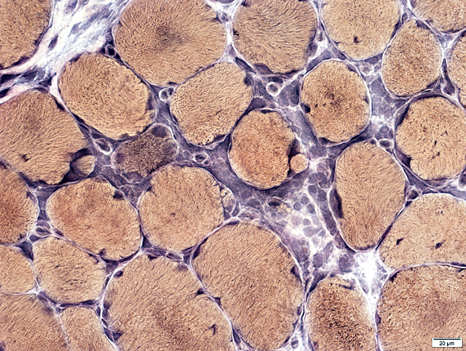 VvG stain |
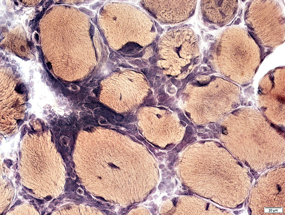 VvG stain |
ENDOMYSIAL IMMUNE CELLS
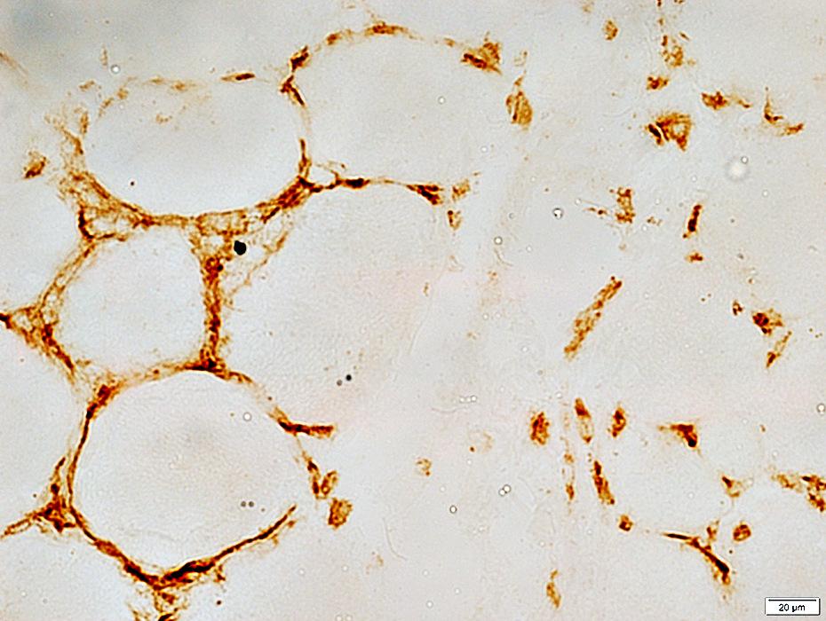 CD4 stain |
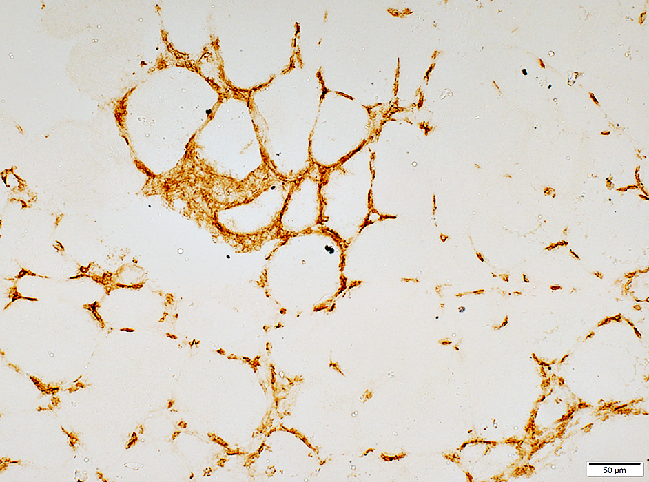 CD8 stain |
PERIMYSIAL PATHOLOGY
 H&E stain |
Immune cells: Large cells with abundant cytoplasm in perimysium
 H&E stain |
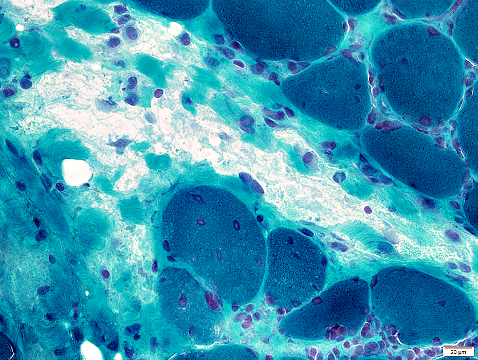 Gomori trichrome stain |
Immune cells: Focus in perimysium
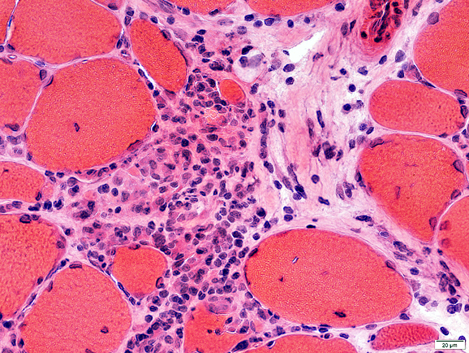 H& E stain |
Perimysial pathology
Abnormal staining of perimysium for alkaline phosphatase
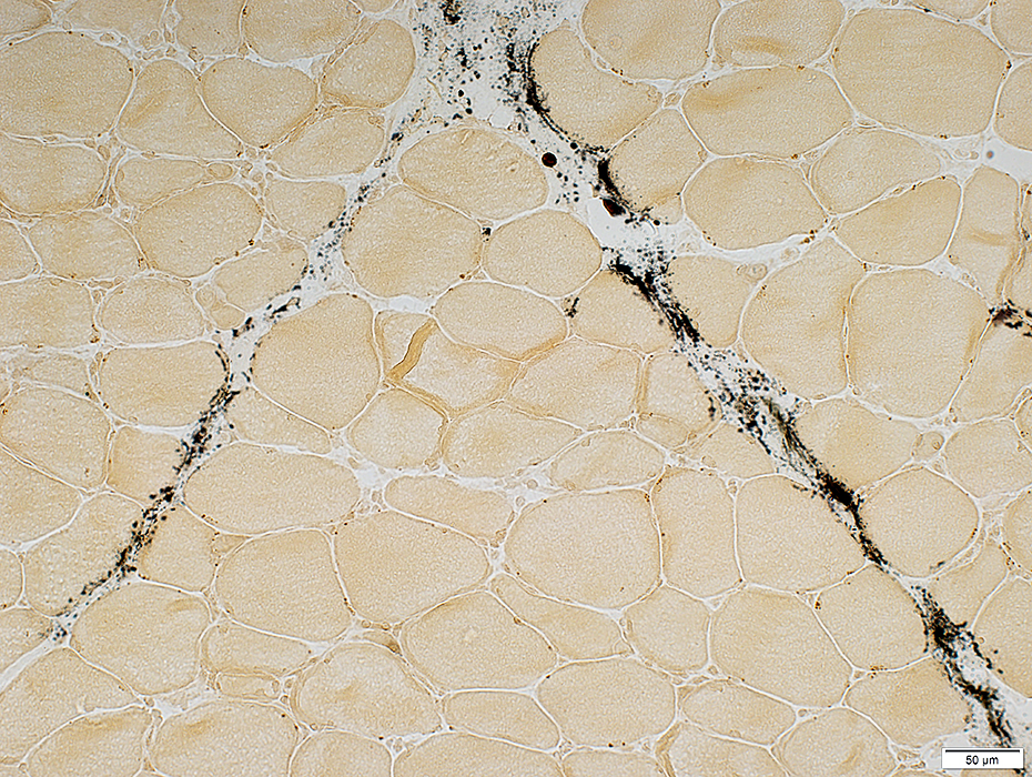 Alkaline Phosphatase stain |
Staining of cells in perimysium, and scattered in endomysium, for acid phosphatase
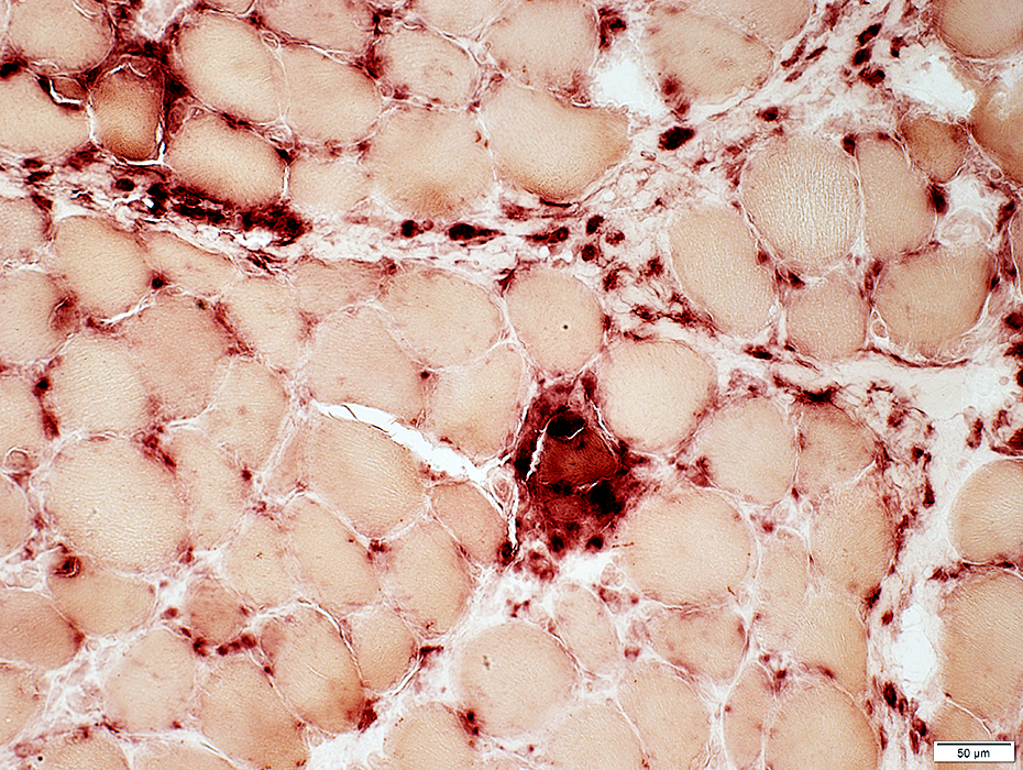 Acid Phosphatase stain |
CD4 cells: In perimysium & endomysium
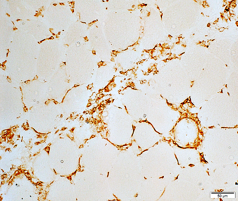 CD4 cell stain |
MUSCLE FIBER PATHOLOGY
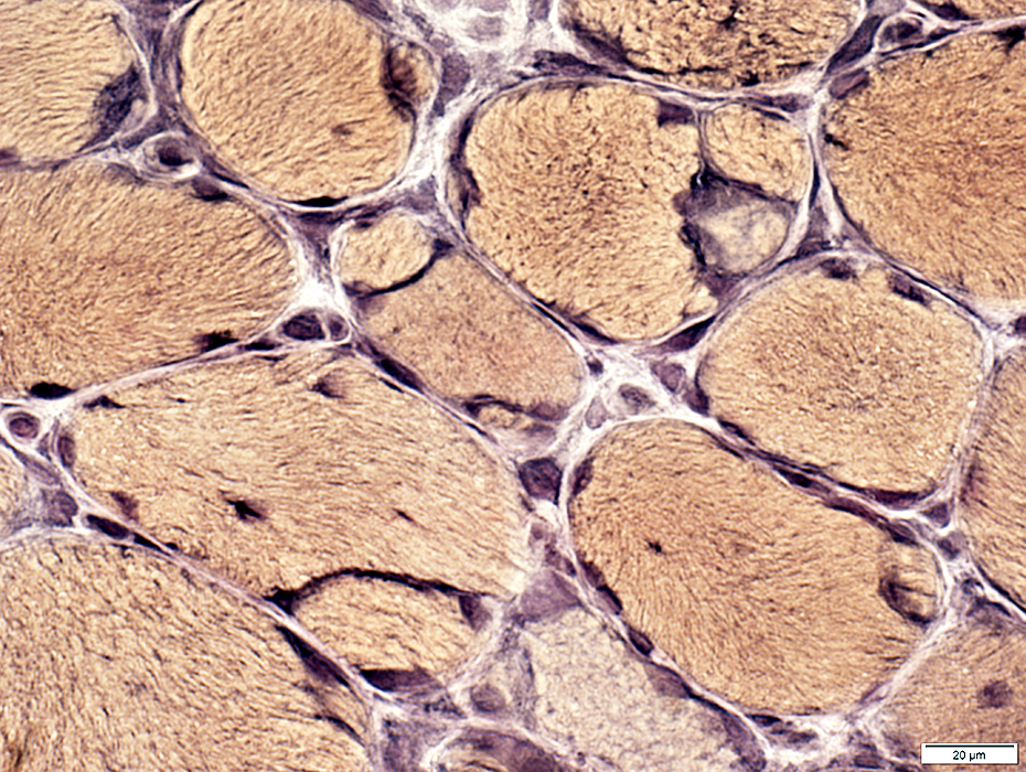 VvG stain |
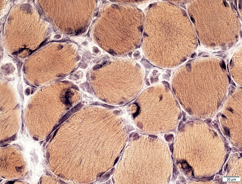 VvG stain |
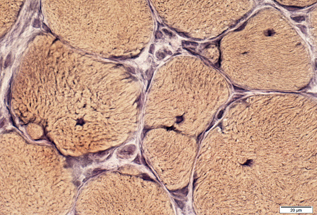 VvG stain |
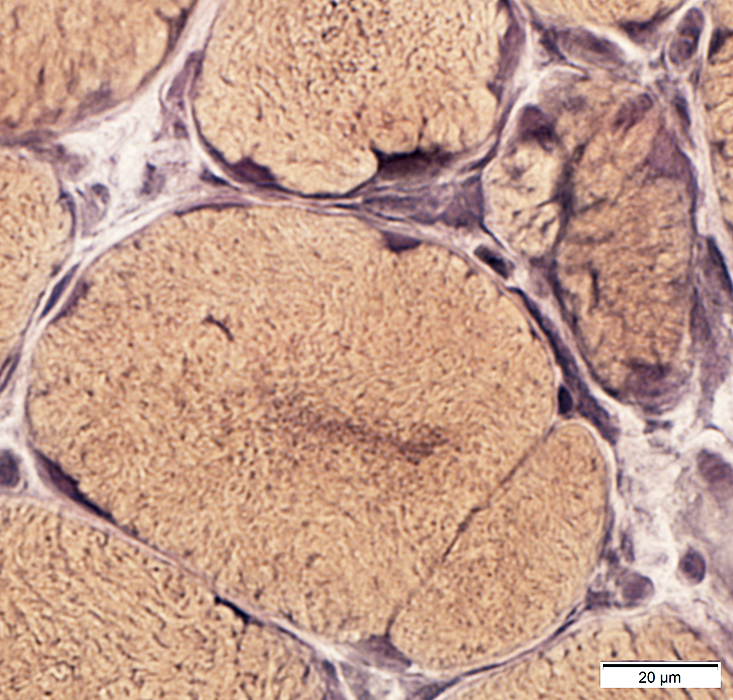 VvG stain |
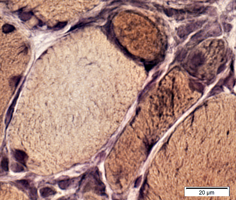 VvG stain |
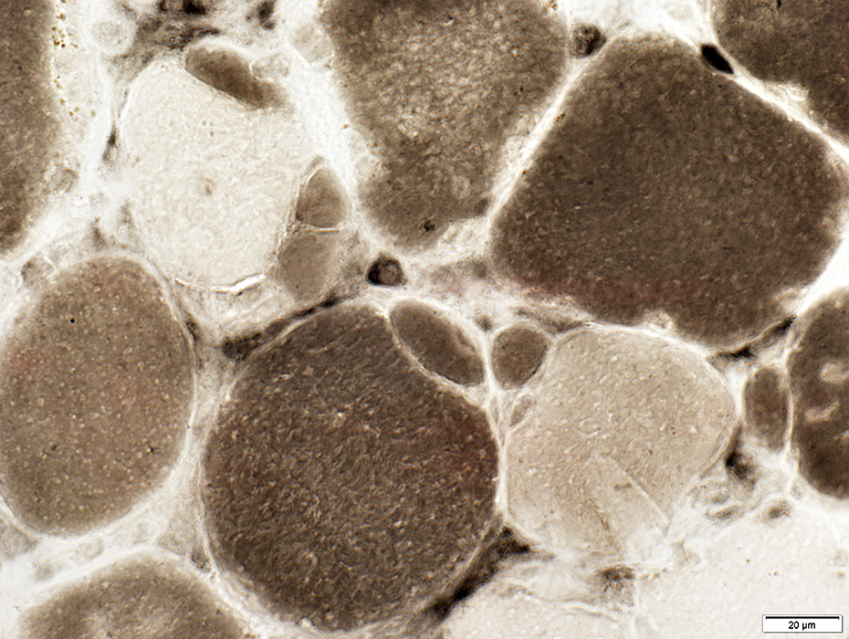 ATPase pH 4.3 stain |
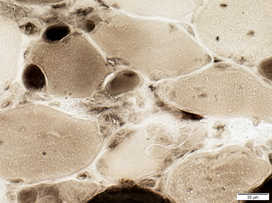 ATPase pH 4.3 stain |
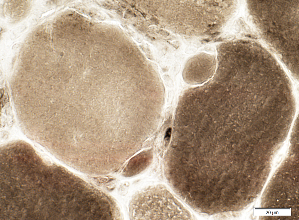 ATPase pH 4.3 stain |
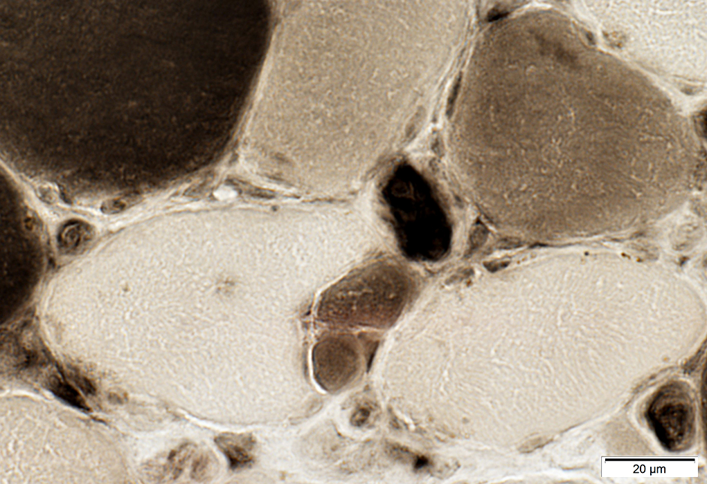 ATPase pH 4.3 stain |
Internal architecture: Irregular; Dark staining in center fo some fibers
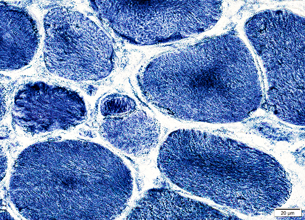 NADH stain |
Immature small unfused muscle fibers: Stain for alkaline phosphatase
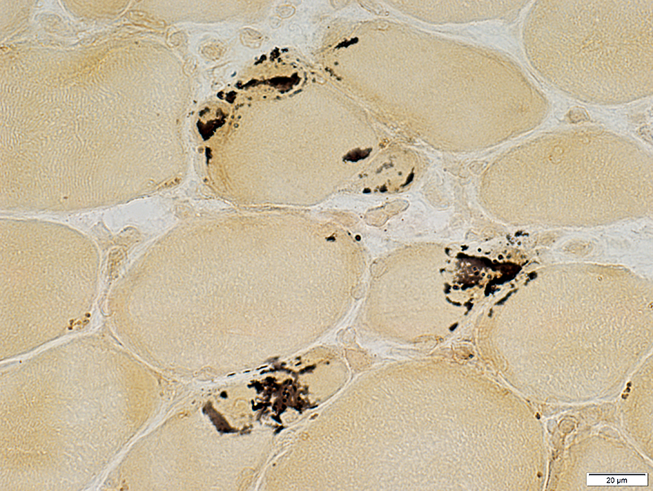 Alkaline phosphatase stain |
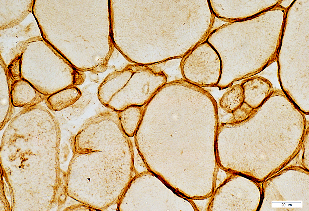 Caveolin-3 stain |
Surface stained for Caveolin-3
Caveolin-3 staining is darkest at region near neighboring larger muscle fiber
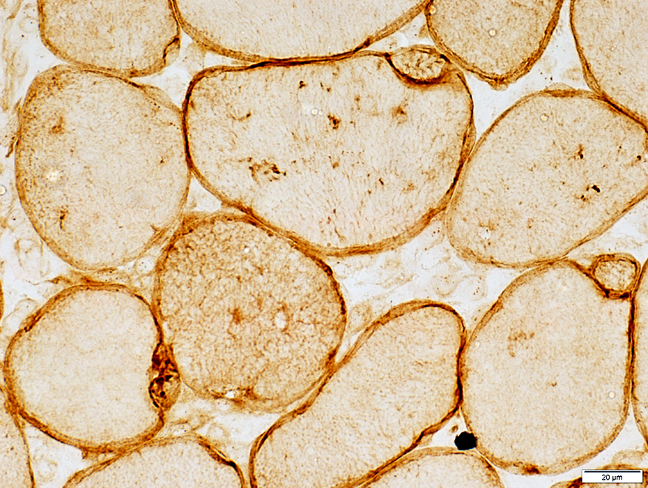 Caveolin-3 stain |
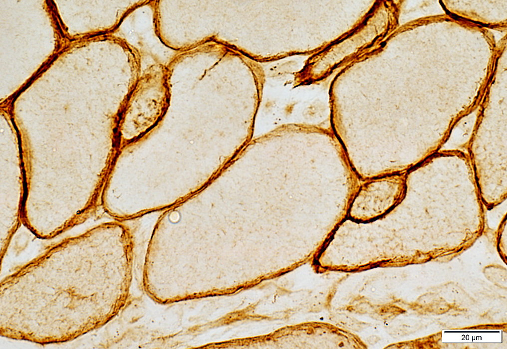 Caveolin-3 stain |
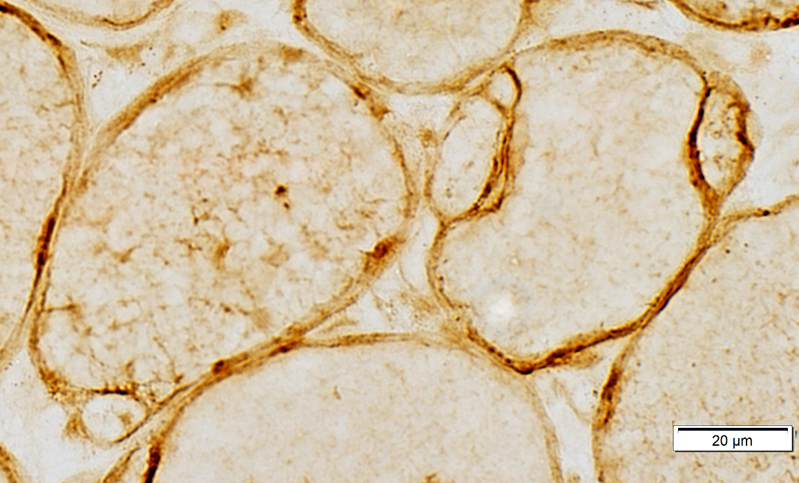 Caveolin-3 stain |
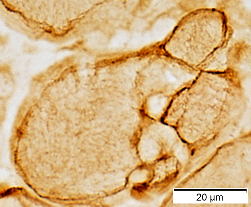 Caveolin-3 stain |
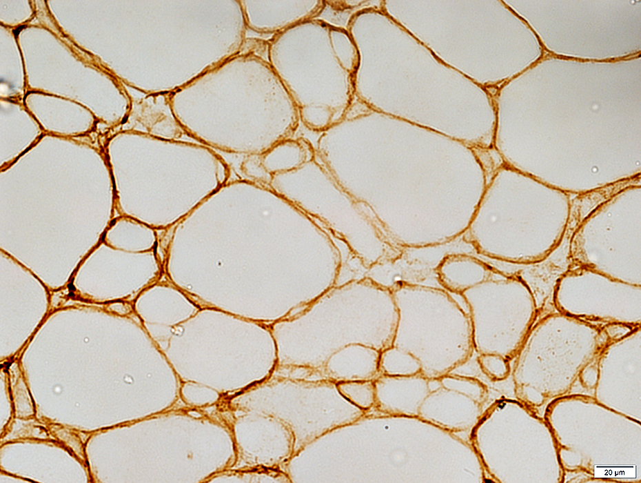 Decorin stain |
Dark surrounding areas of small & large muscle fibers
Pale at region near neighboring larger muscle fiber
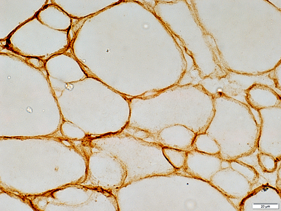 Decorin stain |
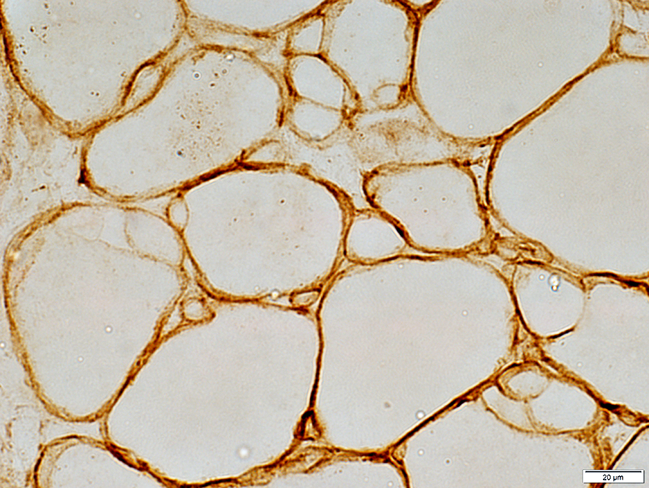 Decorin stain |
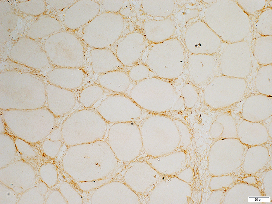 C5b-9 stain |
Muscle fiber surface: Irregular deposits
Necrotic muscle fiber: Cytoplasm
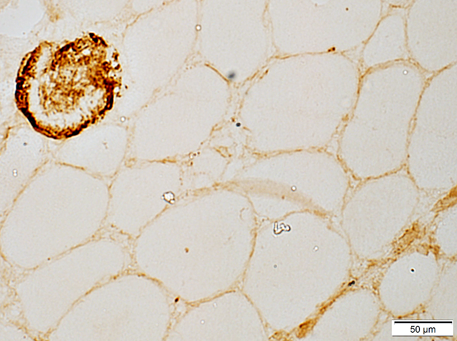 C5b-9 stain |
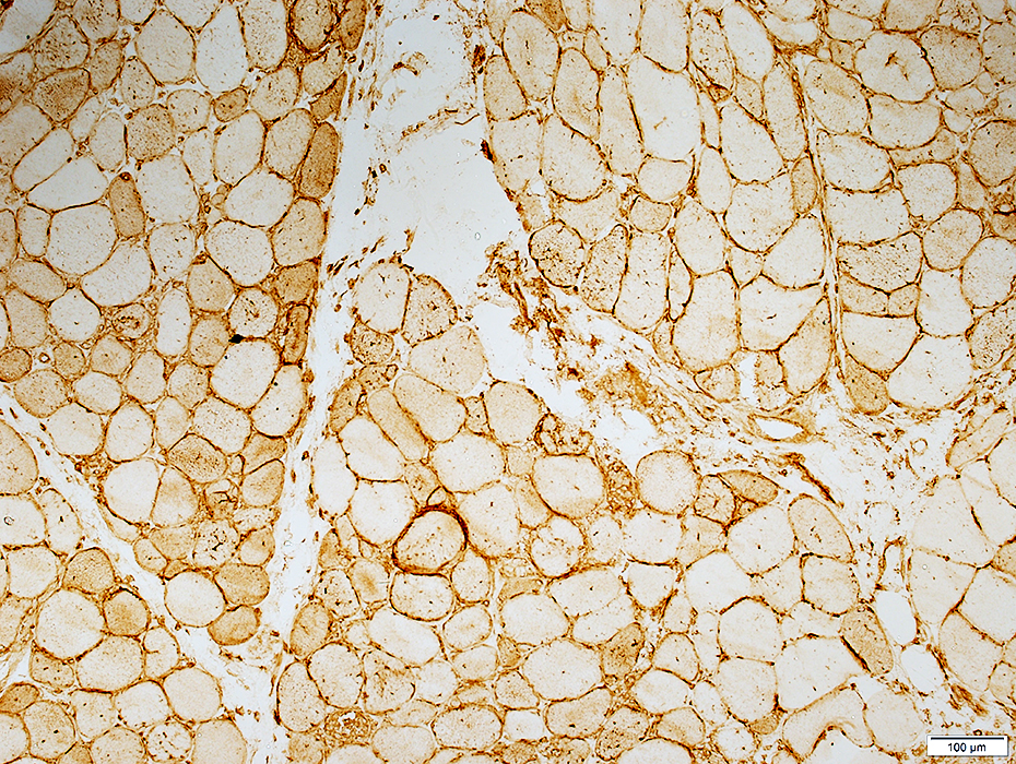 MHC Class I stain |
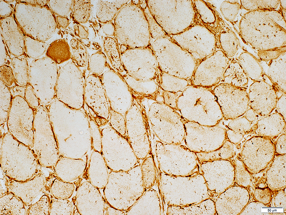 MHC Class I stain |
Muscle fiber necrosis & regeneration
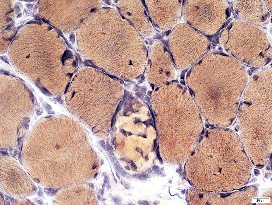 VvG stain |
Replaced by histiocytic cells
Pale on NADH & ATPase stains
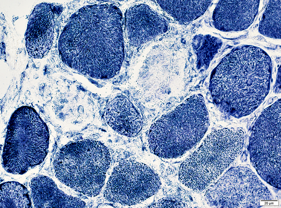 NADH stain |
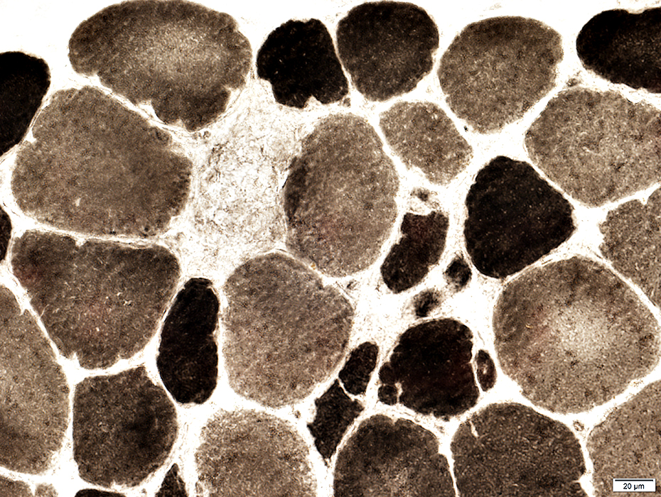 ATPase pH 9.4 stain |
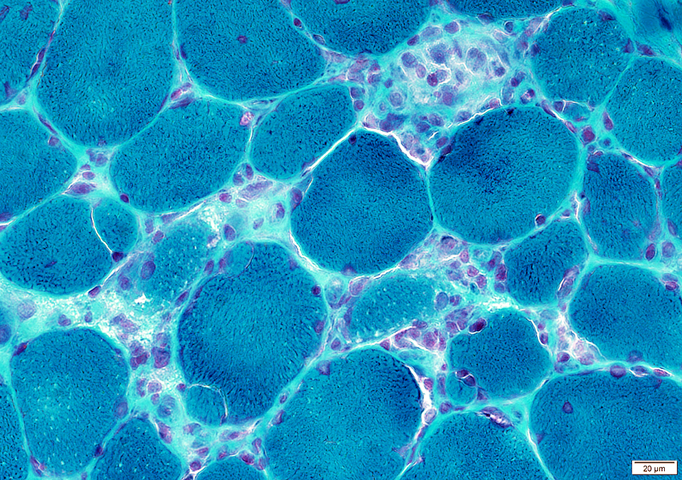 Gomori trichrome stain |
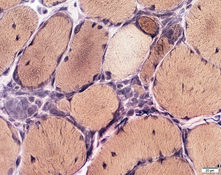 VvG stain |
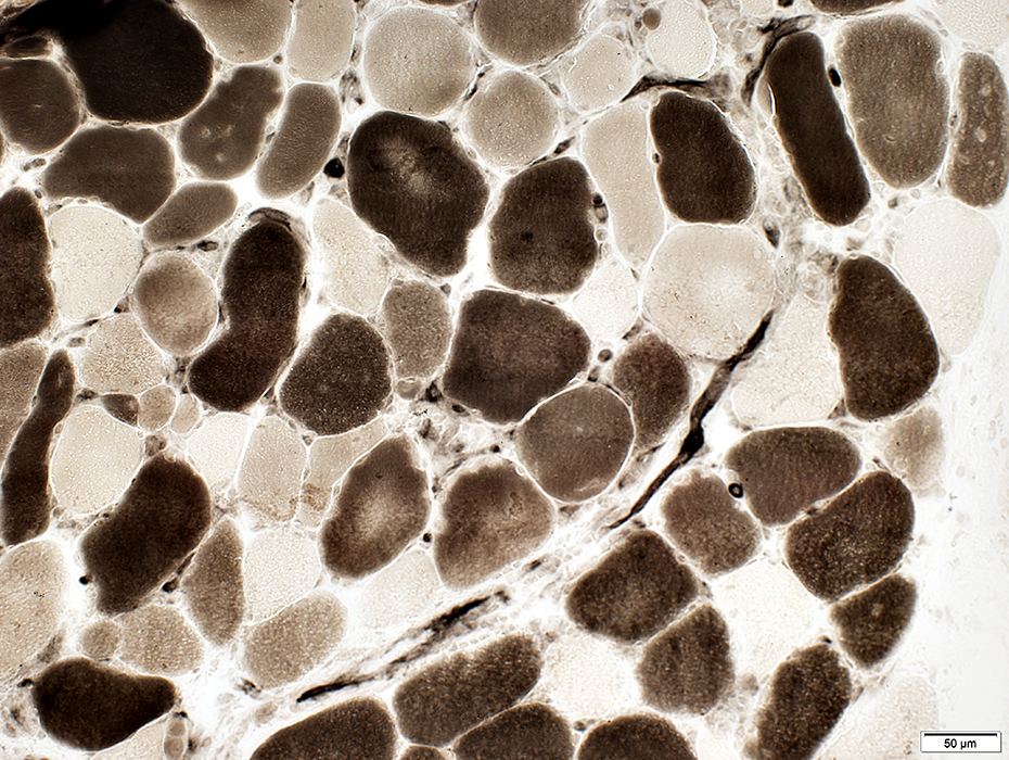 ATPase pH 4.3 stain |
Type 2C: Intermediate-stained on ATPase pH 4.3
Dark-staind on NADH
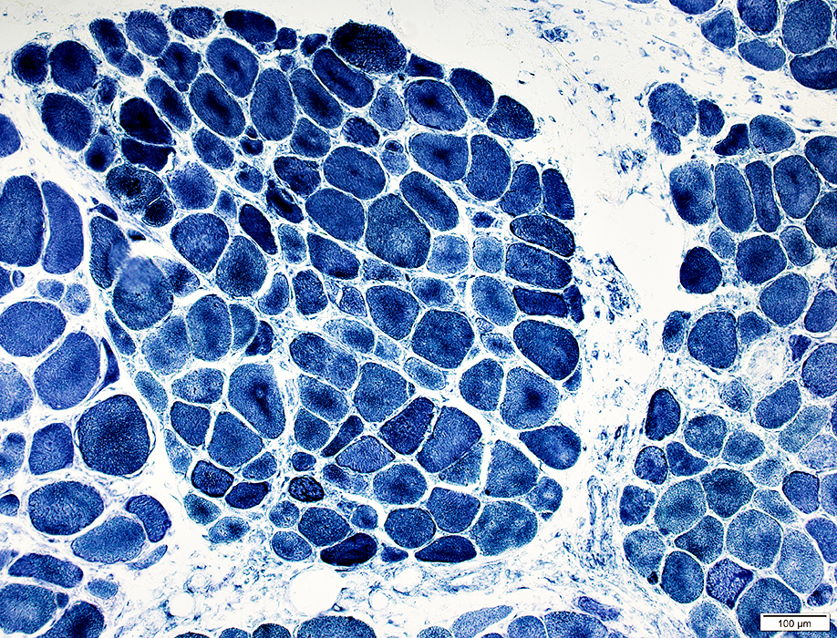 NADH stain |
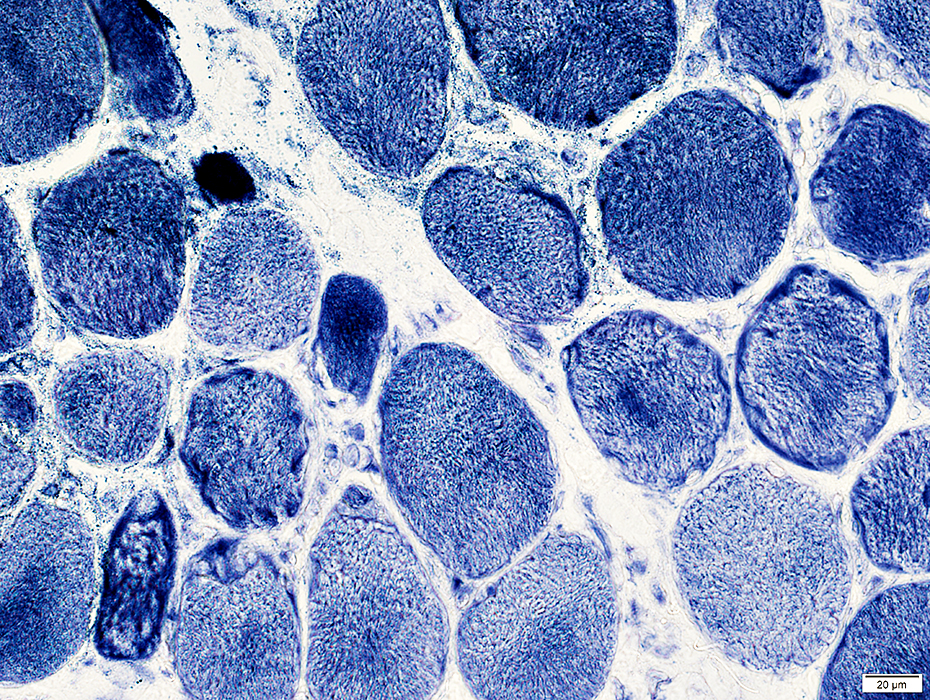 NADH stain |
Immature muscle fibers: Small basophilic fibers atg edge of fascicles
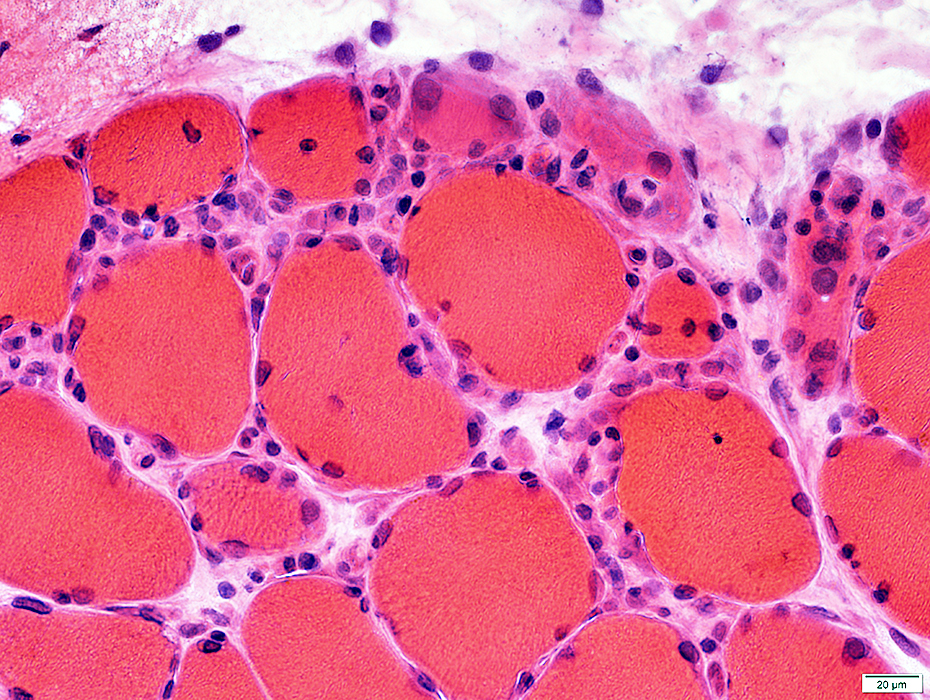 H& stain |
Autophagic aggregates: Muscle fiber cytoplasm
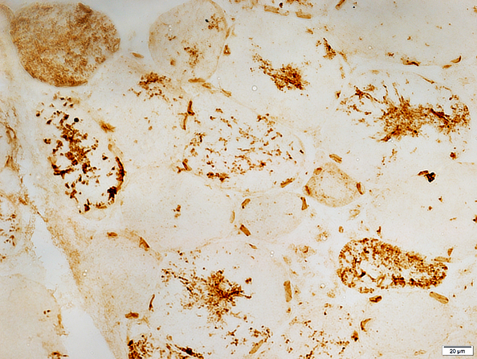 LC3 stain |
Endomysial capillaries: Enlarged
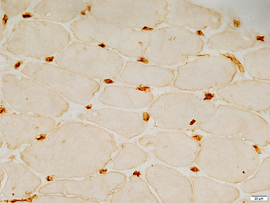 UEA I stain |
Return to Inflammatory myopathies
Return to TRAPS
5/14/2016