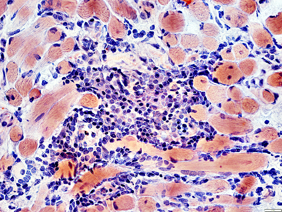
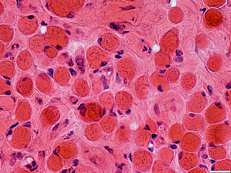
Thyroid Ophthalmopathy: Extraocular Muscle Pathology
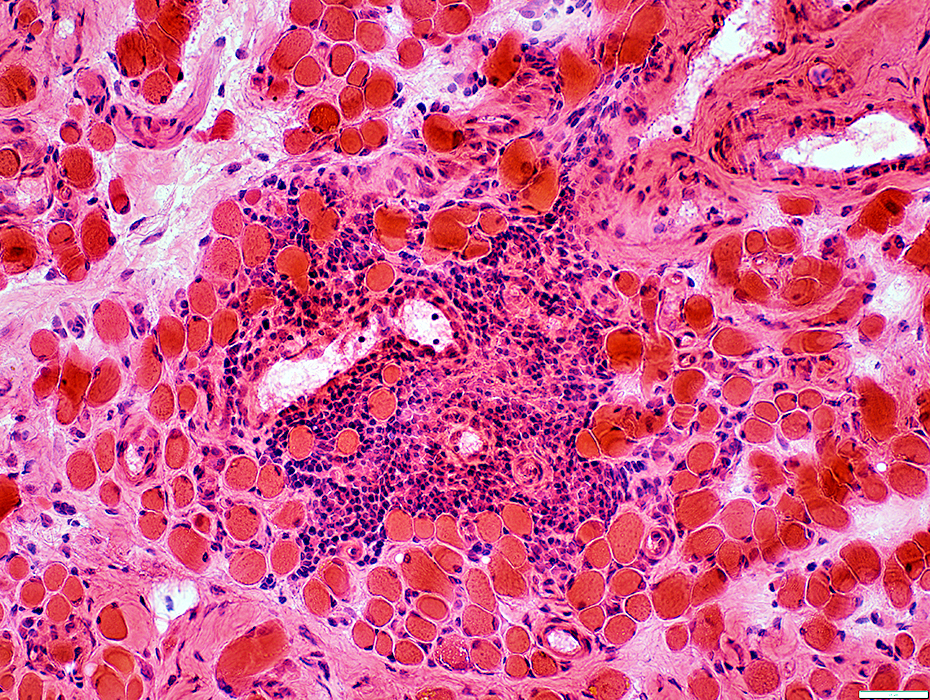
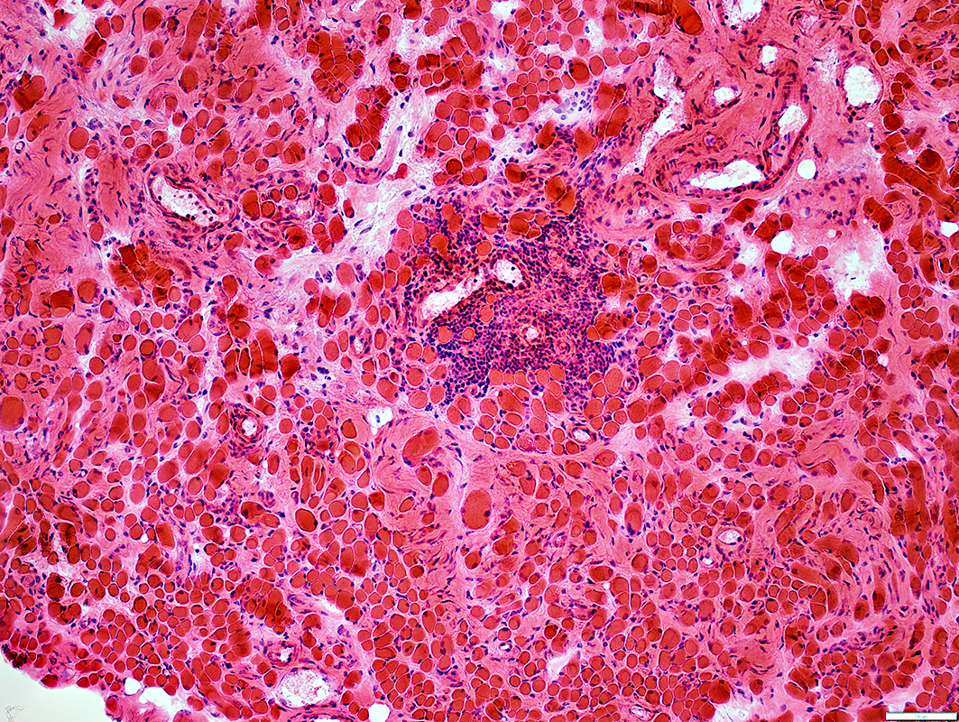 H&E stain |
- Muscle fibers
- Endomysial connective tissue: Increased
- Inflammation: Lymphocyte foci
- Cells: CD4 > CD20; No CD8
- Location: Endomysium or perimysium
 Congo red stain |
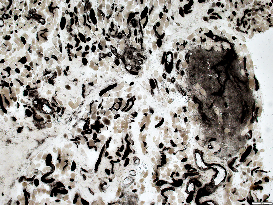
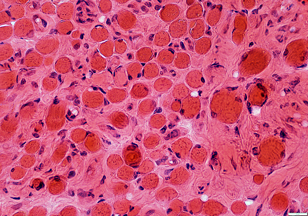
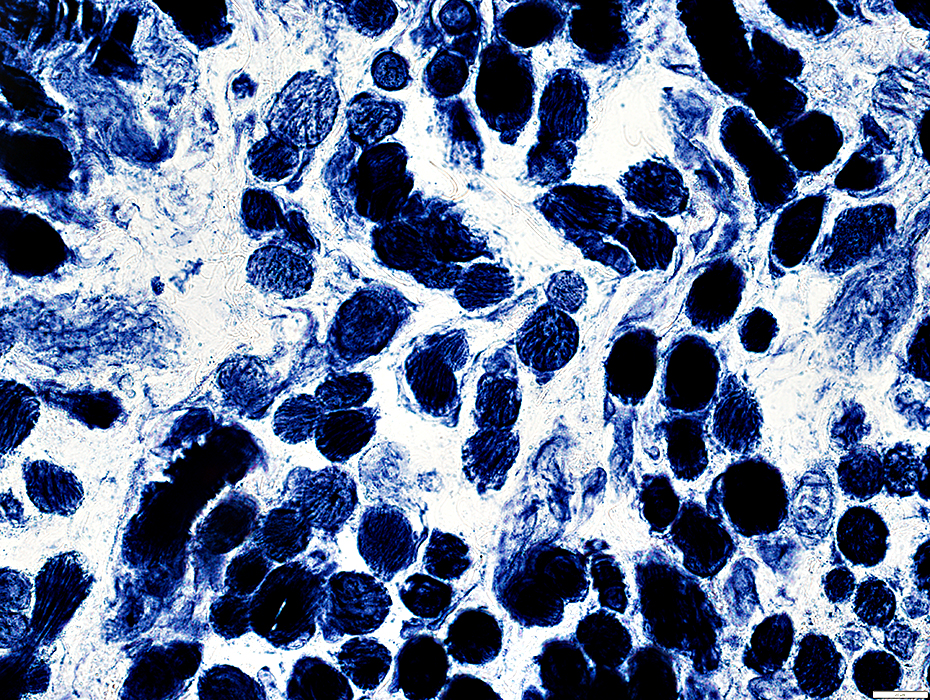
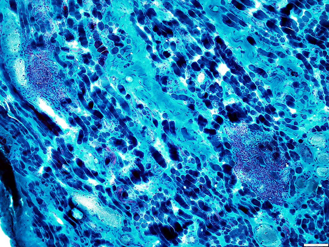 Gomori trichrome stain |
- Muscle fibers
- Varied sizes
- NMJs: Multi-segmented
- Endomysial connective tissue: Increased
- Inflammation: Lymphocyte foci
- Cells: CD4 > CD20; No CD8
- Location: Endomysium or perimysium
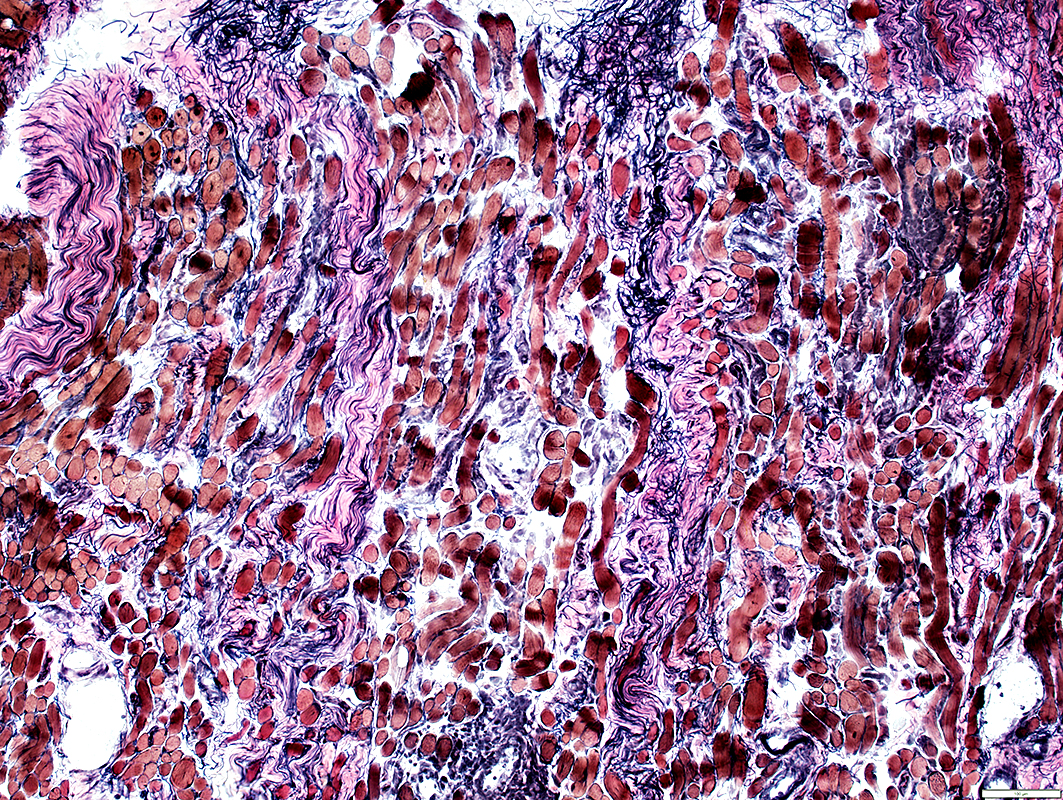 VvG stain |
 ATPase pH 4.3 stain |
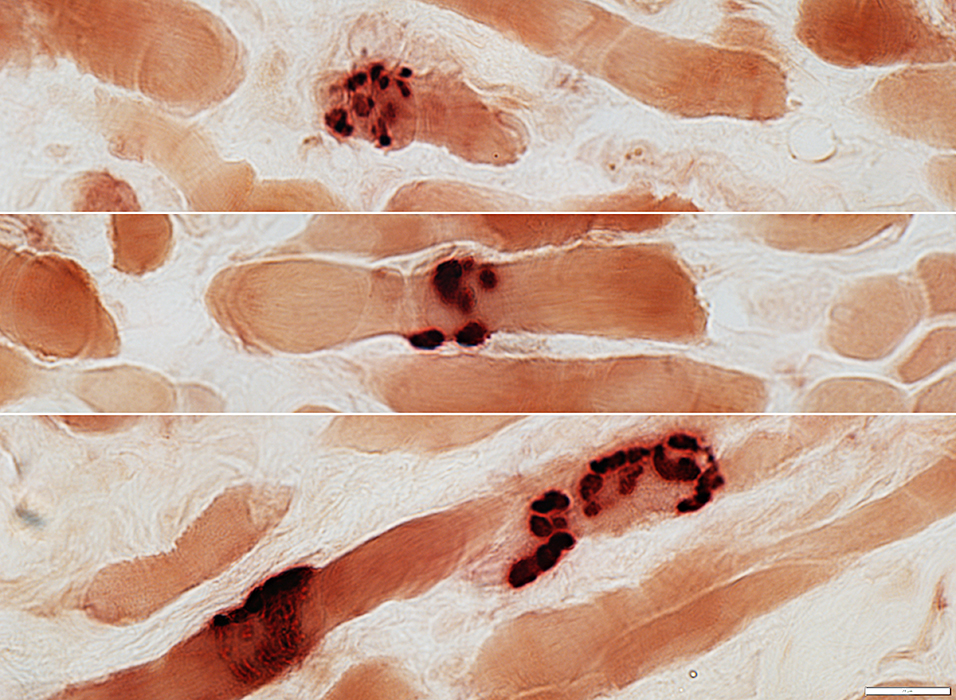
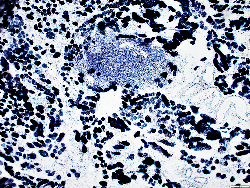
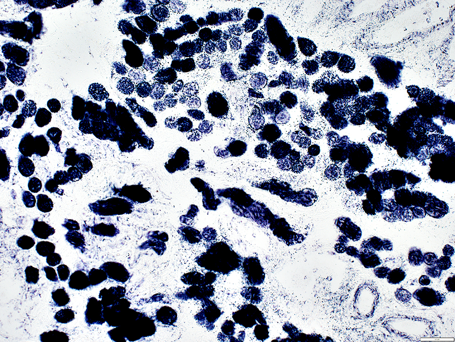
Lymphocyte cell focus (Right)
Muscle fibers: Many scattered, immature (intermediate-stained) muscle fibers
 H&E stain |
Cluster of cells in endomysium
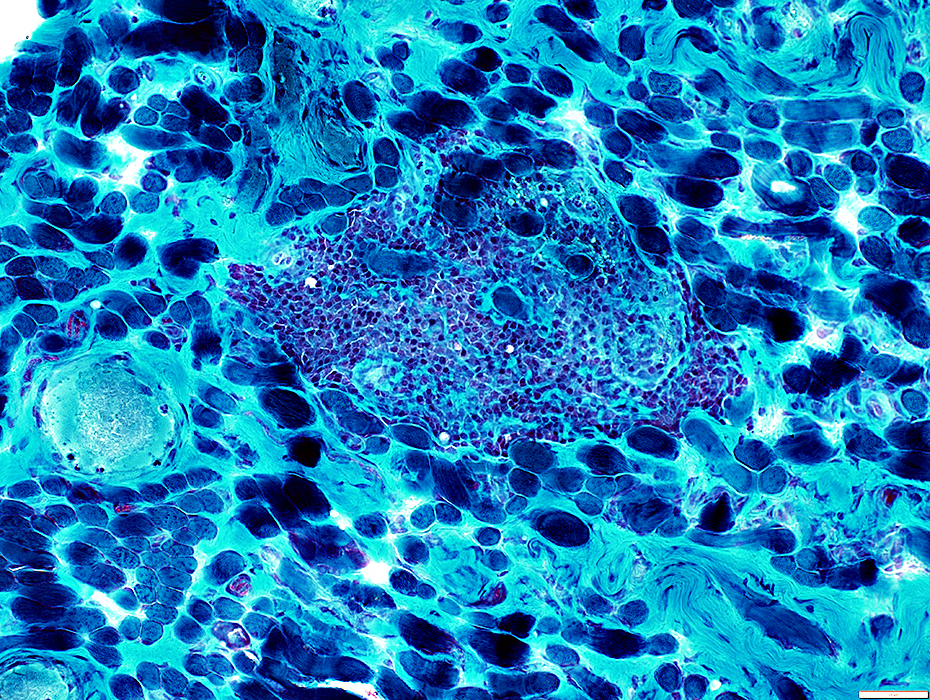 Gomori trichrome stain |
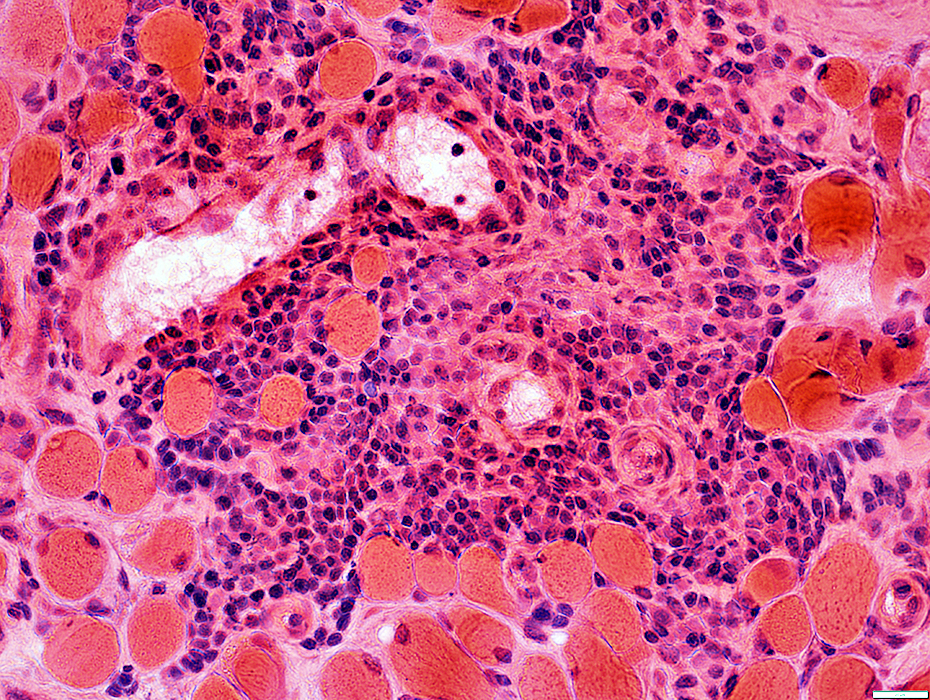 H&E stain |
 Congo red stain |
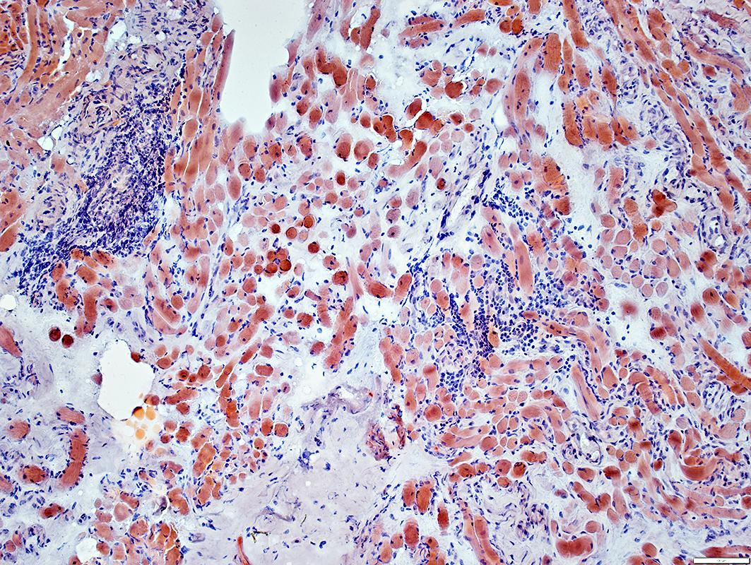
 Esterase stain |
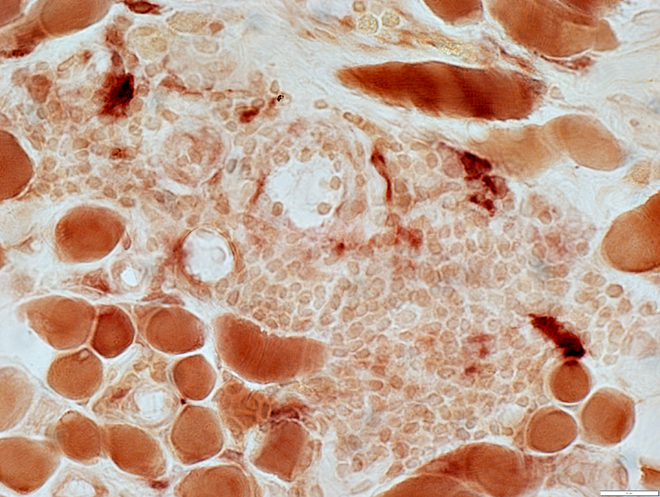
Most cells in foci are not histiocytes: No stain for esterase or acid phosphatase
 Acid phosphatase stain |
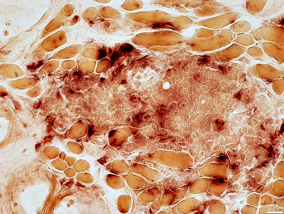
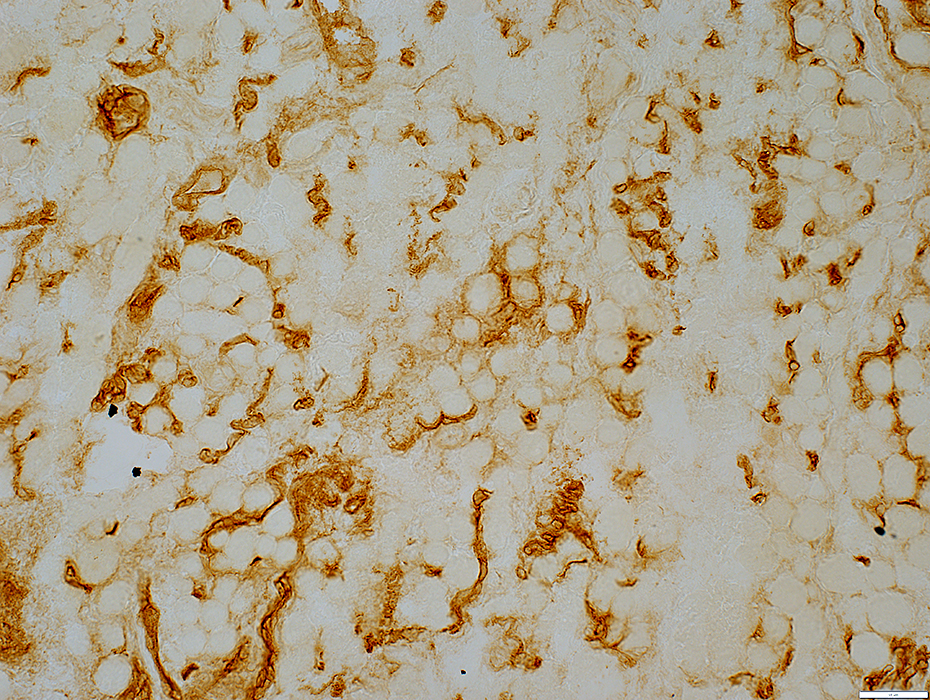
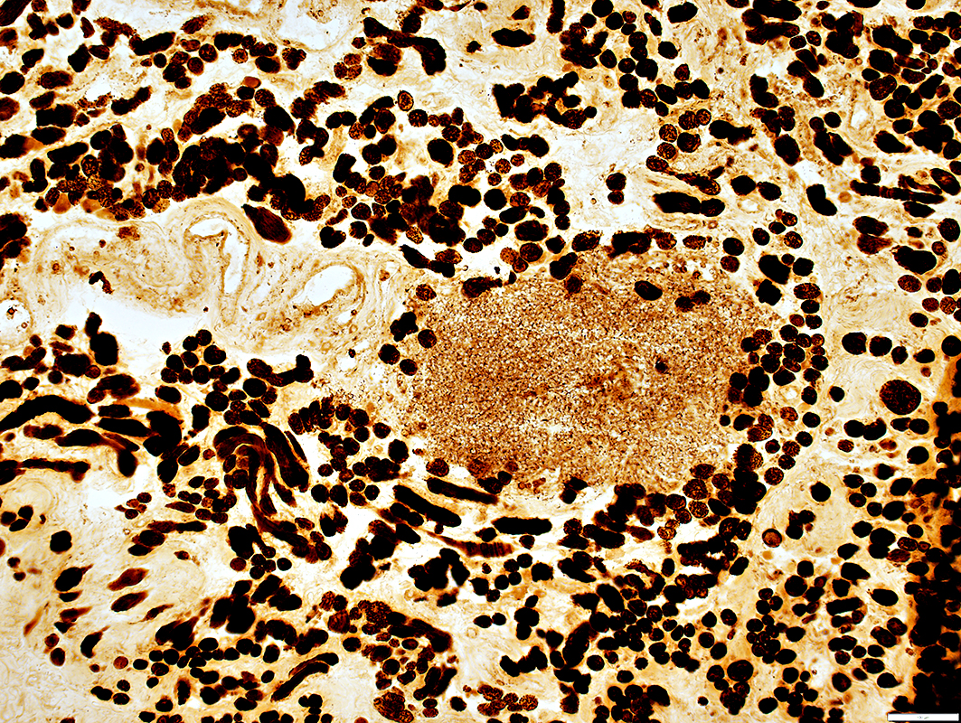
 CD4 stain |
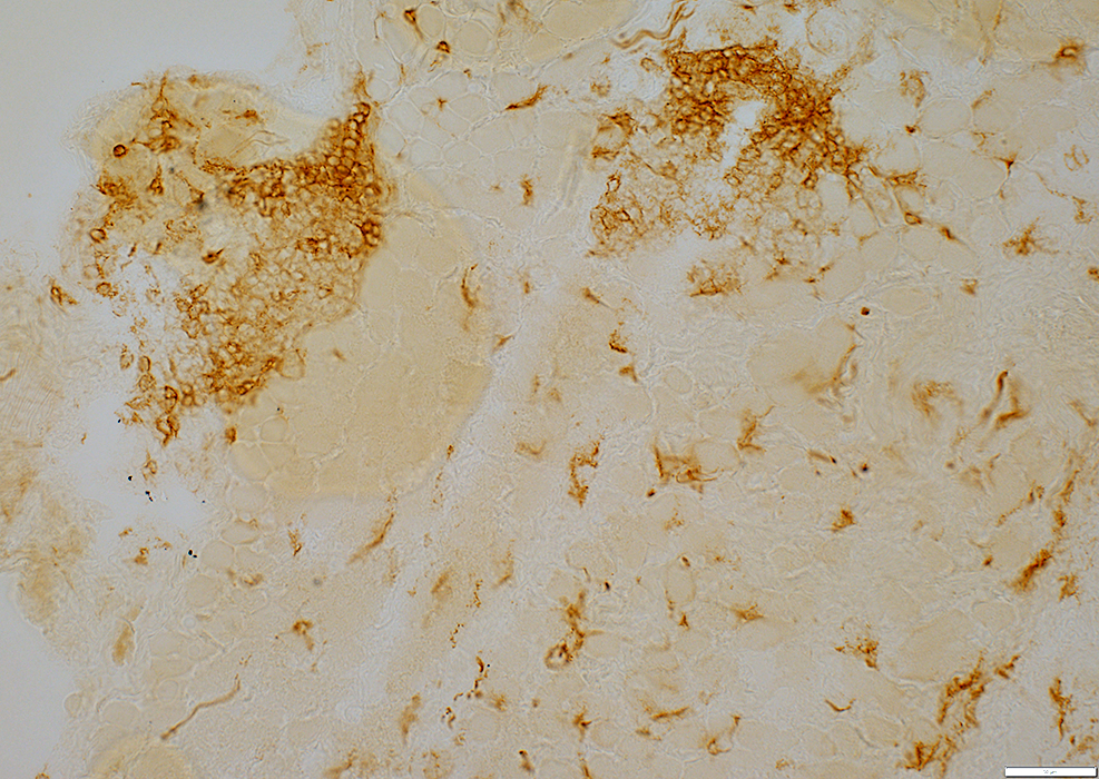
Lymphocyte foci: CD4 & CD20
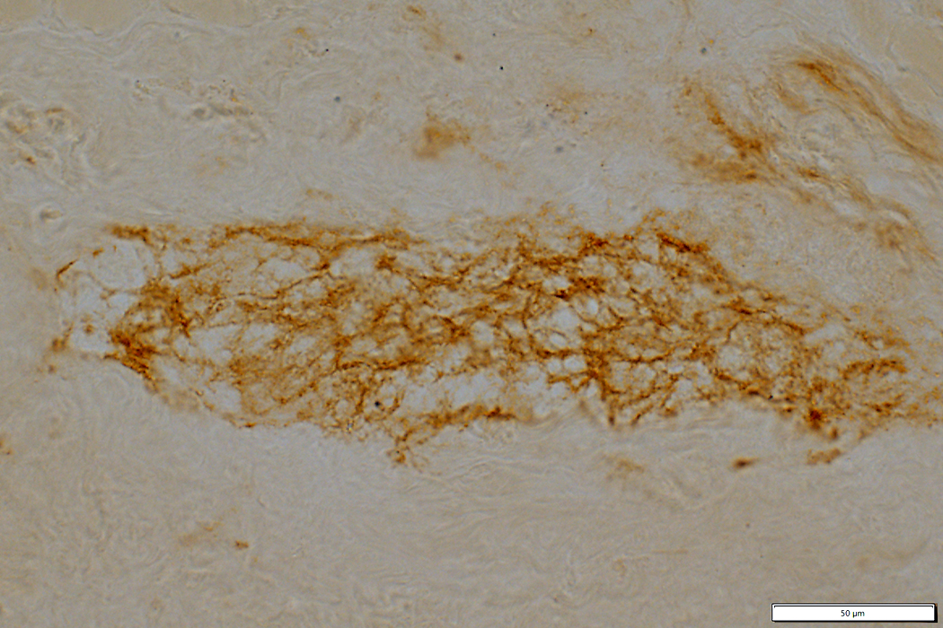 CD4 stain |
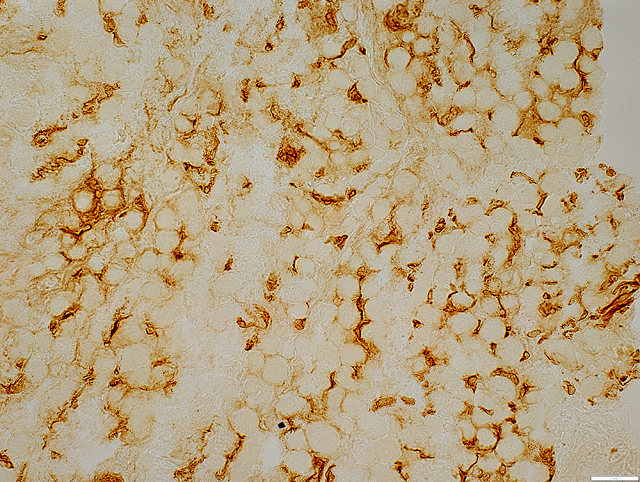
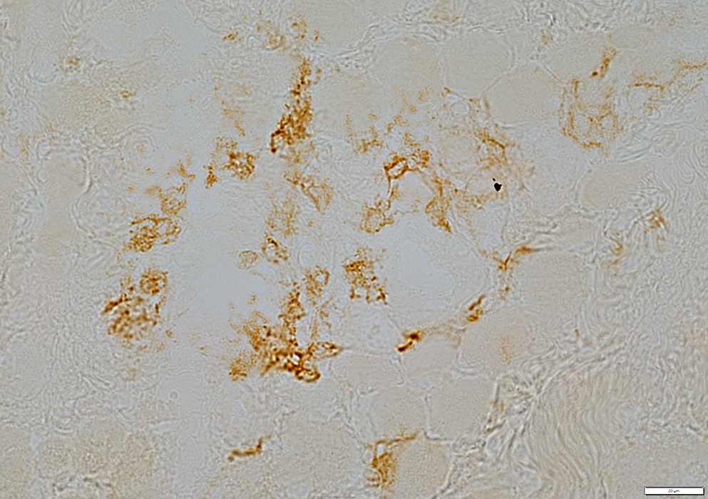 B-cell (CD20) stain |
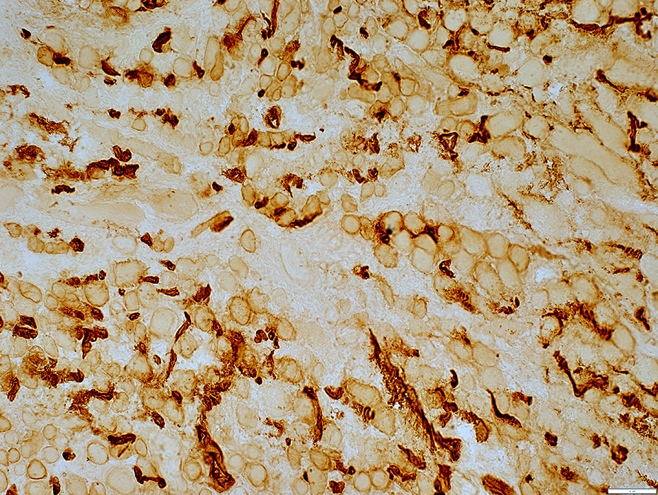
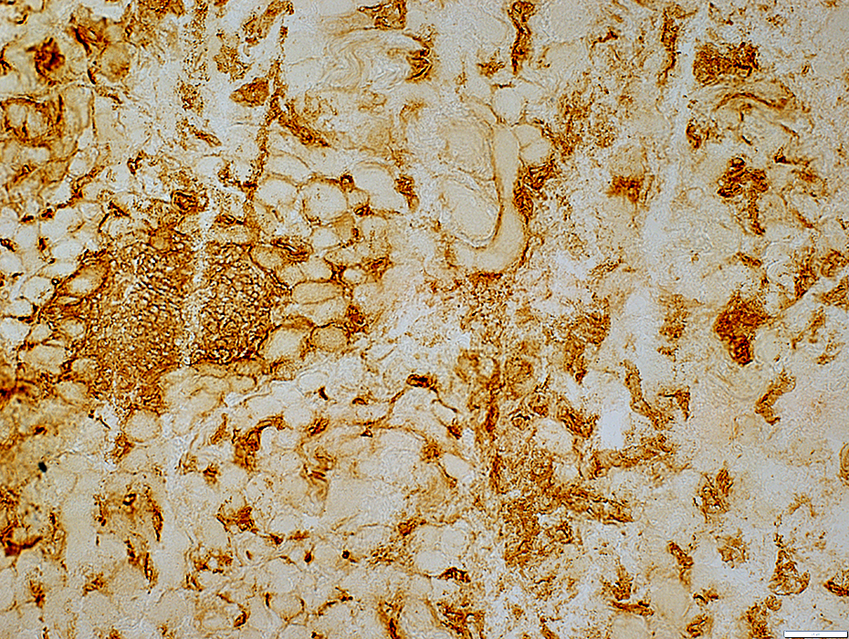 MHC I stain |
Lymphocyte foci
MHC I +
Contain small vessels (ATPase+)
 ATPase pH 9.4 stain |
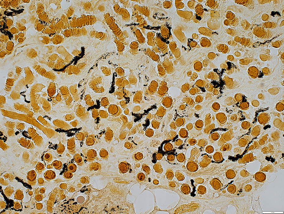
 Acid phosphatase stain |
 H&E stain |
Muscle fibers: Varied sizes
Endomysial connective tissue: Increased
 H&E stain |
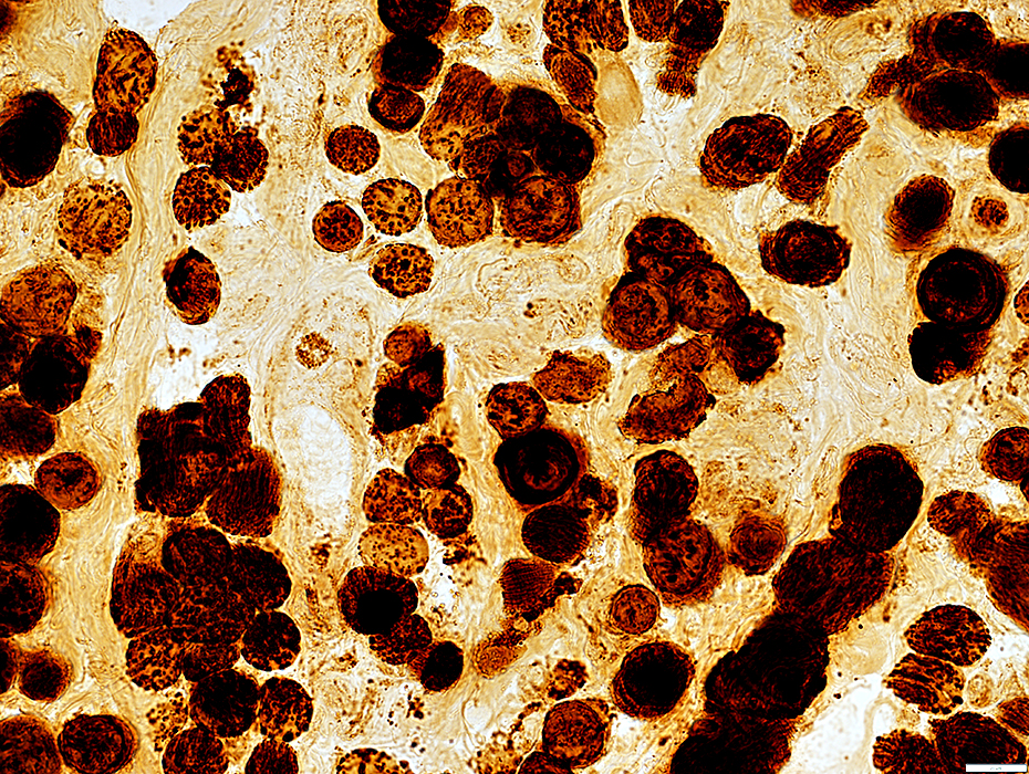
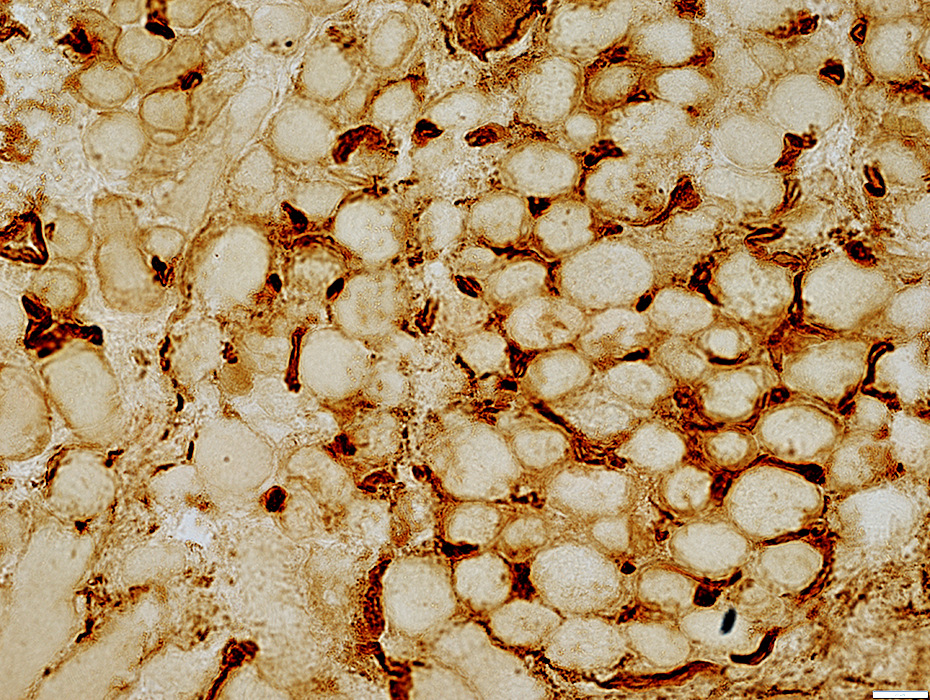
 MHC I stain |
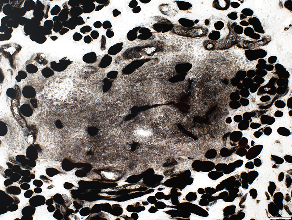
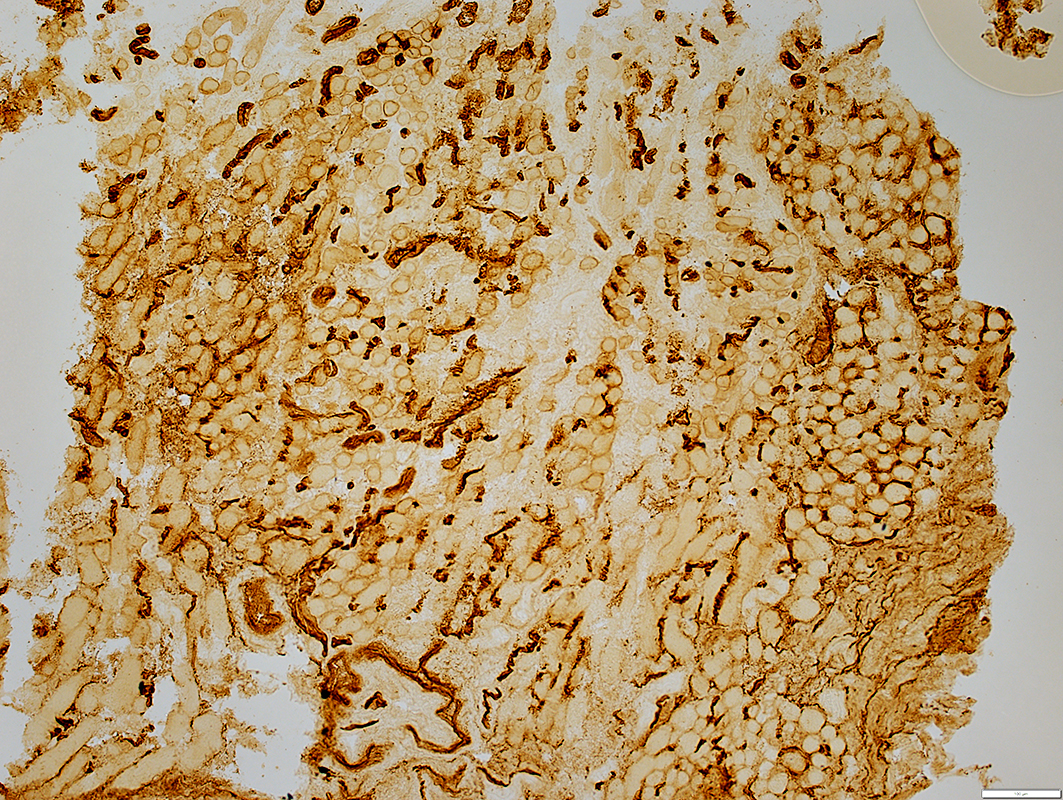
Endomysial capillaries
Sizes: Moderately large
Shape: Sinuous; May be circumferentially oriented
MHC Class I expression by muscle fibers
Some fibers: Mild
 MHC I stain |
Thyroid Ophthalmopathy: Neuromuscular Junctions
Multisegmented
See: Control EOM
 Esterase stain |
 NADH stain |
NADH stain: Dark
Cytochrome oxidase (COX) & Succinate Dehydrogenase (SDH): Dark stain
 Cytochrome oxidase (COX) stain |
 Cytochrome oxidase (COX) stain |
 Succinate dehydrogenase (SDH) stain |
NADH stain: Dark
Cytochrome oxidase (COX) & Succinate Dehydrogenase (SDH): Dark stain
 Succinate dehydrogenase (SDH)stain |
 Alkaline phosphatase stain |
Size: Mildly large
 UEA1 stain |
 UEA1 stain |
Size: Mildly large
 UEA1 stain |
Return to: Lymphoma
Return to: Neuromuscular Home Page
11/14/2022