Spinal Muscular Atrophy
Scapuloperoneal Neuronopathy: TRPV4 mutations
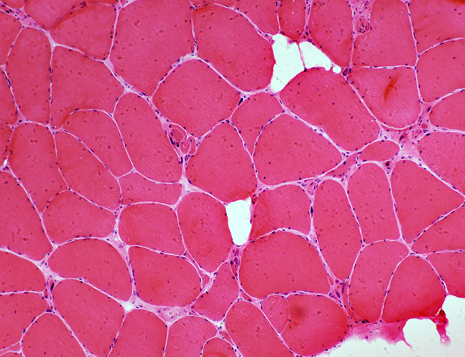 H & E stain |
|
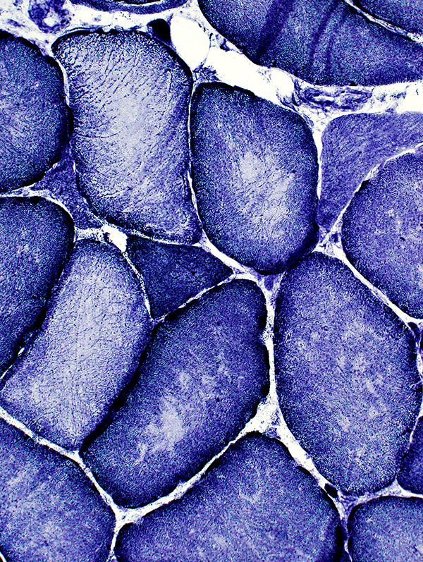
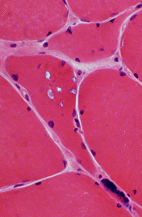
Morphology Muscle fibers Small & Intermediate-sized Angular & Polygonal Hypertrophic Internal nuclei: Often multiple Grouped muscle fiber atrophy |
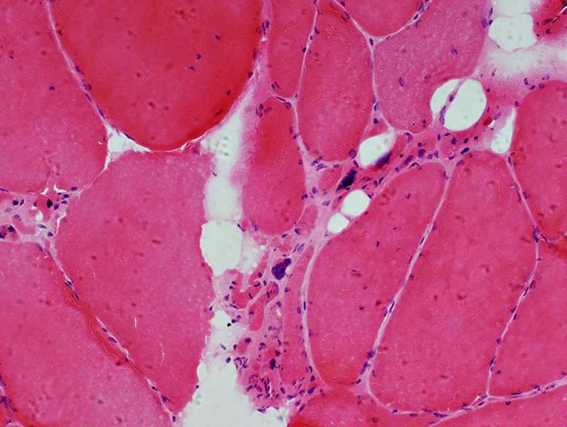 H & E stain |
Internal nuclei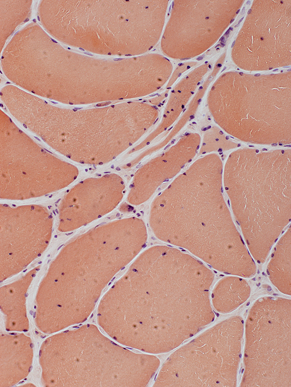 Congo red stain |
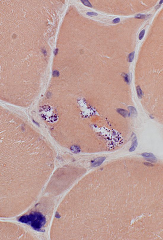
Pyknotic nuclear clumps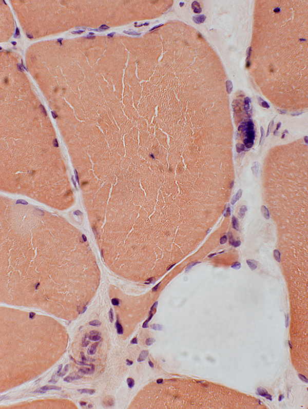 Congo red stain |
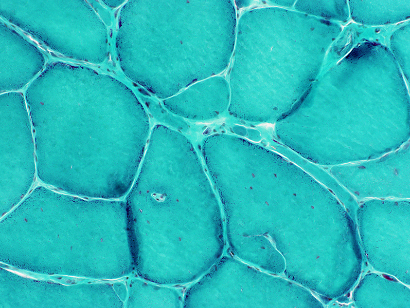
Internal architecture: Irregular in larger muscle fibers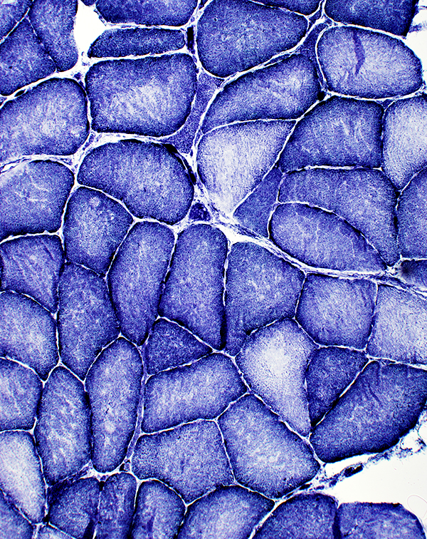 NADH stain |
 NADH stain |
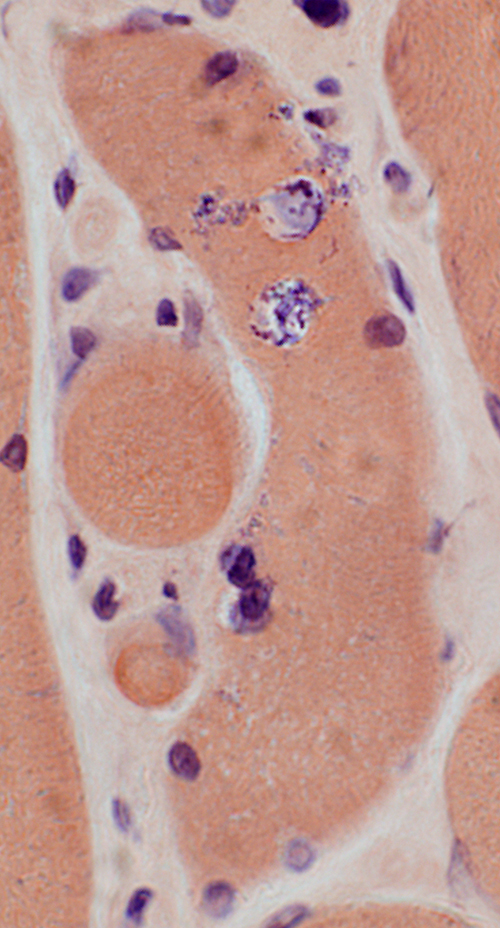
 VvG stain Chronic changes Internal nuclei Partial fusion of muscle fibers Capillary inside muscle fiber Pyknotic nuclear clumps |
 Gomori trichrome stain |
Cytoplasmic bodies in a small muscle fiber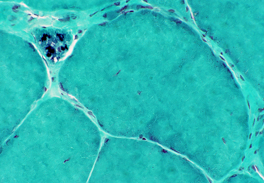 Gomori trichrome stain |
Muscle fiber types: Type 1 predominance; No fiber type grouping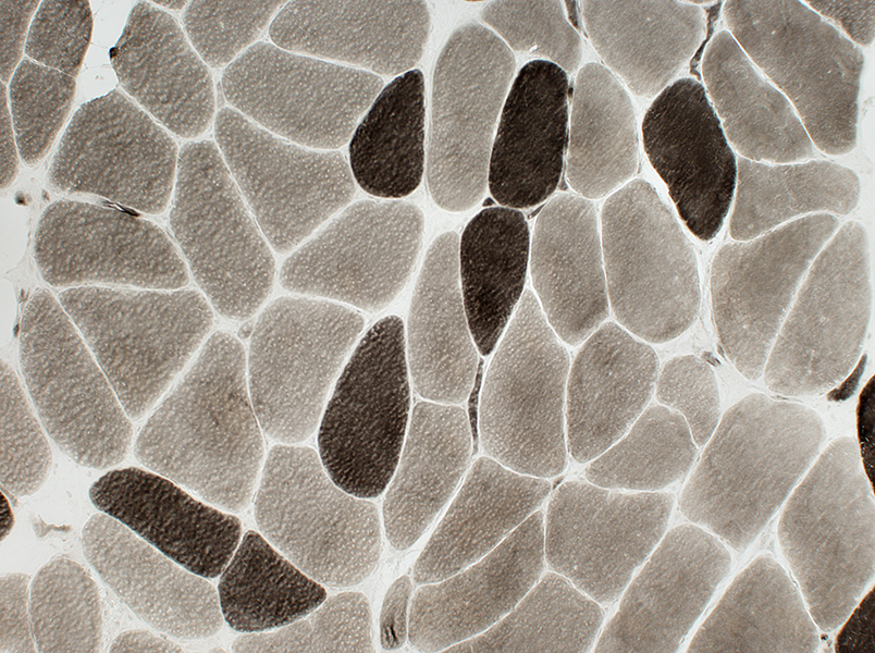 ATPase, pH 9.4 |
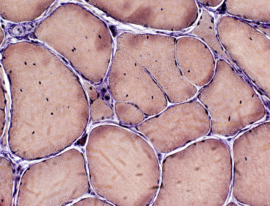
Vacuoles in Muscle fibers H & E stain |
 H & E stain |
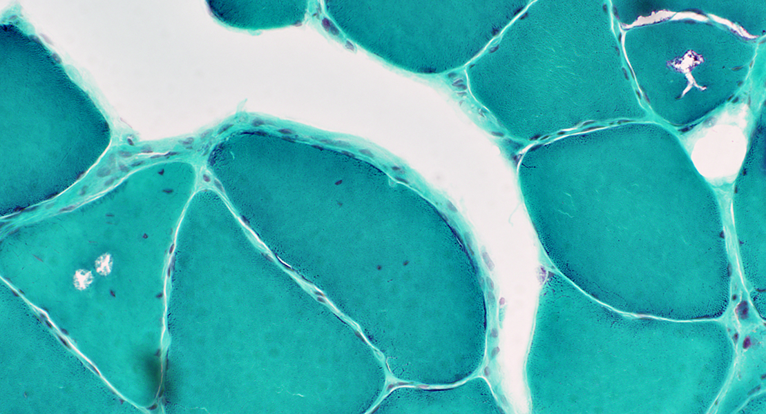 Gomori trichrome stain |
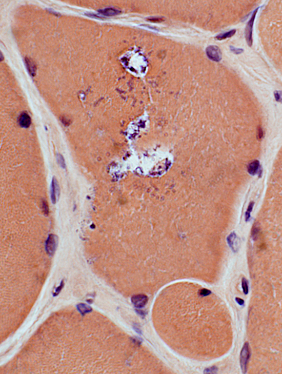
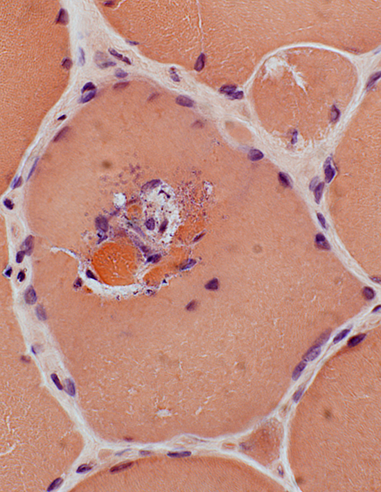
 Congo red stain Vacuoles Contain basophilic granular debris May be near eosinophilic cytoplasmic bodies |
 Congo red stain |
 Congo red stain |
 Congo red stain |
|
Aggregates Irregular; Dark; Cytoplasmic May be near or around internal nuclei 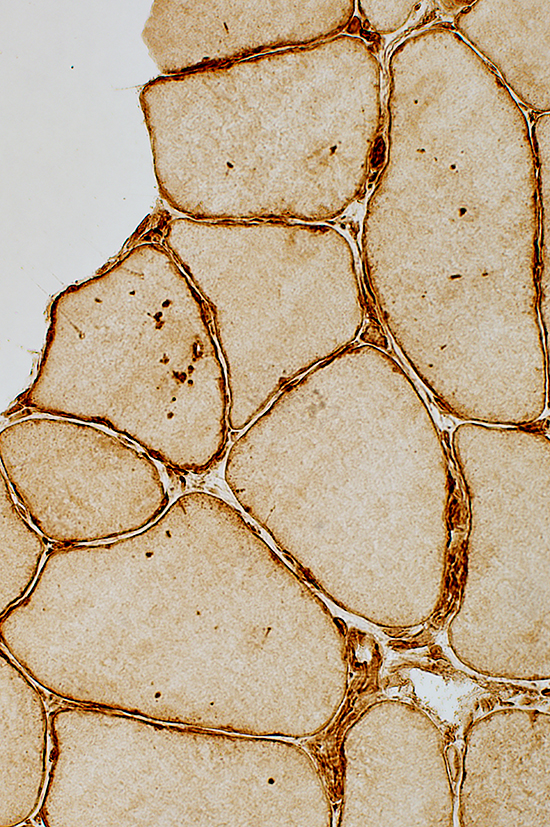 Caveolin-3 stain |
 Caveolin-3 stain |
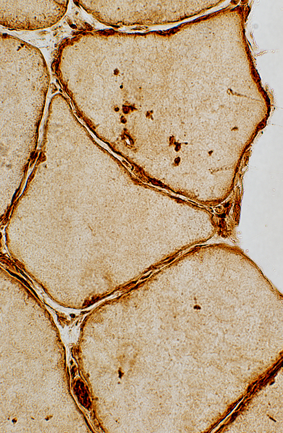
Caveolin-3 may also stain rim of vacuoles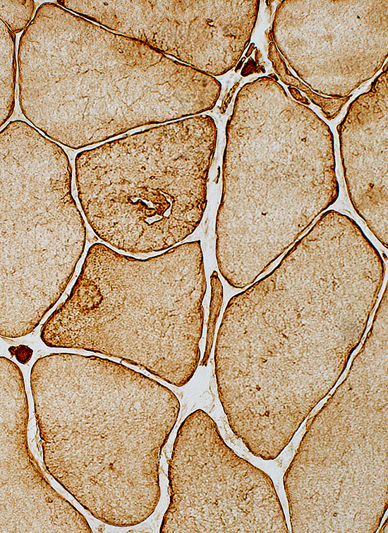 Caveolin-3 stain |
Caveolin-3 staining around internalized capillary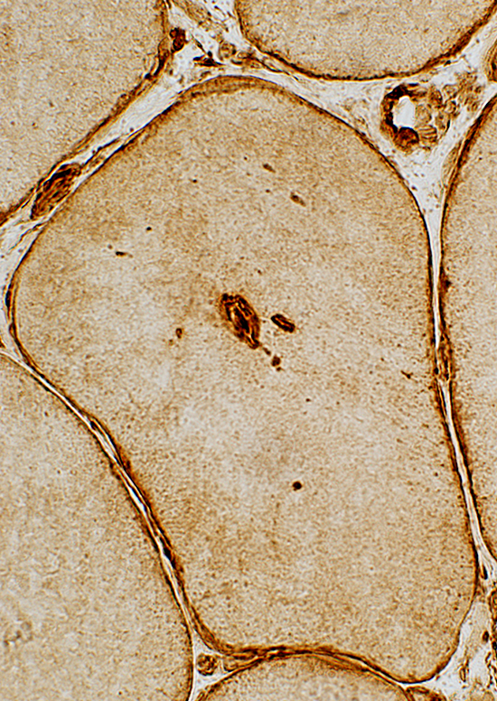 Caveolin-3 stain |
Return to Neuromuscular Home Page
Return to Pathology index
Return to Scapuloperoneal neuronopathy (SPSMA)
8/23/2023