SPINAL MUSCULAR ATROPHY: Lower Extremity, Dominant
SMA-LED: Early pathology (2 year-old patient)
Denervation in muscle
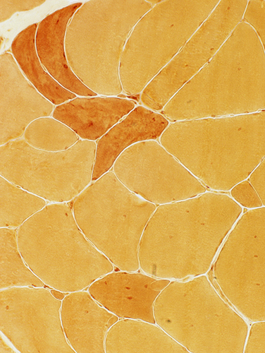
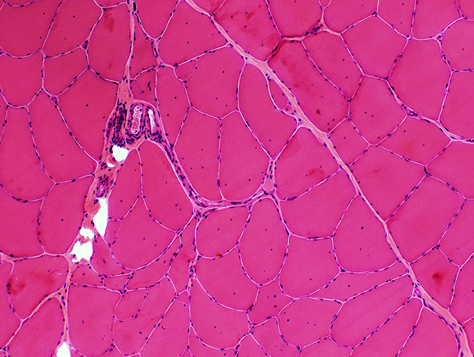 H & E stain |
|
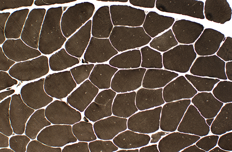
Morphology Small & intermediate-sized angular & polygonal muscle fibers Grouped muscle fiber atrophy Hypertrophic muscle fibers Internal nuclei |
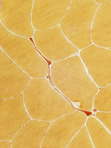
 H & E stain |
 H & E stain |
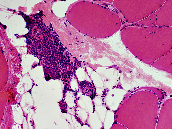
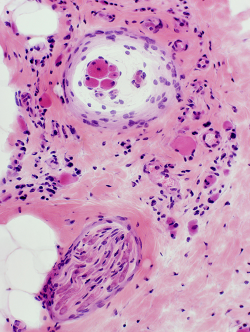
Inflammation: Mononuclear cells surround Intermediate-sized perimysial vessels |
|
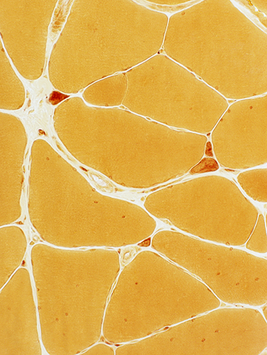
 H & E stain |
|
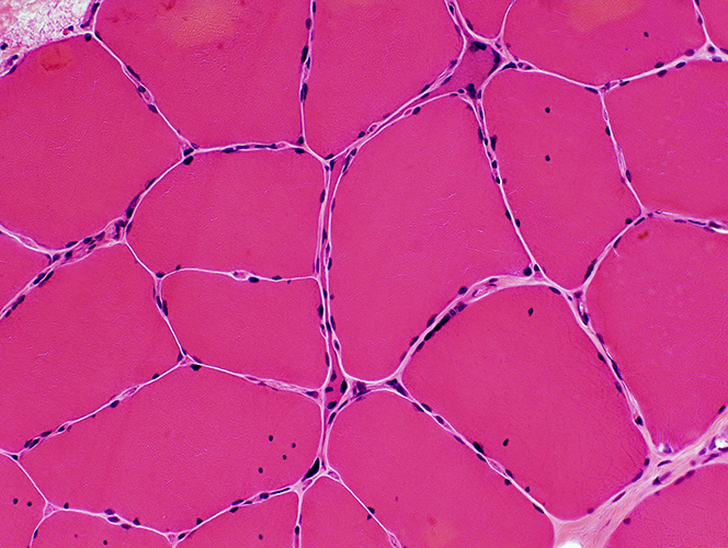
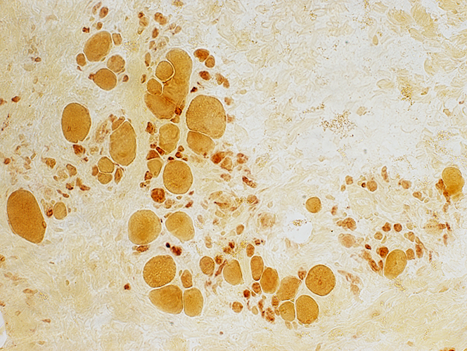
Muscle fibers: Intermediate-sized & Small; Polygonal, Angular or Very small, Rounded (Pyknotic clunps)
 Esterase stain |
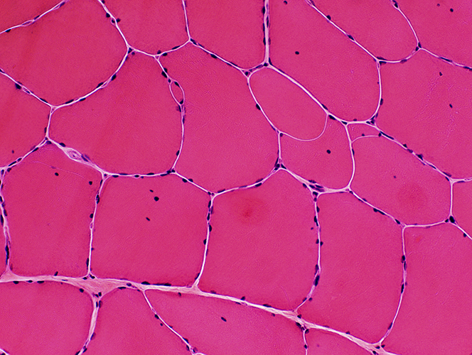
Muscle fibers: Internal architecture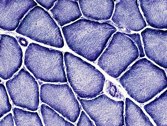 NADH stain |
|
Large muscle fibers Coarse internal architecture Pale centers |
Muscle: Fiber types
 ATPase, pH 9.4 |
|
Large muscle fibers: Type 2 predominance |
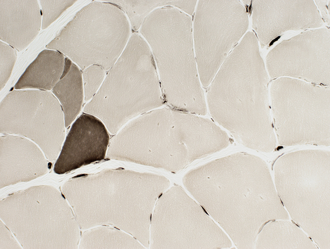 ATPase, pH 4.3 |
SMA-LED: Late stage muscle (22 years-old)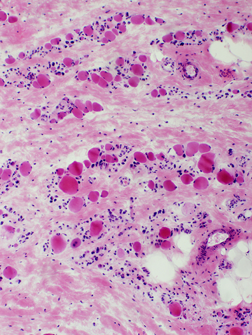 H & E stain |
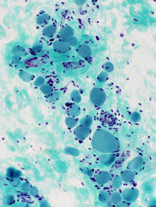 GT stain |
Muscle fibers: Small, Rounded
Endomysial connective tissue: Greatly increased
Small muscle fibers: Esterase stained
 Esterase stain |
Muscle spindles: Preserved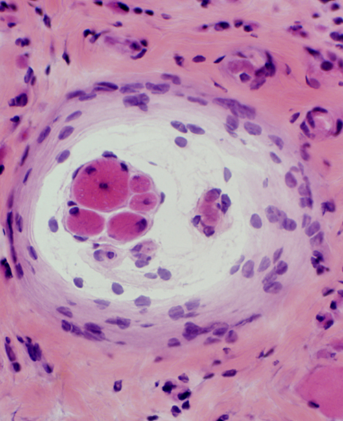 H&E stain |
 H&E stain |
Intramuscular nerve: Reduced staining on VvG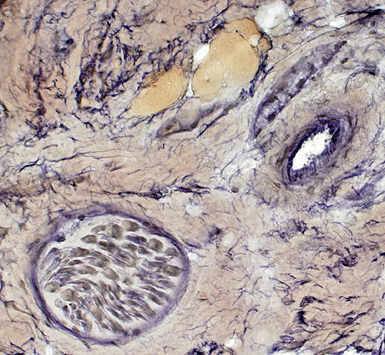 VvG stain |
Return to Neuromuscular Home Page
Return to Pathology index
Return to SMA-LED
12/21/2012