Reducing Body & FHL1 Myopathy
|
FHL1: Emery Dreifus 6 |
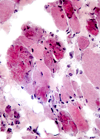
 H & E stain From: CG Bonnemann MD |
|
|
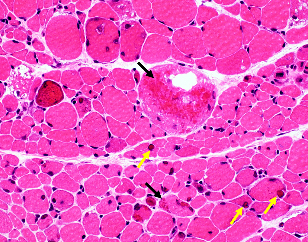
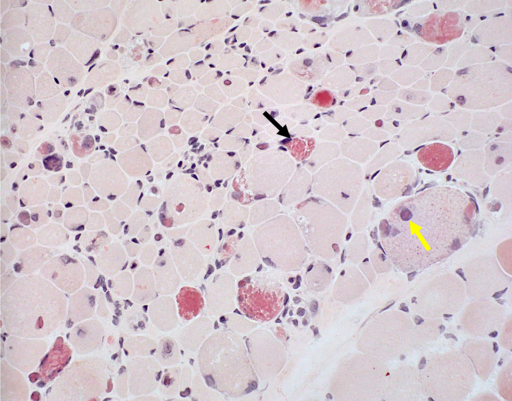
Reducing bodies: Irregularly shaped, cytoplasmic inclusions Muscle fiber sizes: Varied |
|
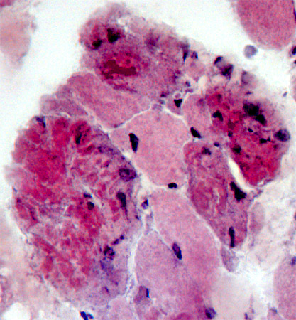
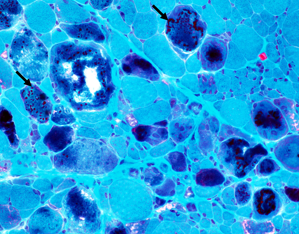
 H & E stain From: C Cai FHL1 myopathy (Heterozygous, de novo, c.369C>G (p.H123Q) mutation) Fiber size variation: Marked Sarcoplasmic inclusions (Black arrow) Intranuclear inclusions (Yellow arrows) |

|
 Gomori trichrome stain From: CG Bonnemann MD |
|
Reducing bodies: Darkly stained on Gomori trichrome |
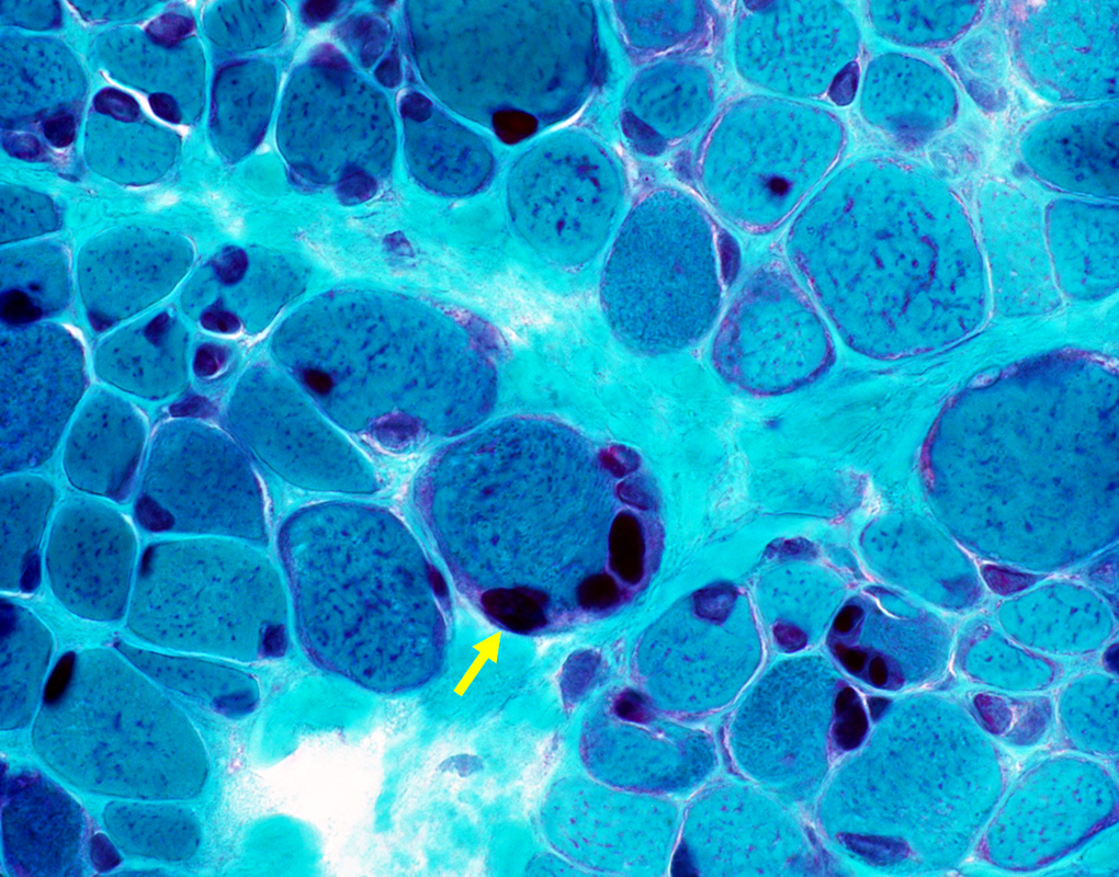
 Gomori trichrome stain From: C Cai FHL1 myopathy Aggregates: Cytoplasmic bodies & Reducing Bodies (Arrows) |
 Gomori trichrome stain From: C Cai FHL1 myopathy Intranuclear inclusions |
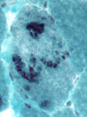
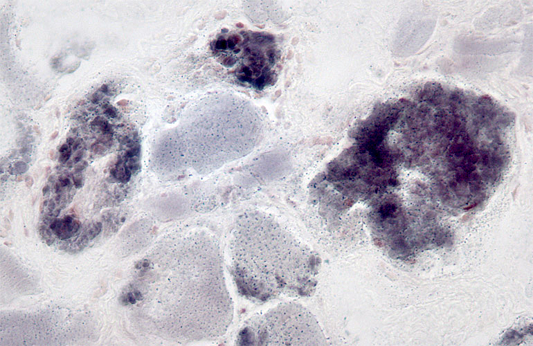 Menadione-NBT stain From: CG Bonnemann MD |
|
|
Reducing bodies: Stain with Menadione-NBT |
|
 Congo red stain From: C Cai FHL1 myopathy Inclusions: Cytoplasmic (Black arrow) & Nuclear (Yellow arrow) |
 From: CG Bonnemann MD |
 From: CG Bonnemann MD |
|
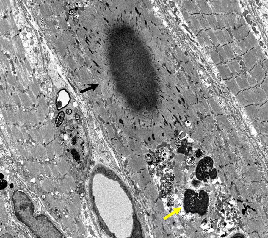
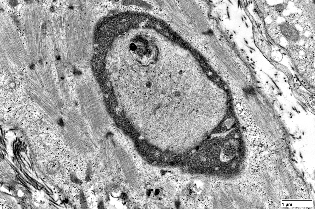
Reducing bodies: Ultrastructure Structure: Granular tubulo-filamentous material (17 nm in diameter) Location: At periphery of sarcoplasm & around nucleus |
|
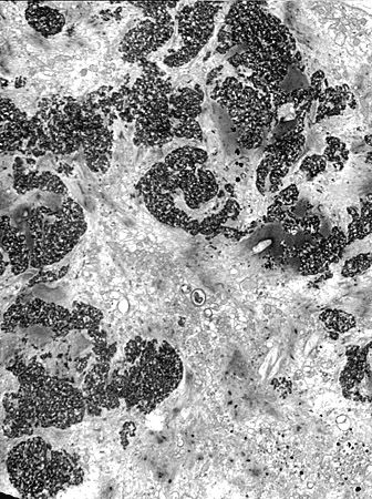
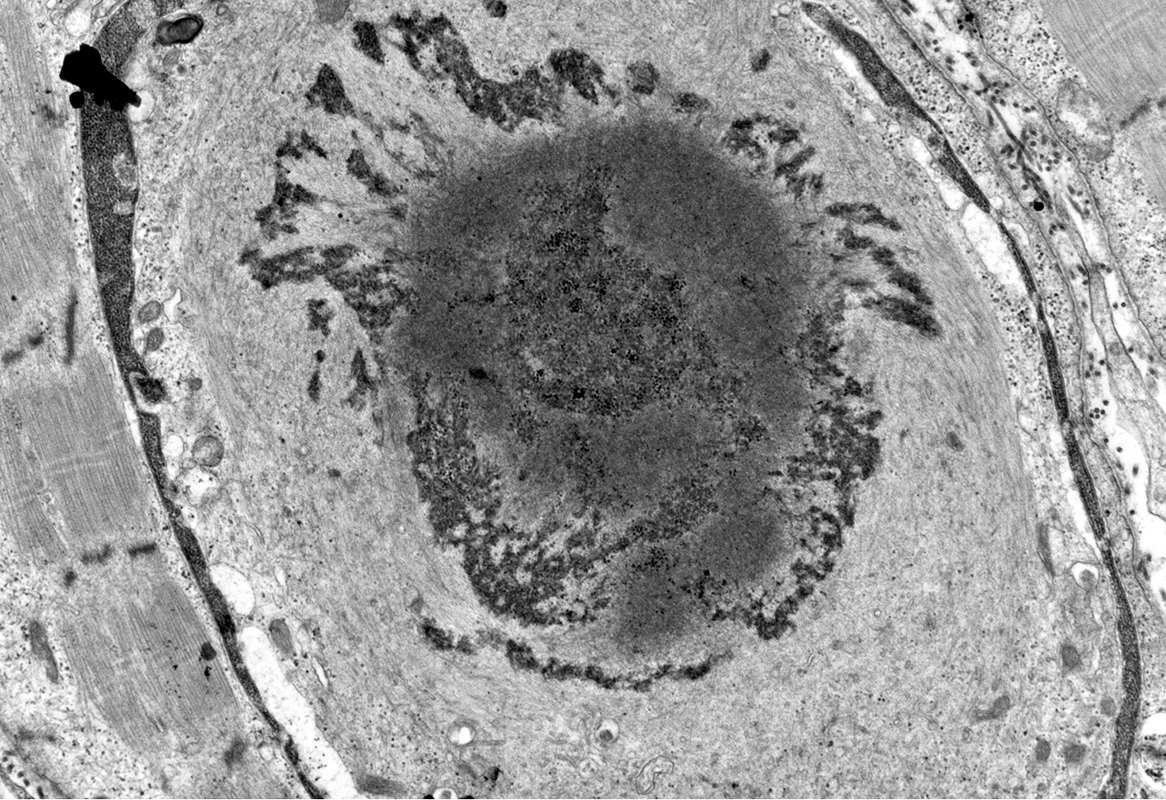
 From: C Cai FHL1 myopathy Inclusions: Cytoplasmic body (Black arrow) & Granule, electron dense (Yellow arrow) |
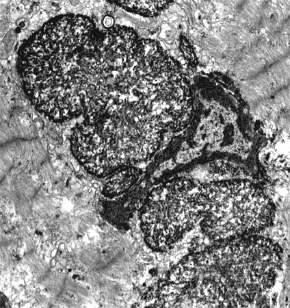
 From: C Cai FHL1 myopathy Inclusion: Granule, electron dense, composed of 20-30 nm spherical particles |
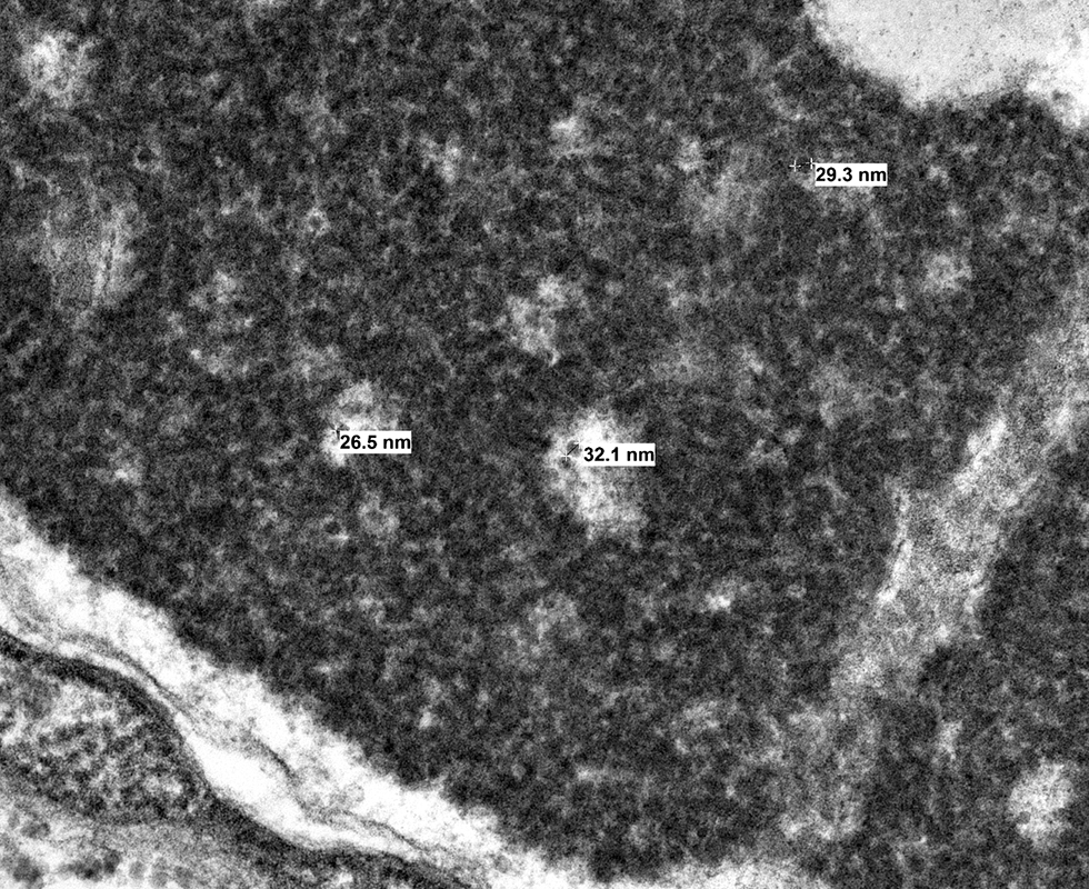
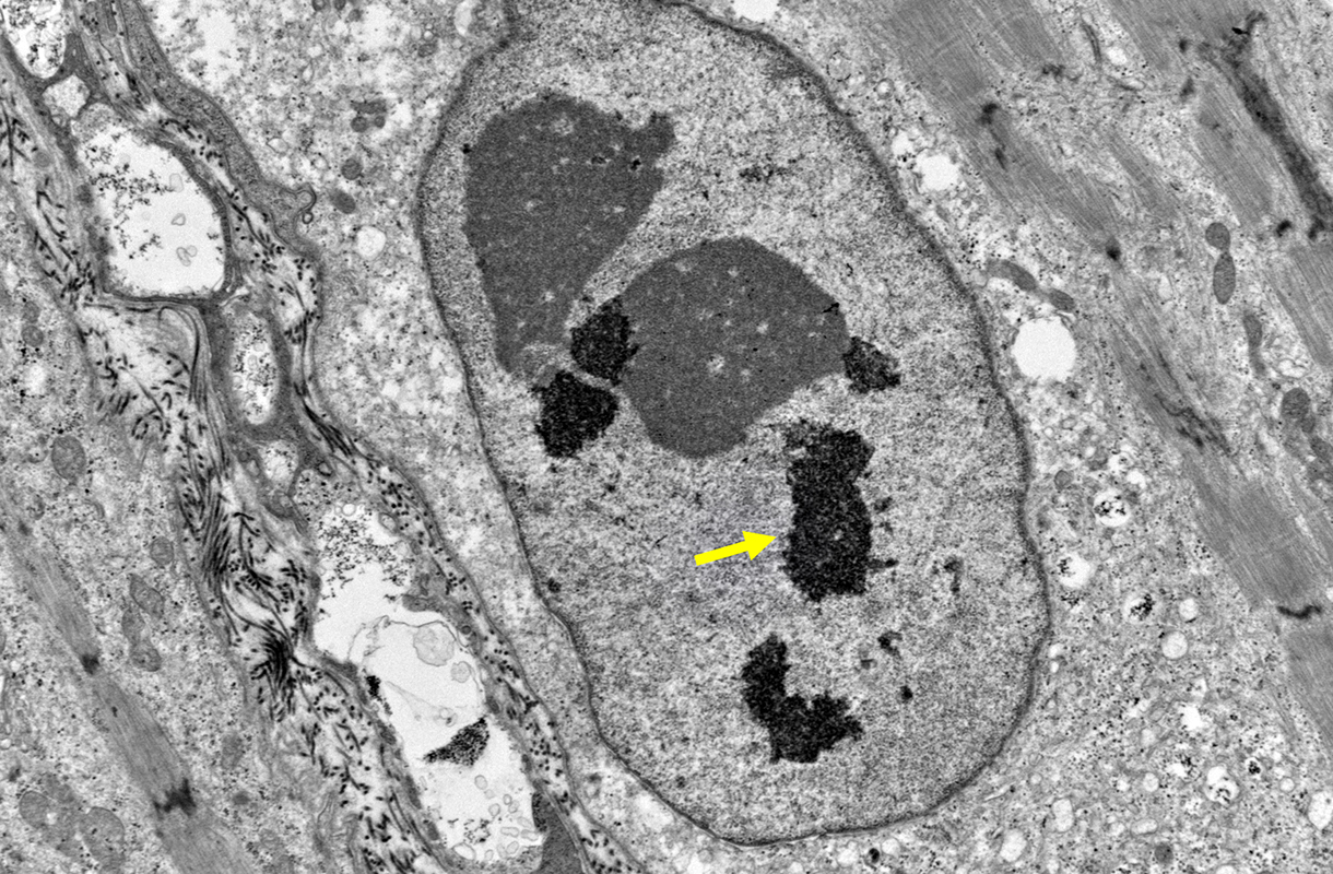 From: C Cai FHL1 myopathy Inclusions, Intranuclear (Yellow arrow): Electron dense, granular aggregates |
 From: C Cai FHL1 myopathy Inclusion, Intranuclear: Thin filaments & Electron dense, granular aggregates |
 From: C Cai FHL1 myopathy Inclusion, Intranuclear: Filamentous material |
Return to Neuromuscular Home Page
Return to Pathology index
Return to Reducing body myopathy
9/16/2024