FHL1 myopathy: Emery-Dreifuss phenotype
|
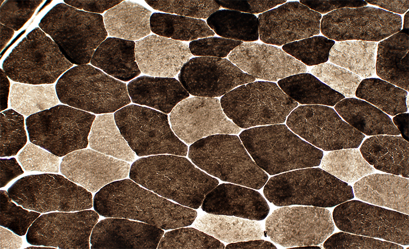
Muscle Mild pathology Myopathy FHL1: Reducing Body |
 H & E stain |
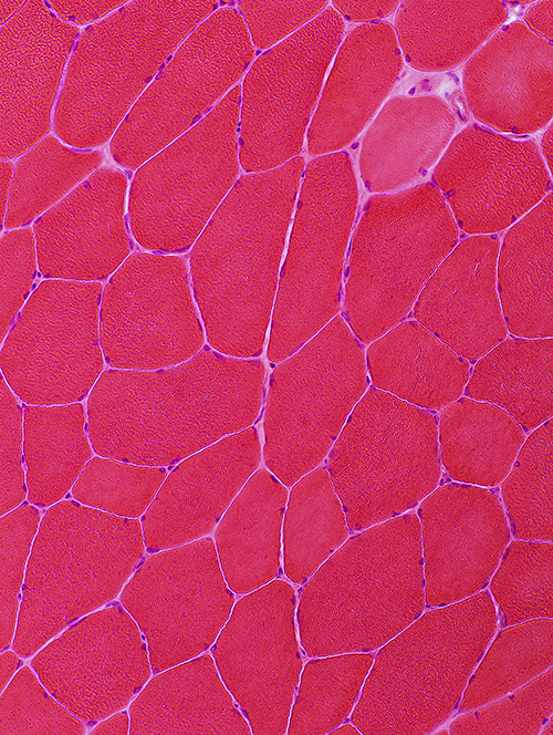 H & E stain |
Small polygonal & angular: Scattered
Very small, basophilic: Few
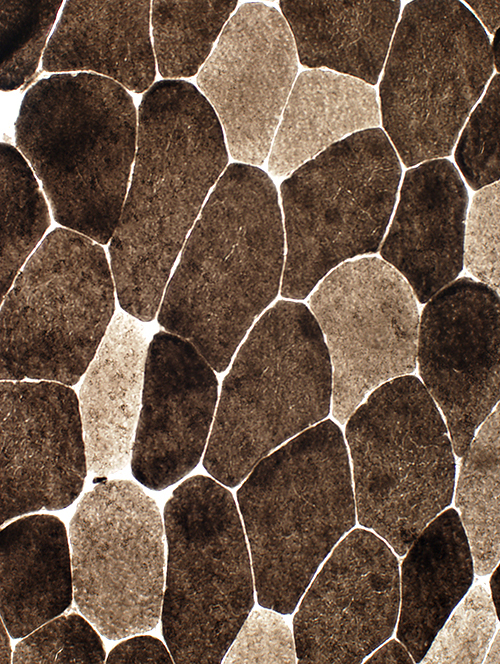
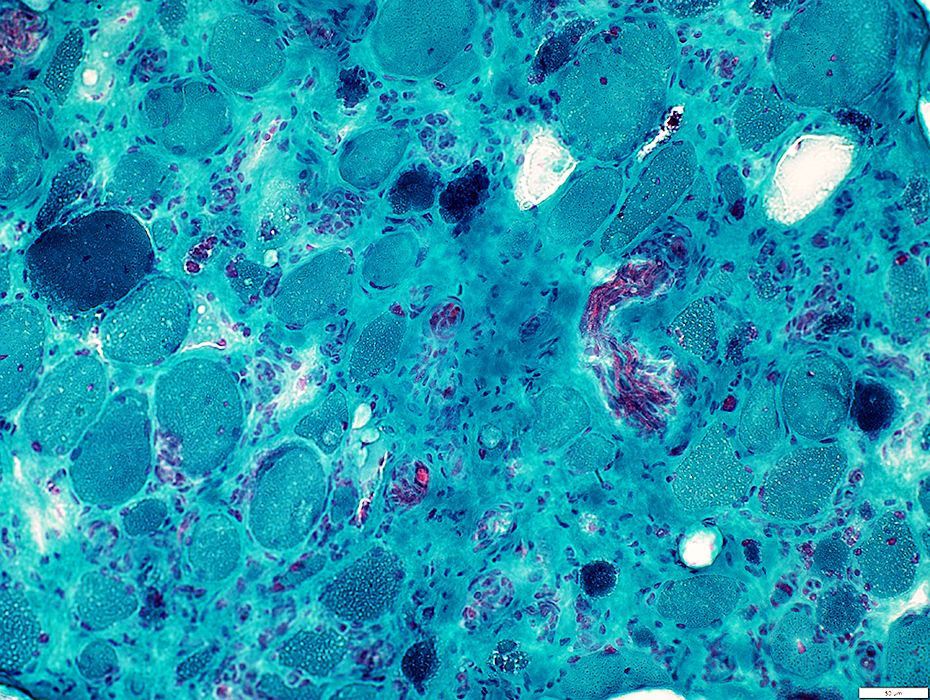
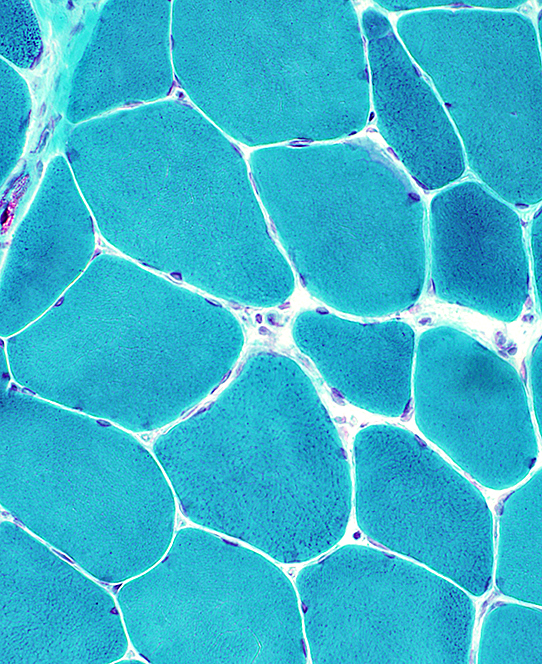 Gomori trichrome stain |
 Gomori trichrome stain |
Small polygonal & angular: Scattered
Very small: Few
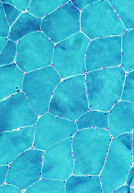
 NADH stain |
Internal architecture: Coarse or irregular
 NADH stain |
 NADH stain |
Internal architecture: Coarse or irregular
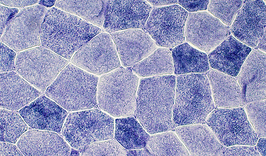
 COX stain |
 COX stain |
Internal architecture: Irregular
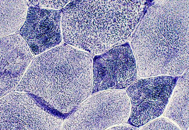
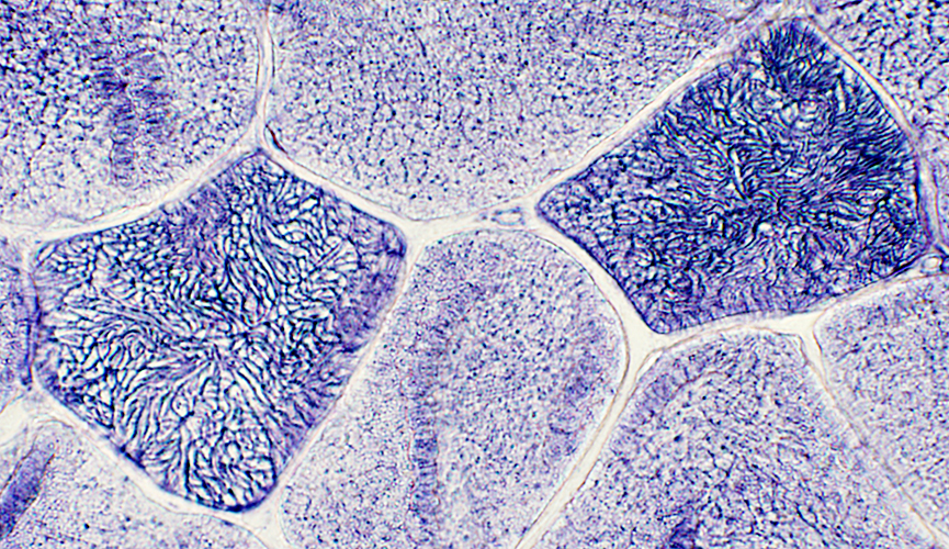
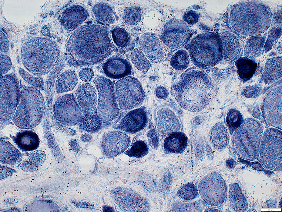
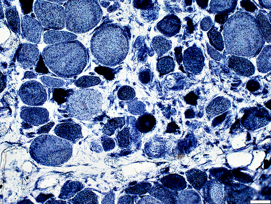
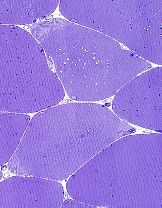 Toluidine blue stain |
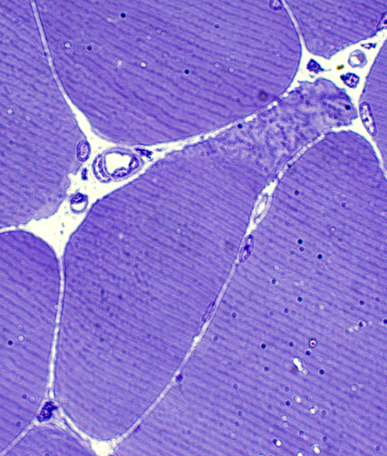 Toluidine blue stain |
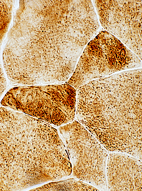
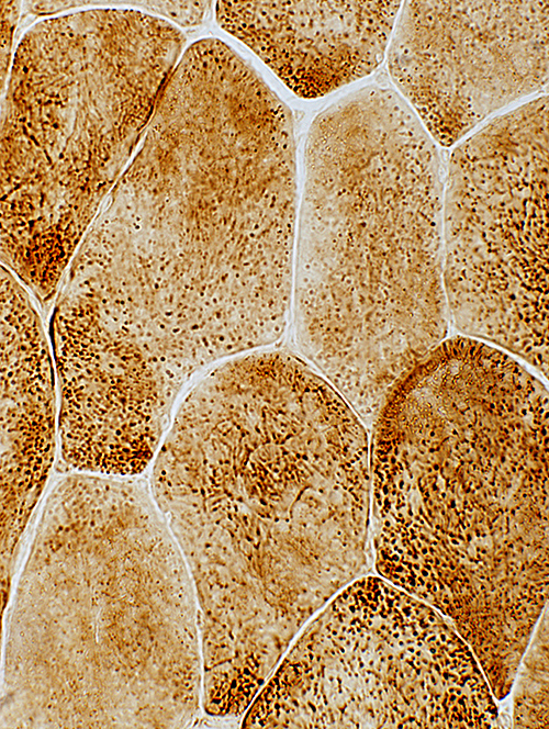
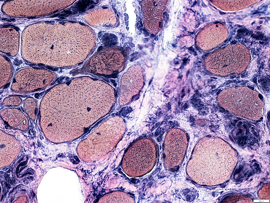
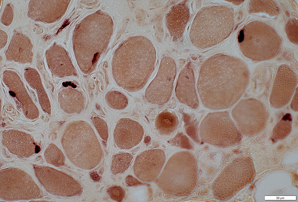
 Esterase stain |
 Esterase stain |
Cytoplasm of Small muscle fibers: Dark
Neuromuscular Junctions (Right): Small or Multisegmented
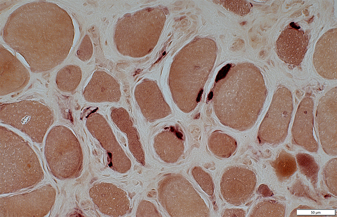
 ATPase pH 9.4 stain |
 ATPase pH 9.4 stain |
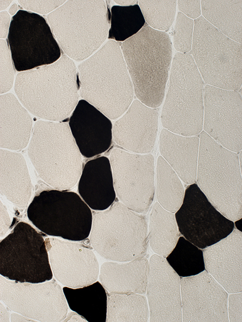 ATPase pH 4.3 stain |
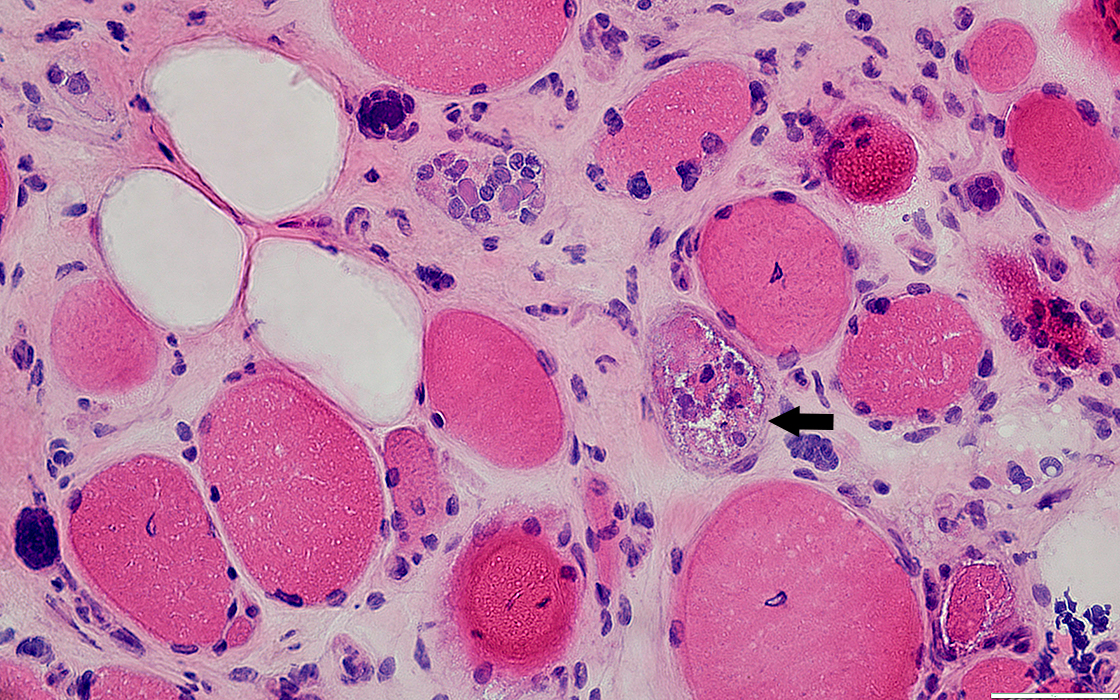
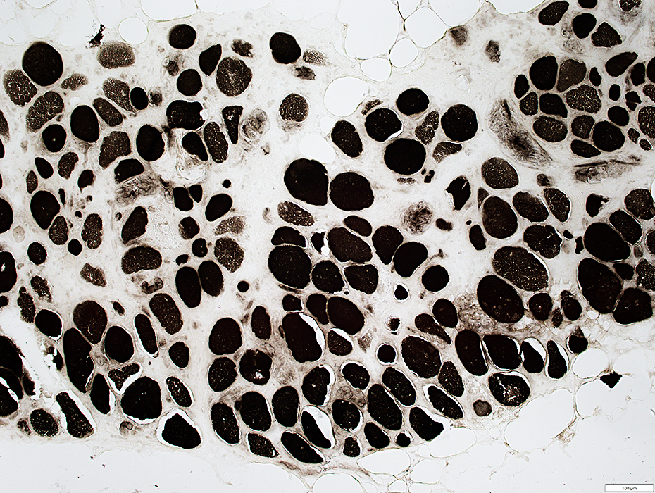
FHL1: Myopathy (C276Y heterozygous mutation)
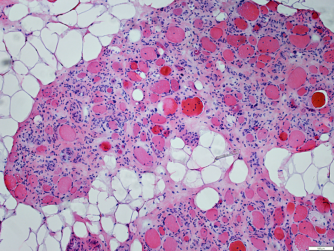 H&E stain |
Muscle Fibers
Sizes: Varied
Internal nuclei
Connective tissue
Endomysium: Markedly increased
Perimysium: Marked fat replacement
 Gomori trichrome stain |
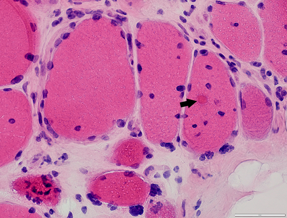 H&E stain |
Myonuclei: Large; Irregular shapes
Cytoplasmic aggregate (Arrow)
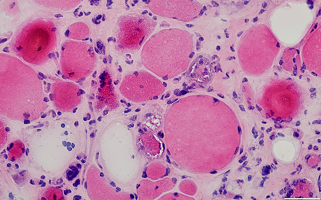
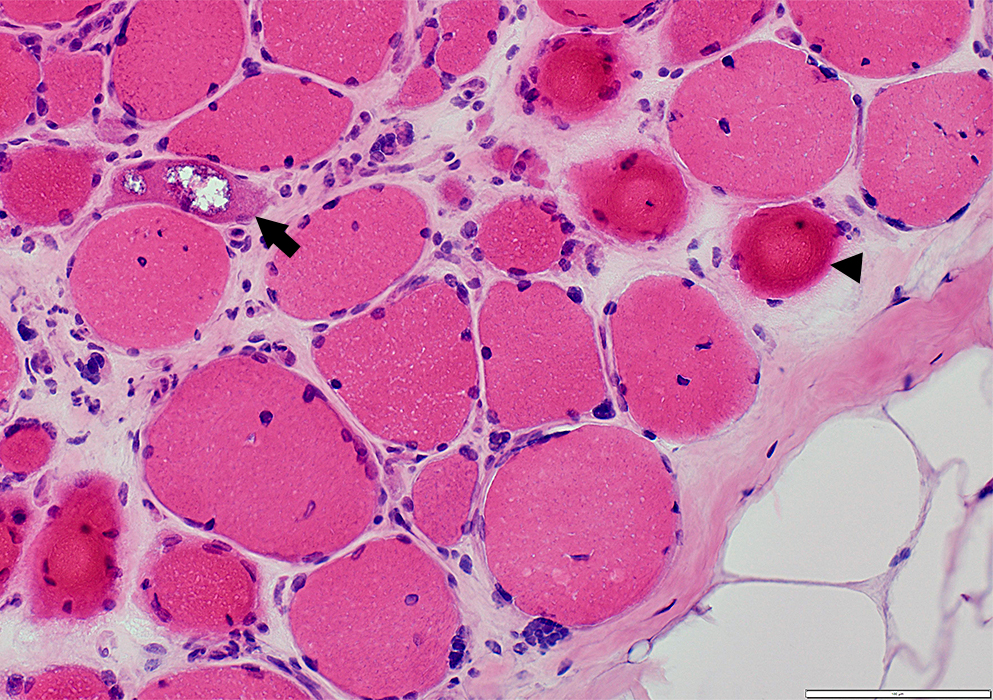 H&E stain |
Vacuole: Irregular shape (Arrow)
Ring fibers + Sarcoplasmic pads (Arrowhead)
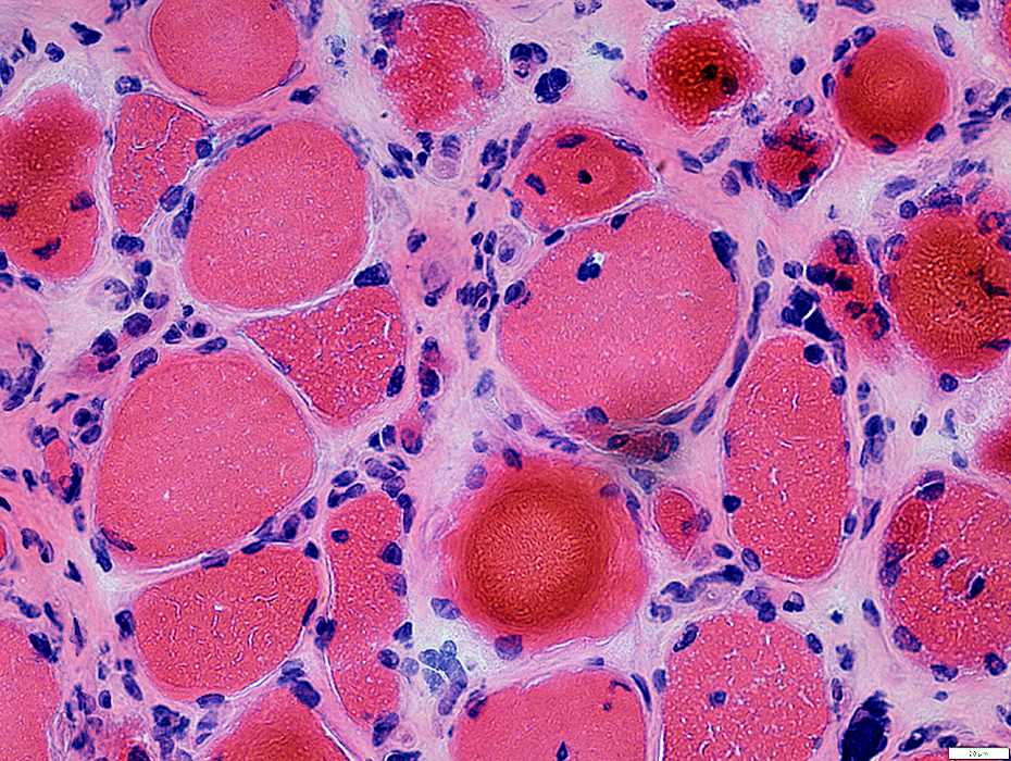
 H&E stain |
Sizes: Mostly large
Shapes: Irregular
Number: Some fibers have increased numbers, scattered or clustered
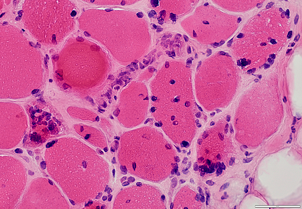
 H&E stain |
Irregular cytoplasm structure (Arrow)
Large nuclear clumps
 H&E stain |
 H&E stain |
Ring fibers + Sarcoplasmic pads
 VvG stain |
 AMPDA stain |
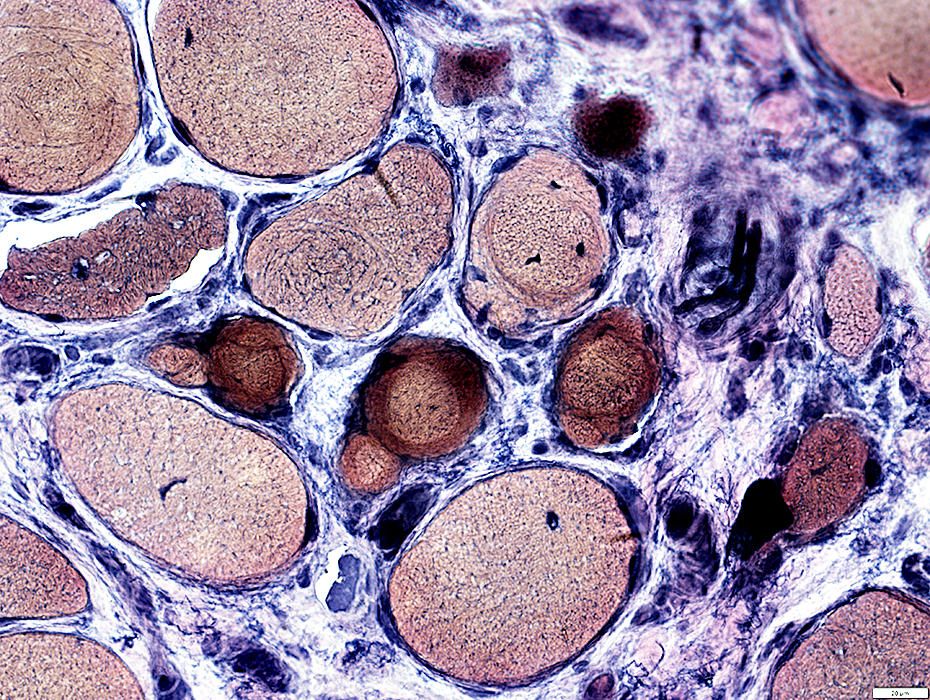
 NADH stain |
Scattered fibers are small & dark stained
Internal architecture: Coarse
 VvG stain |
 Esterase stain |
 Esterase stain |
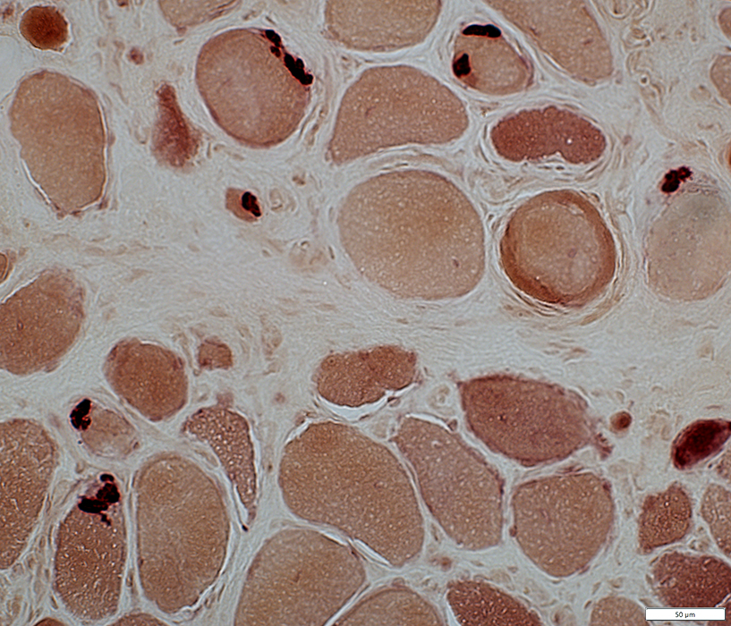 Esterase stain |
 Acid phosphatase stain |
Scattered in endomysial connective tissue
Intramuscular nerves: Myelin has increased staining
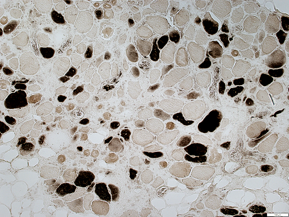 ATPase pH 4.3 stain |
Type 2 predominance
Scattered Immature (type 2C) fibers
 ATPase pH 9.4 stain |
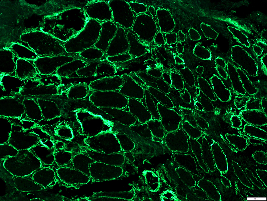
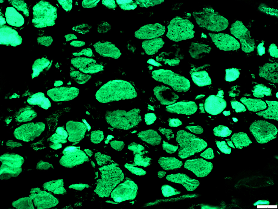 Phalloidin stain |
Actin in muscle fiber cytoplasm is often reduced & irregularly distributed.
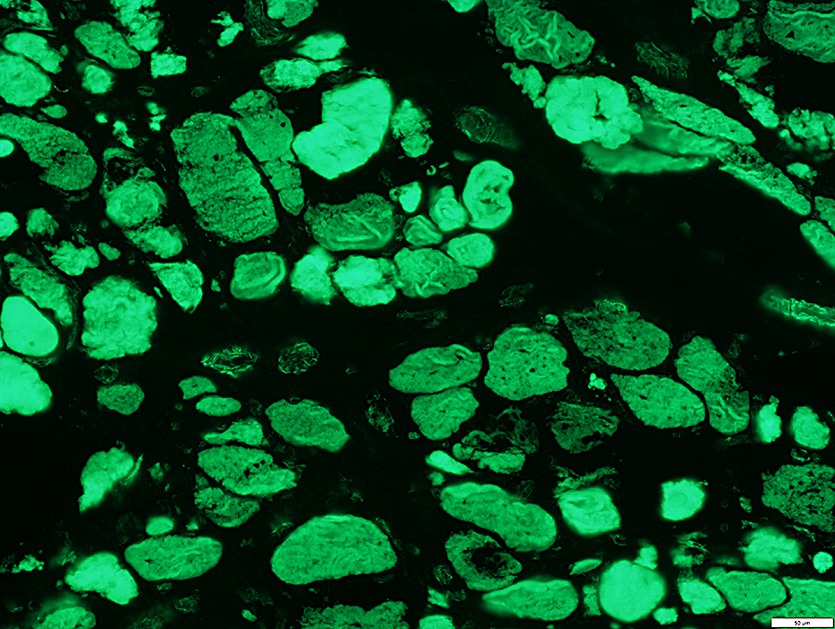 Phalloidin stain |
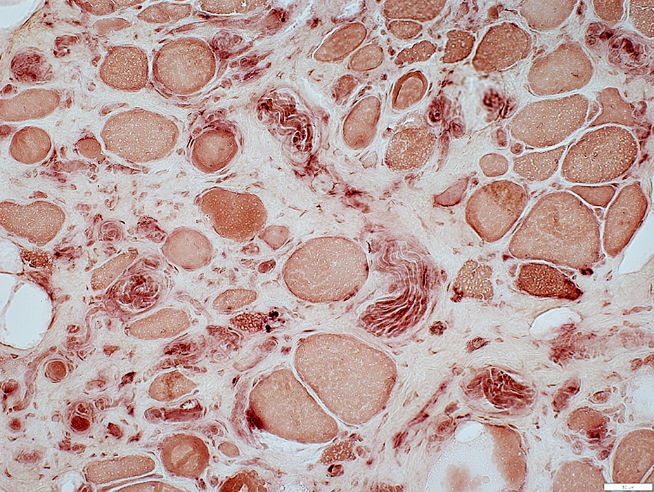
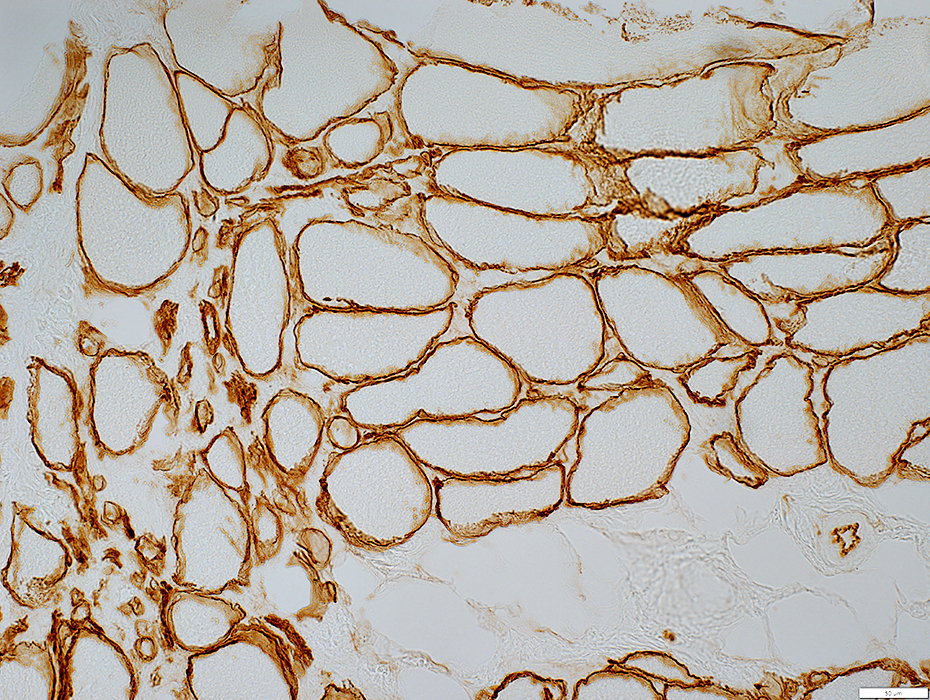 Laminin-α2 stain |
Thick & Irregular around muscle fibers
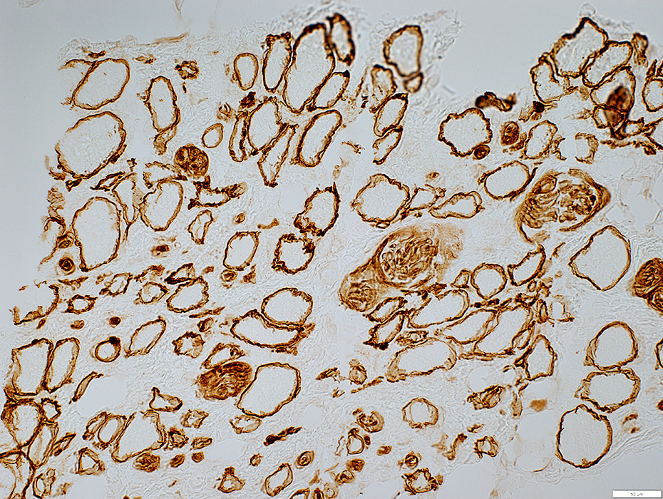 Laminin-α2 stain |
 α-Dystroglycan stain |
α-Dystroglycan is irregularly distributed on muscle fiber surfaces
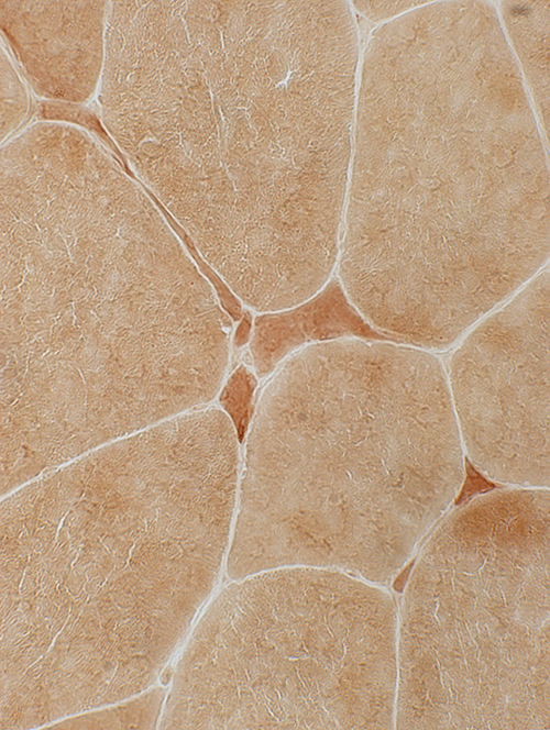
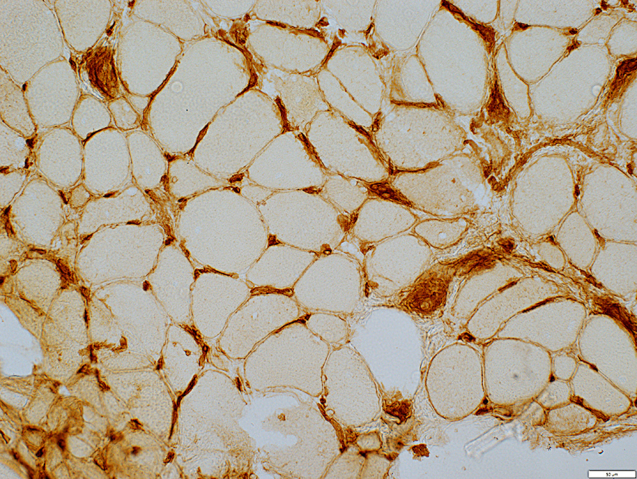 MHC I stain |
MHC Class I: Mildly upregulated on muscle fiber surfaces
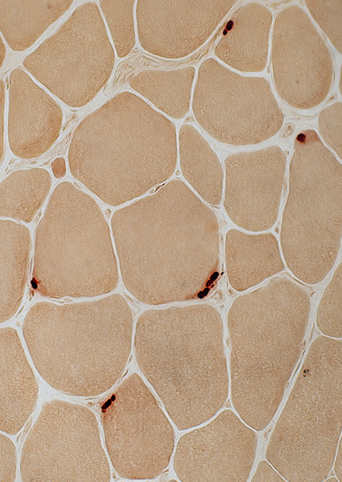
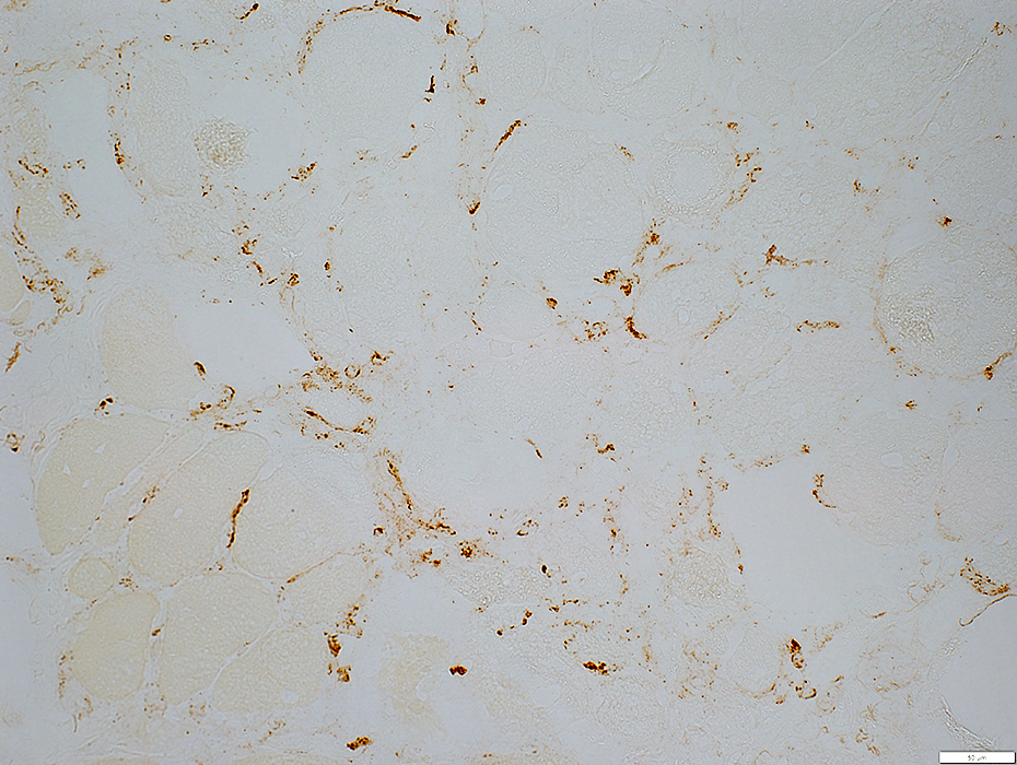 C5b-9 stain |
Stains endomysial capillaries
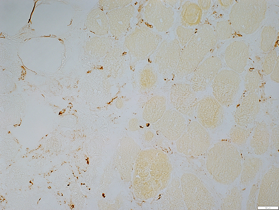 C5b-9 stain |
Return to FHL1.
Return to Neuromuscular syndromes
Return to Neuromuscular home page
5/8/2023