Orbital Myositis
EOM Pathology
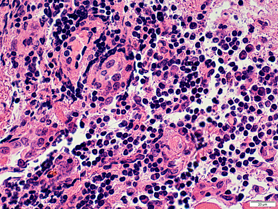
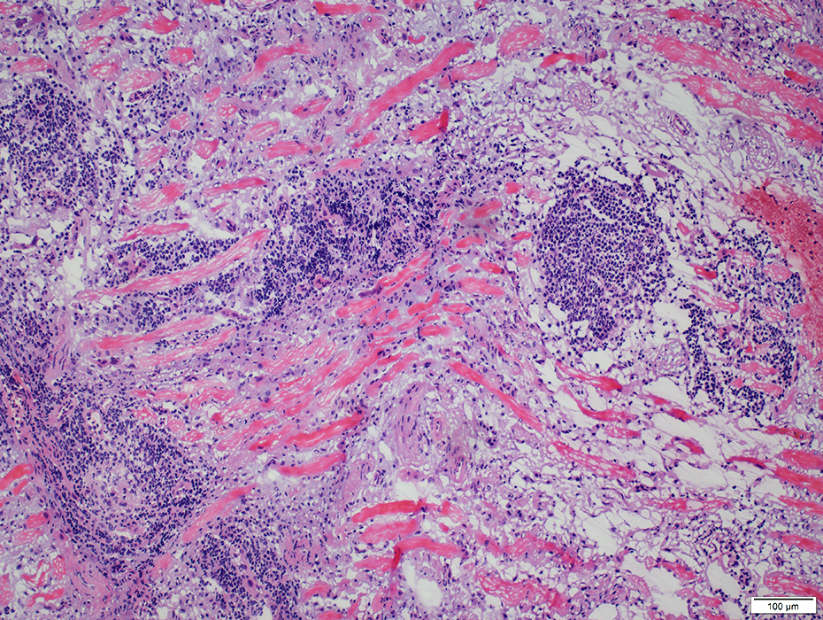 H&E stain |
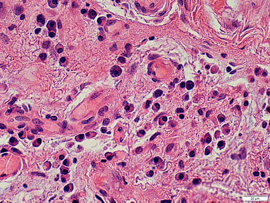
Multiple scattered foci of lymphocytes
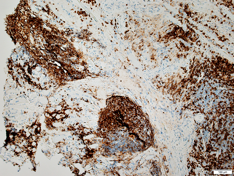 CD3 (T-cell) stain |
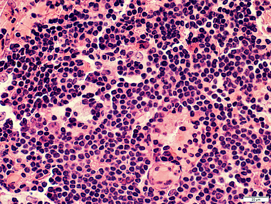
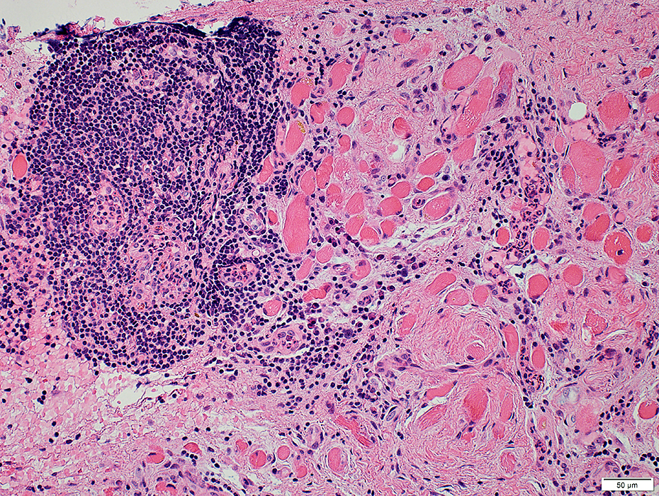 H&E stain |
Contain
Smaller vessels (High endothelial venules)
B-cells and T-cells
Locations
Endomysium: Between muscle fibers
Perimysium: Between fascicles
 H&E stain |
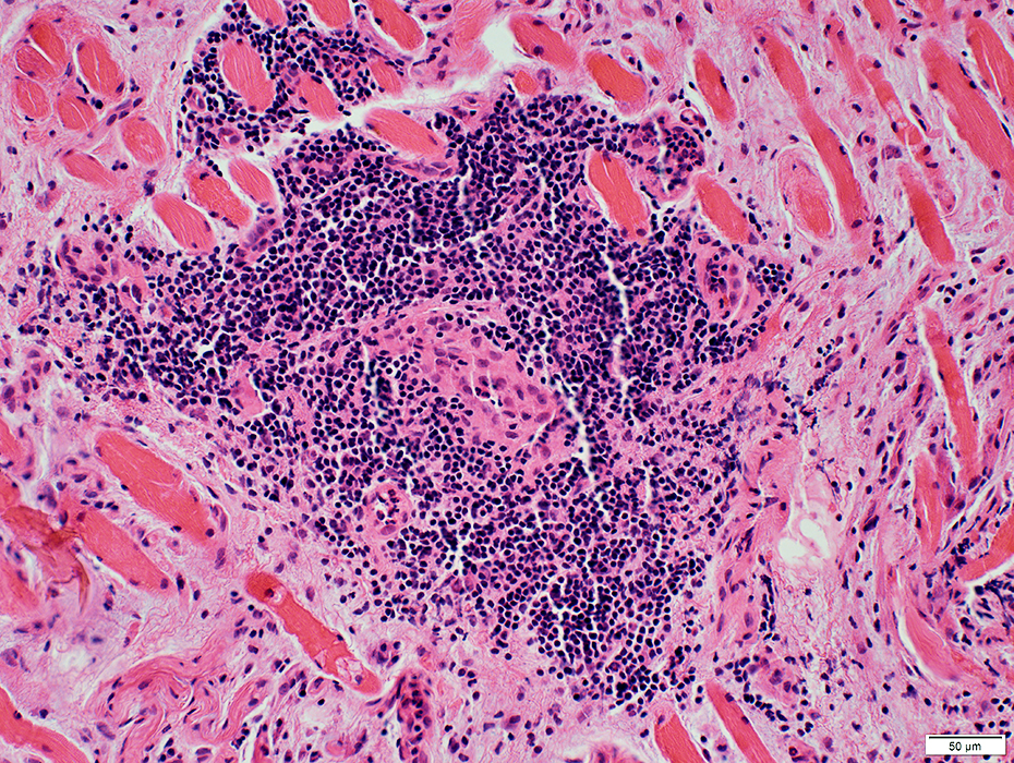
 H&E stain |
In foci
Common: B-cells & T-cells
Scattered: Plasma cells
Few: Eosinophils
 H&E stain |
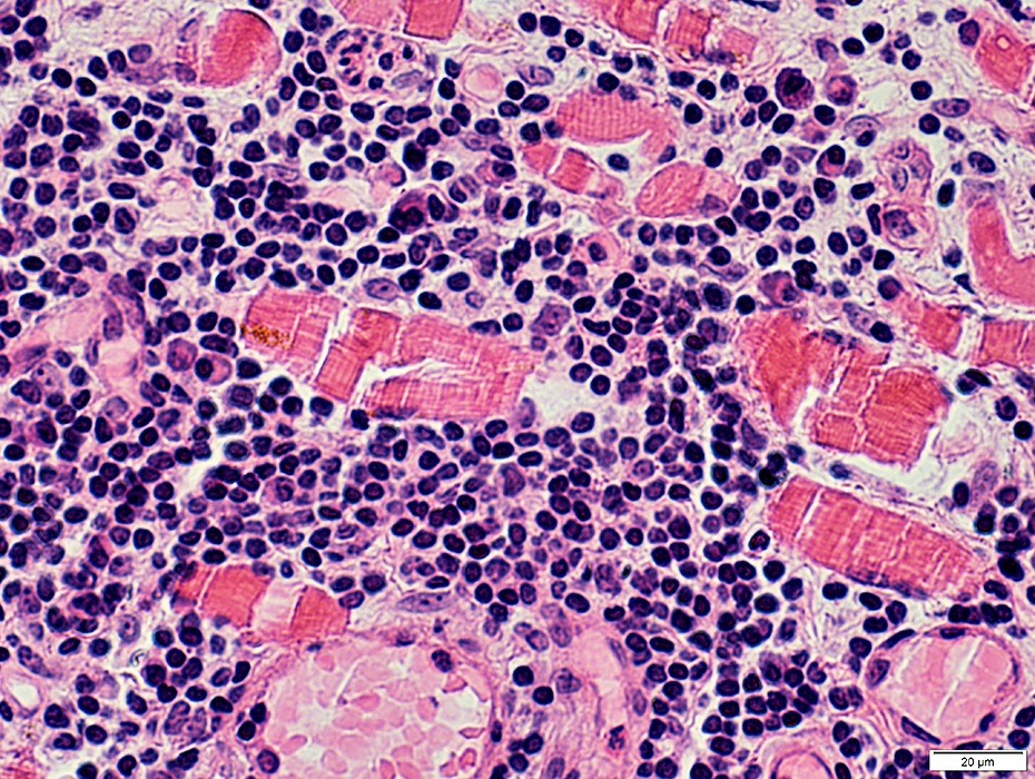 H&E stain |
T/B Cell Separation
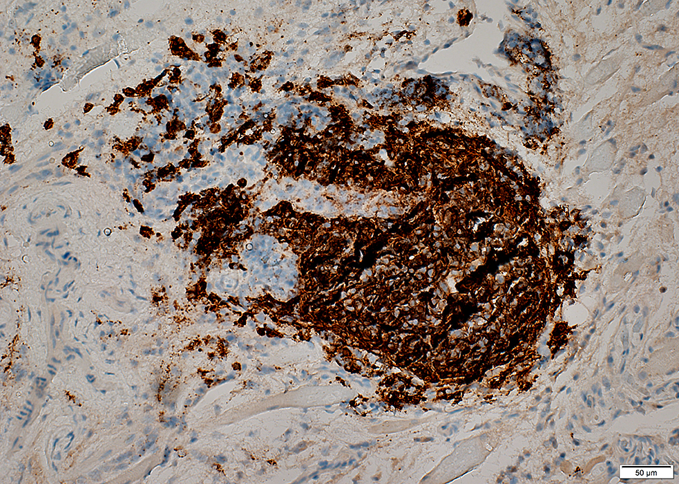 CD20 (B-cell) stain |
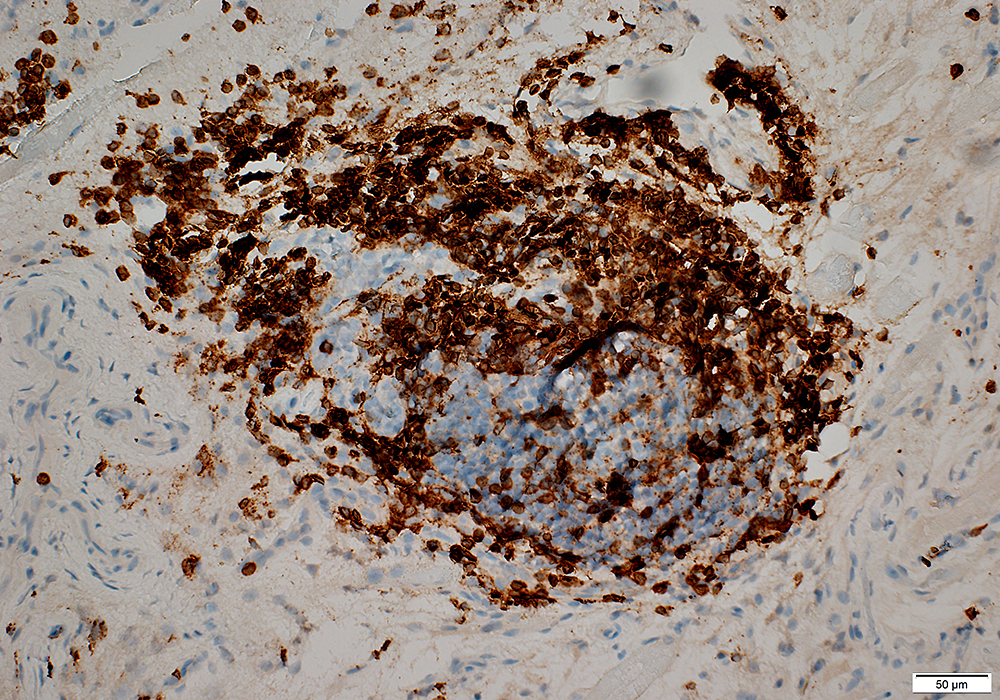 CD3 (T-cell) stain |
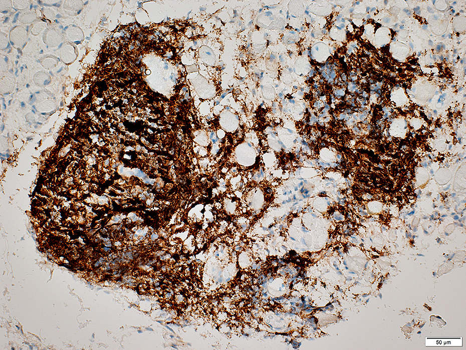 CD20 (B-cell) stain |
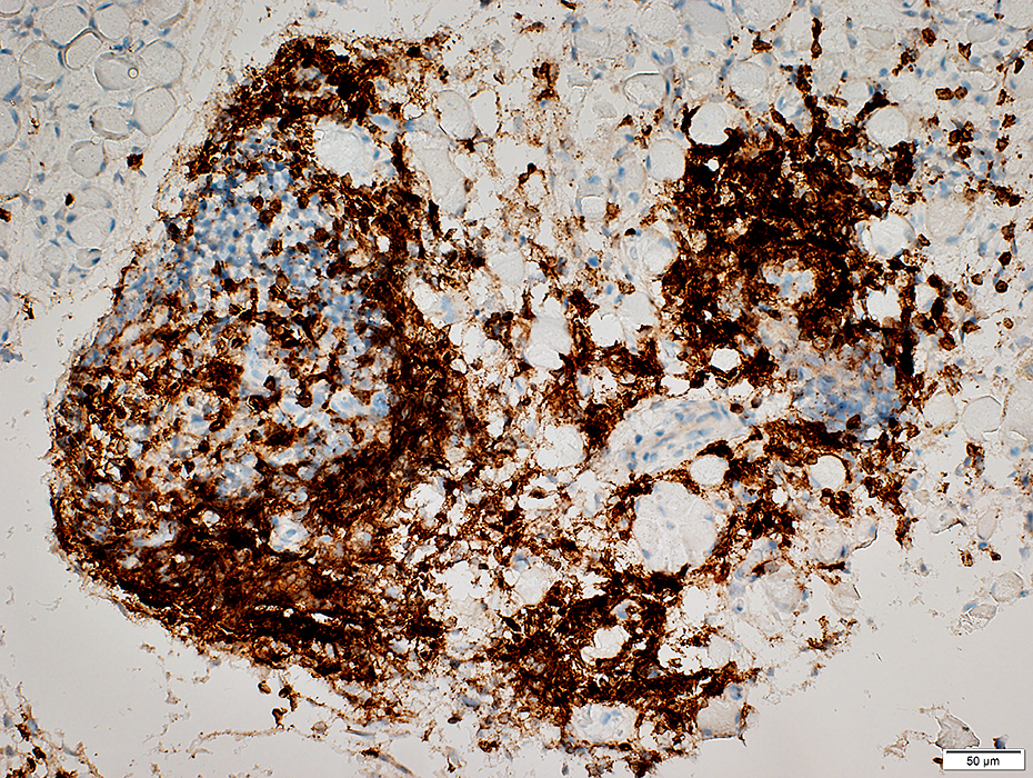 CD3 (T-cell) stain |
 H& E stain |
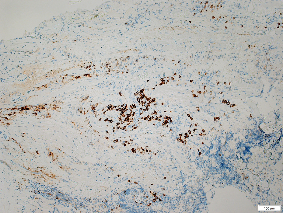 CD138 (Plasma cell) stain |
Return to: Normal EOM
Return to Orbital Myositis
Return to Neuromuscular home page
References
1. Nat Rev Immunol 2014;14:447-462, Front Immunol 2016;7:430
11/10/2022