Immune Myopathies with Perimysial Pathology (IMPP): OJ antibody associated 1
|
Perimysial connective tissue pathology Histiocytic cells Structure damage Muscle fiber pathology Necrosis & Regeneration MHC I Aggregates Also see IMPP Dermatomyositis: Adult, IMPP type Jo-1 muscle pathology |
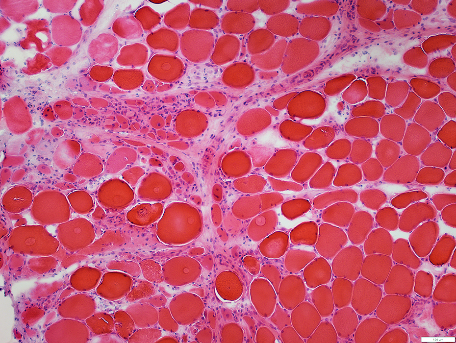
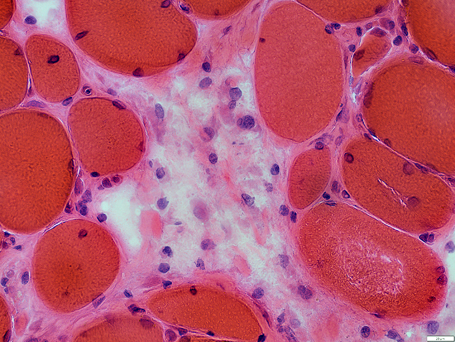
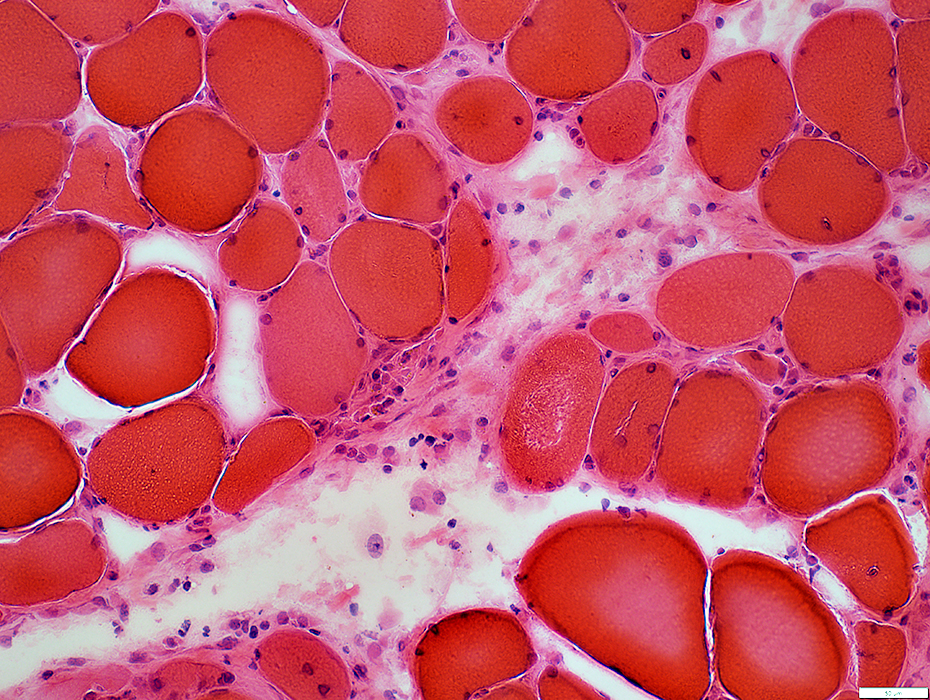
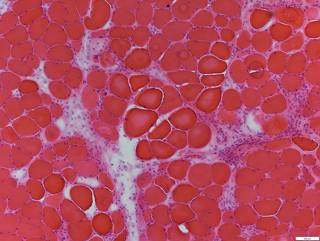 H&E stain |
- Perimysial Cells: Histiocytic
- Large nuclei
- Prominent cytoplasm
- Stain for: Acid Phosphatase, Esterase and CD-68
- Structure
- Fragmented
- Pale staining
- Loss of fibrils
- Fiber sizes
- Perifascicular fibers: May be small with necrosis or regeneration
- Other fibers: Varied sizes, Intermediate or Normal
- Fiber damage: More in perifascicular regions
- Necrosis & Regeneration
- MHC Class I: Upregulated
- Autophagy: LC3 aggregates in muscle fiber cytoplasm
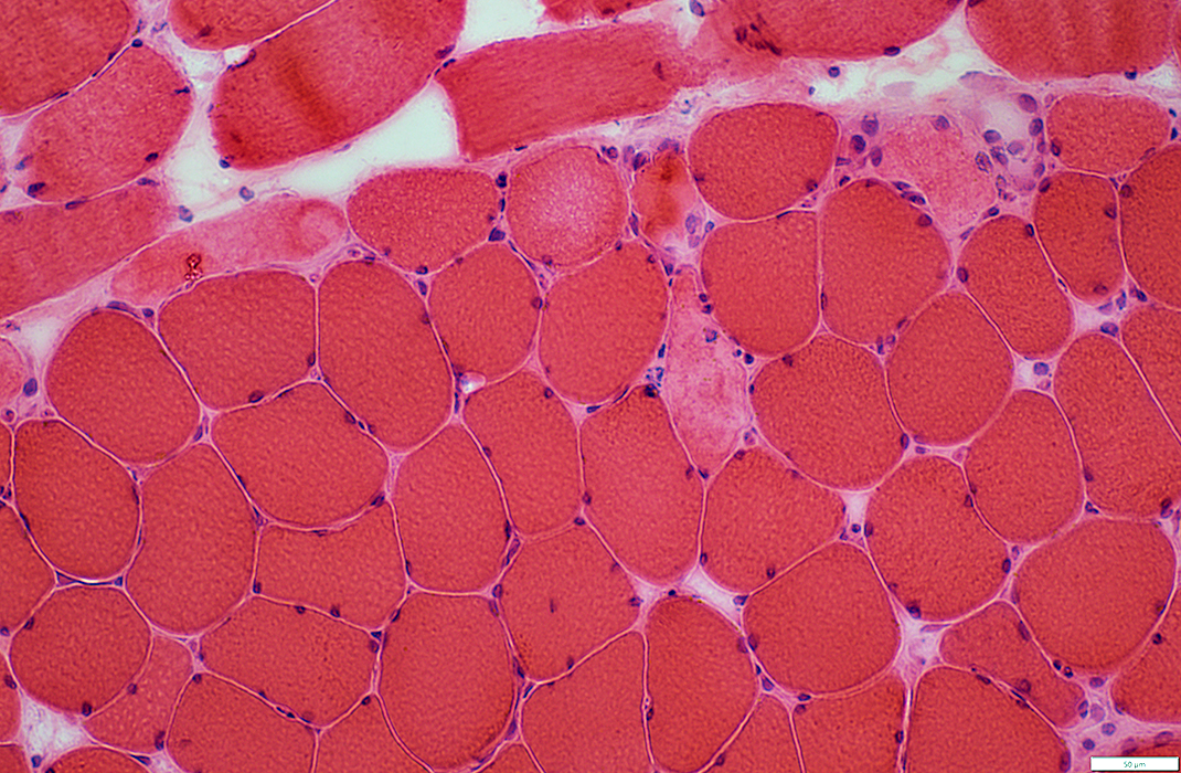
 H&E stain |
OJ antibodies: Perimysial pathology
 Esterase stain |
 Esterase stain |
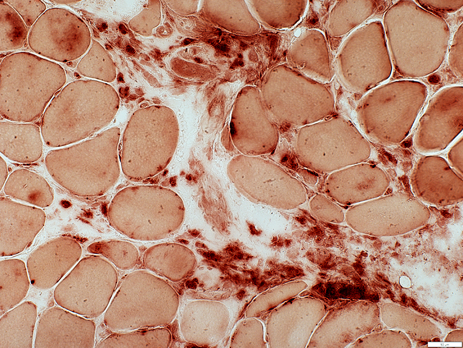 Acid phosphatase |
- Large nuclei
- Prominent cytoplasm
- Stain for: Acid Phosphatase, Esterase, HAM56 & CD-68
 H&E stain |
 HAM56 stain |
 Gomori trichrome stain |
- Fragmented
- Pale staining
- Loss of fibrils
 H&E stain |
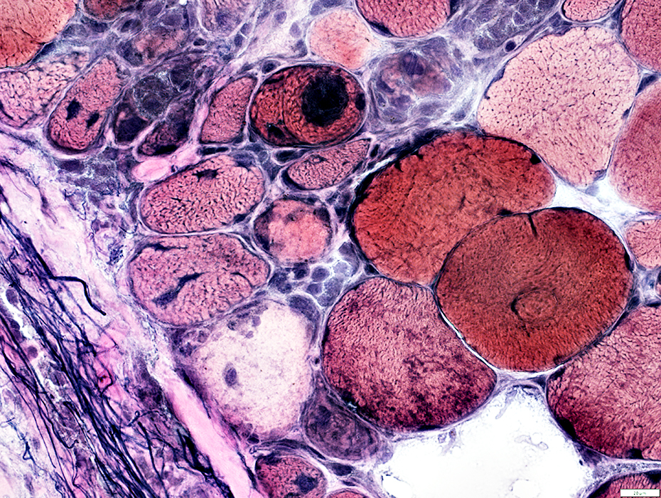
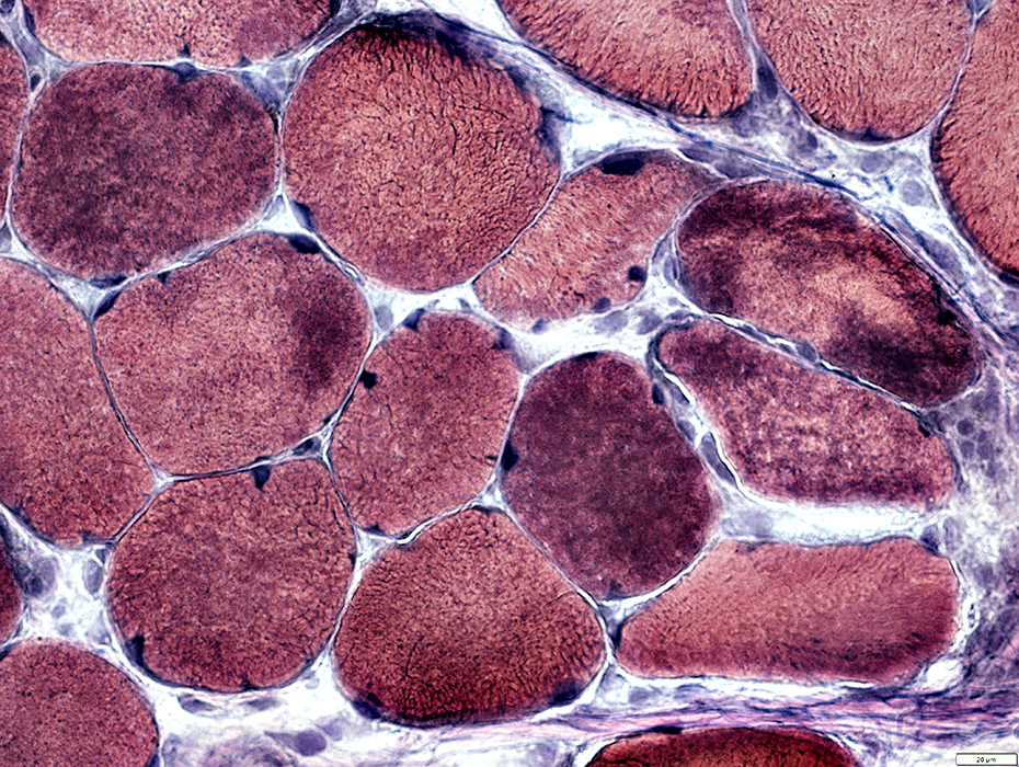
 VvG stain |
 VvG stain |
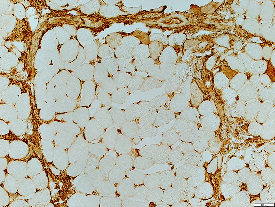
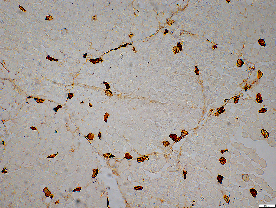
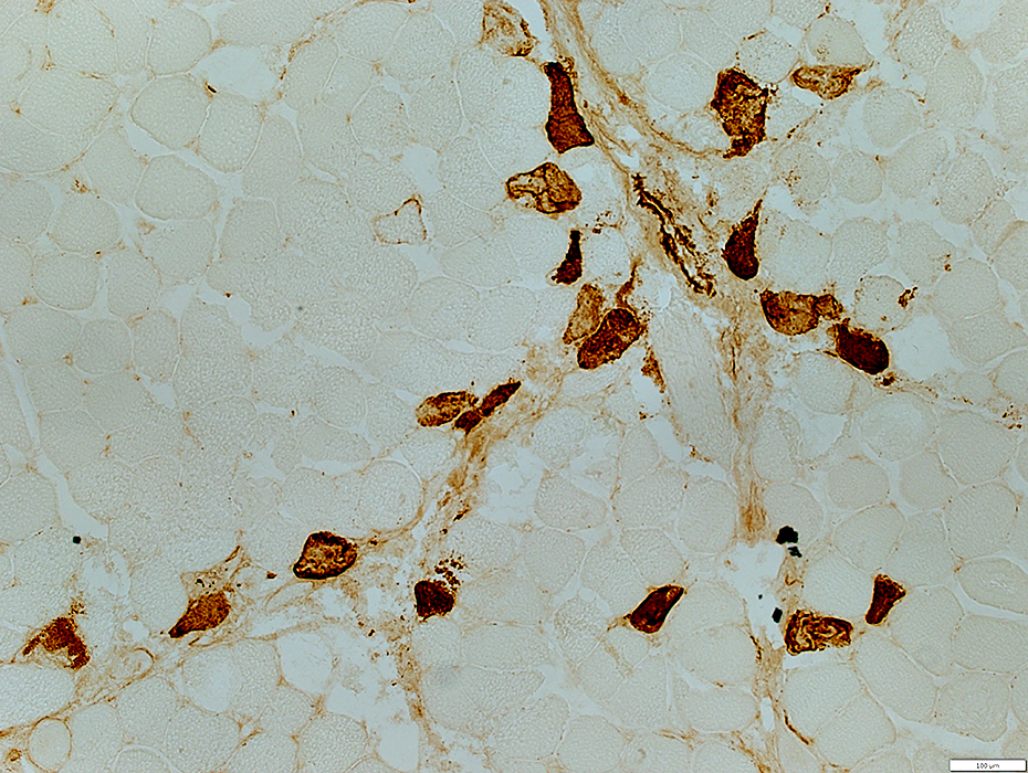
OJ-antibody related myopathy: Perimysial pathology
Alkaline phosphatase stains
Perimysial connective tissue
Cytoplasm of many neighboring, small regenerating, muscle fibers
 Alkaline phosphatase |
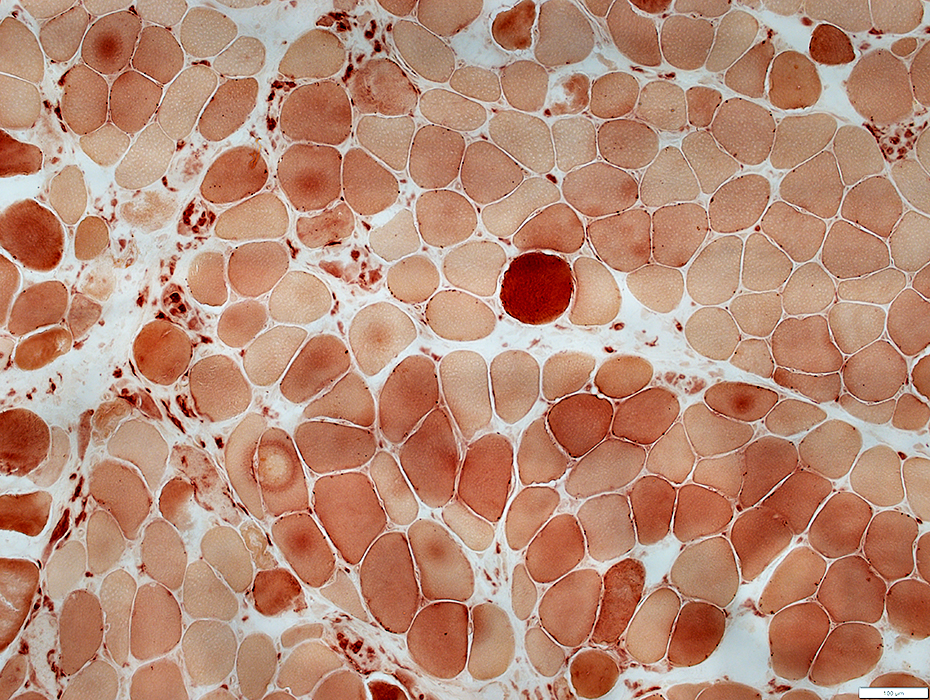
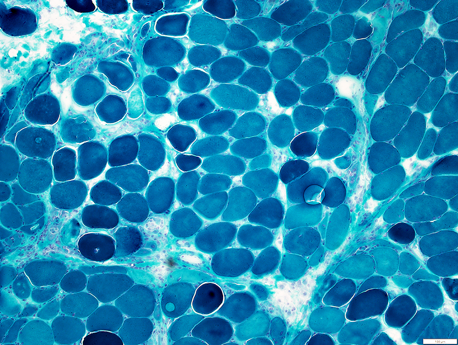
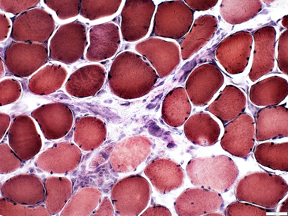
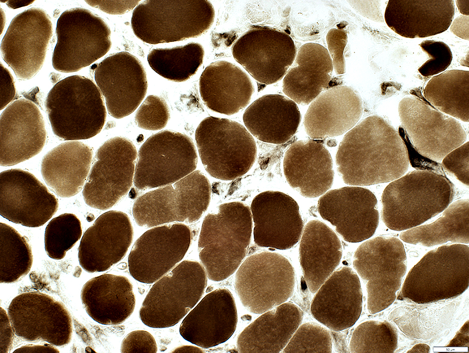
OJ-antibody related myopathy: Muscle fibers
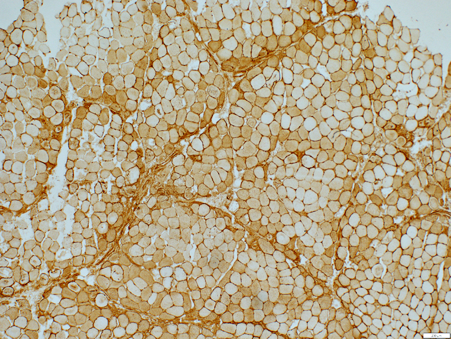 MHC Class I stain |
- Fiber sizes
- Perifascicular fibers: May be small with necrosis or regeneration
- Other fibers: Varied sizes, Intermediate or Normal
- Fiber damage: More in perifascicular regions
- Necrosis & Regeneration
- MHC Class I: Upregulated
- Autophagy: LC3 aggregates in muscle fiber cytoplasm
 C5b-9 stain |
 C5b-9 stain |
 H&E stain |
 VvG stain |
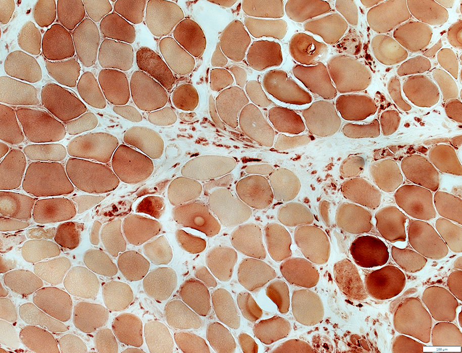
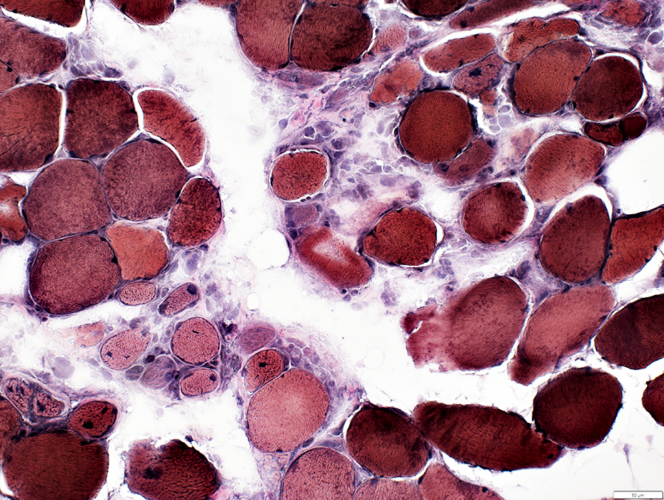
OJ-antibody related myopathy: Muscle fiber aggregates
 VvG stain |
Location: Cytoplasm
Stain for: LC3; VvG
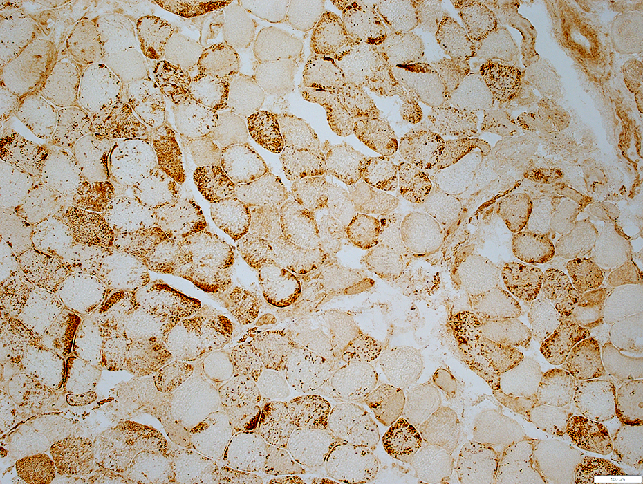 LC3 stain |
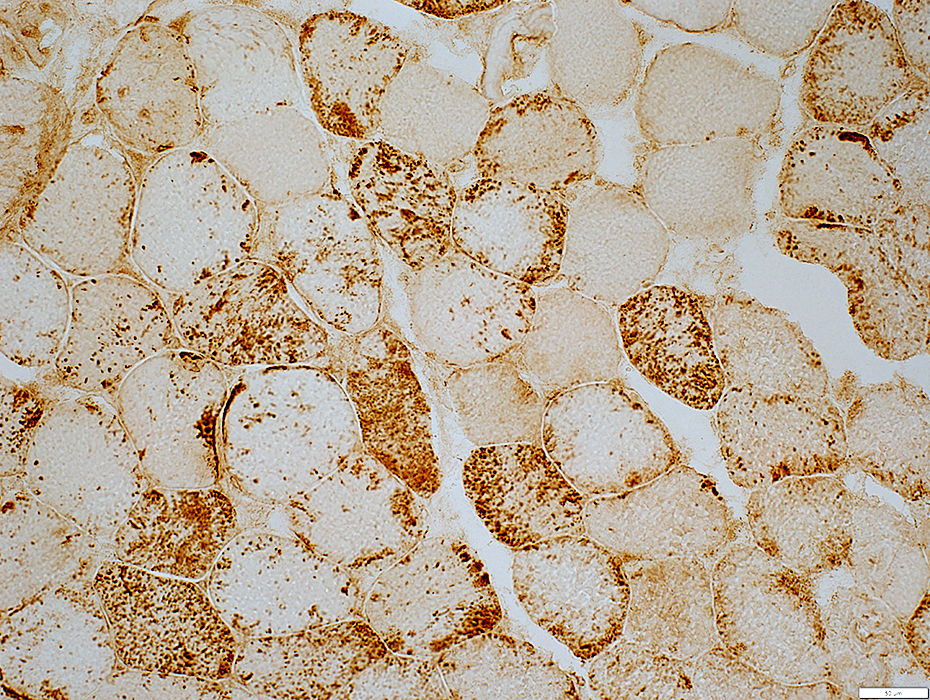 LC3 stain |
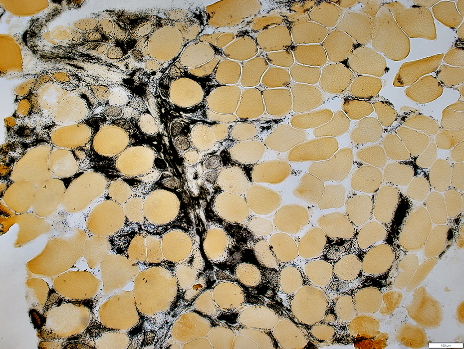
Capillaries
Staining by ATPase
 ATPase pH 4.3 stain |
Return to Inflammatory myopathies
Return to IMPP
References
1. Brain Pathol 2023 Mar 7:e13155
3/20/2023