Nerve Specific (Non-Systemic) Inflammatory Vasculopathic Neuropathy (NSVN)
Pathology features- Nerve
- Epineurium: Small Vessels (< 75 μM)
- Inflammation
- Location: Surrounds, & within walls of, small epineurial vessels
- Type: Lymphocytes & Histiocytes
- Structure: Damaged wall & Smoooth muscle layer
- Neovascularization
- Inflammation
- Axons
- Wallerian degeneration
- Loss: Differential fascicular
- Endoneurium
- Loss of ATPase stain
- Edema: Subperineurial & Endoneurial
- Epineurium: Small Vessels (< 75 μM)
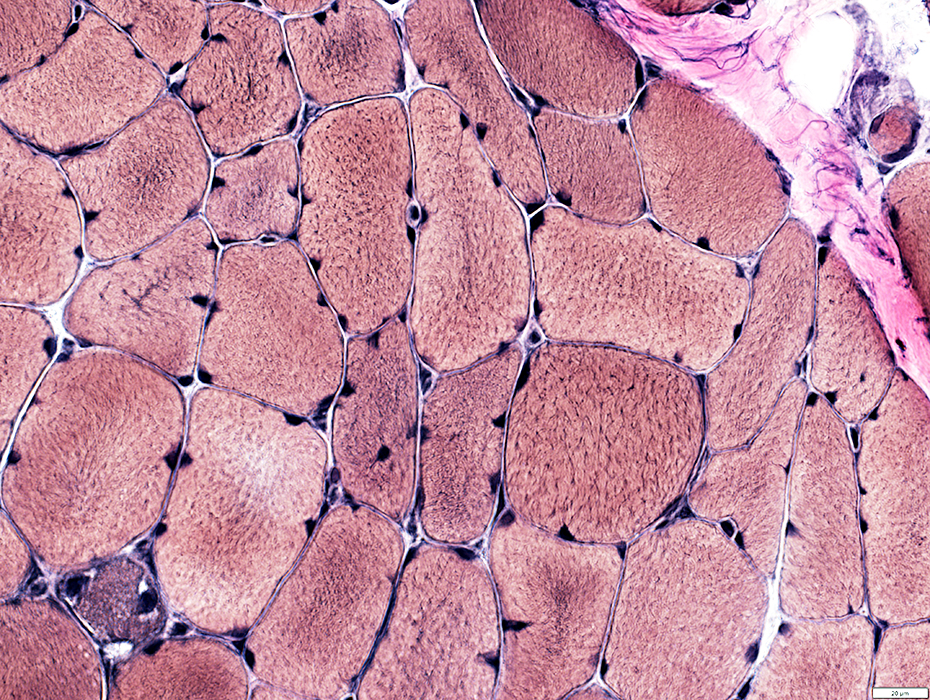
- Muscle
- General
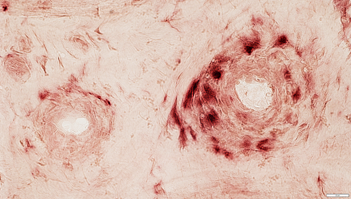
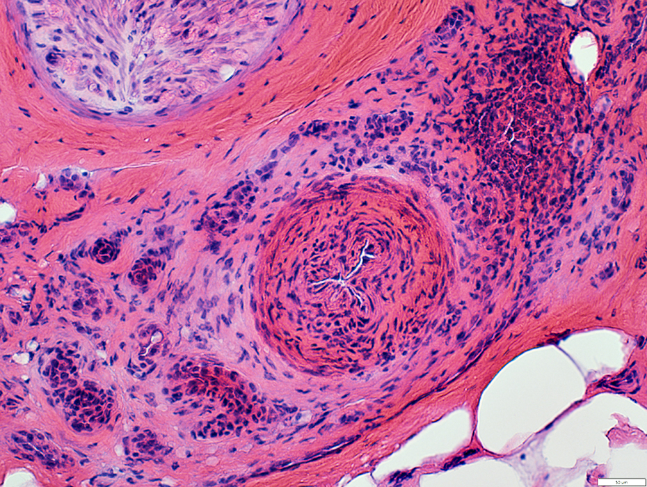
Perivascular Inflammation
Surrounds small epineurial vessels (Below; Arrow)
Larger epineurial Arteries & Veins: Varied pathology
May be Normal (Below) or Damaged (Above)
Epineurium: May have neovascularization (Above; White Arrow))
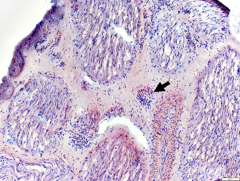 Congo red stain |
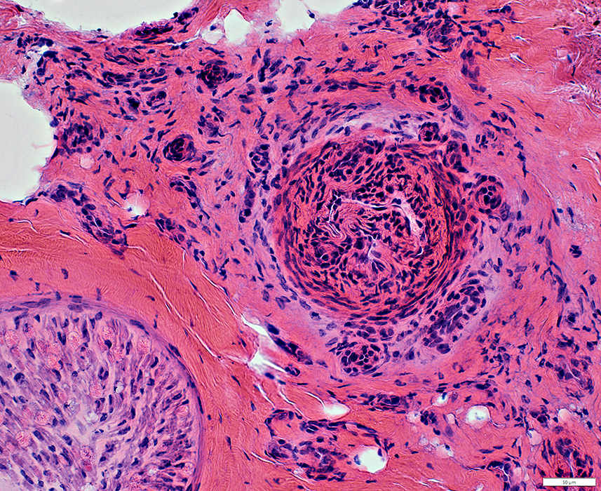
Perivascular Inflammation
Surrounds small epineurial vessels (Below; Arrow)
Larger epineurial arteries & veins: Varied pathology
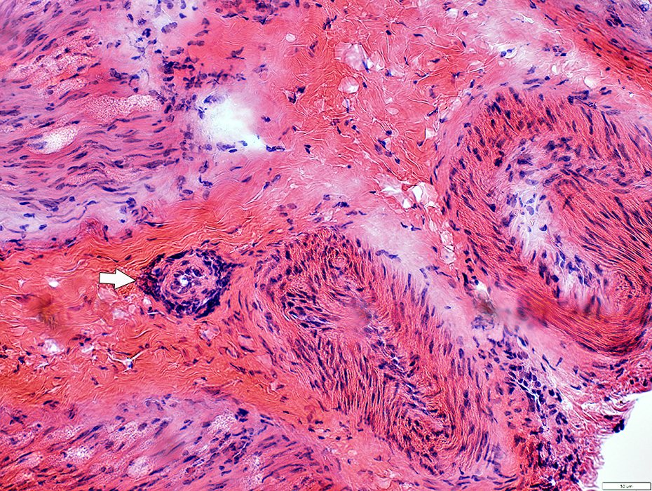 H&E stain |
 H&E stain |
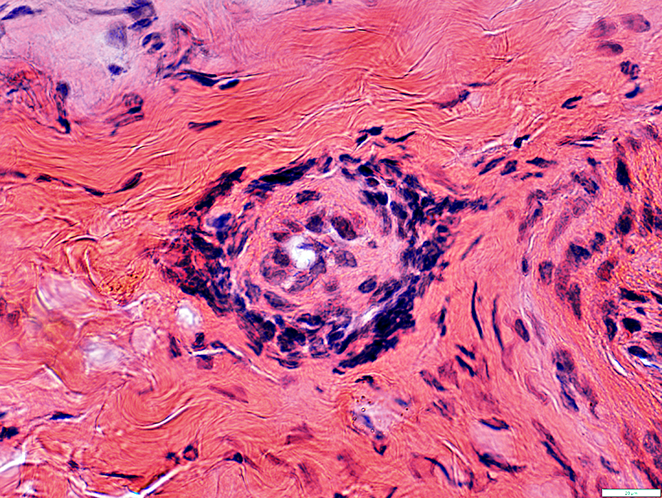
Surrounds small epineurial vessels
Contains CD4 & CD8 lymphocytes
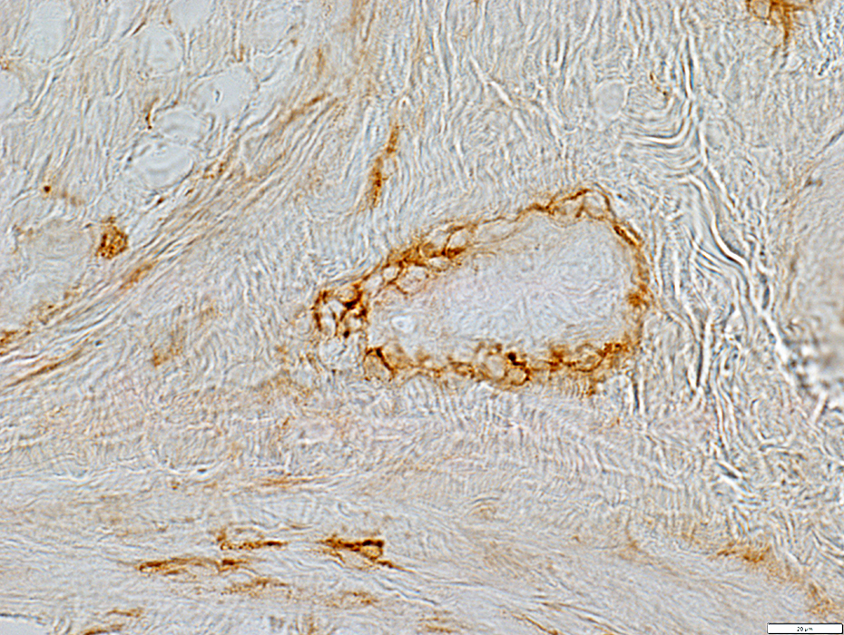 CD4 stain |
 CD8 stain |
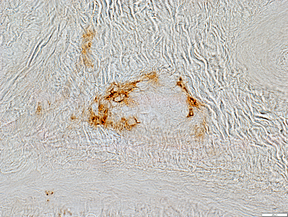
Histiocytes in & near Epineurial Vessel walls
 Acid Phosphatase stain |
MHC Class I
Expressed by epineurial cells & connective tissue
 MHC Classs I stain |
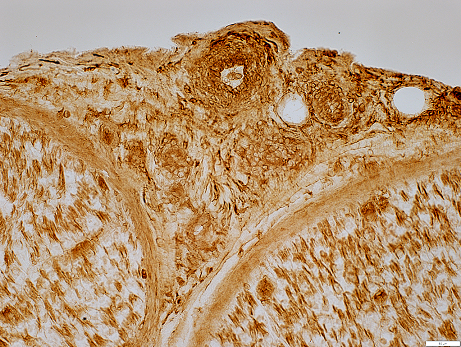
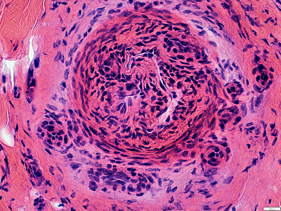
NSVN Vessel Damage: Smoooth Muscle Layer
Varied at different levels
Irregular width & Damaged (Above; Arrow)
More normal: Dark stained in vessel wall (Below)
Inflammation
Cells surround epinerial vessels
Neovascularization
Below damaged vessel
 ATPase pH 4.3 stain |
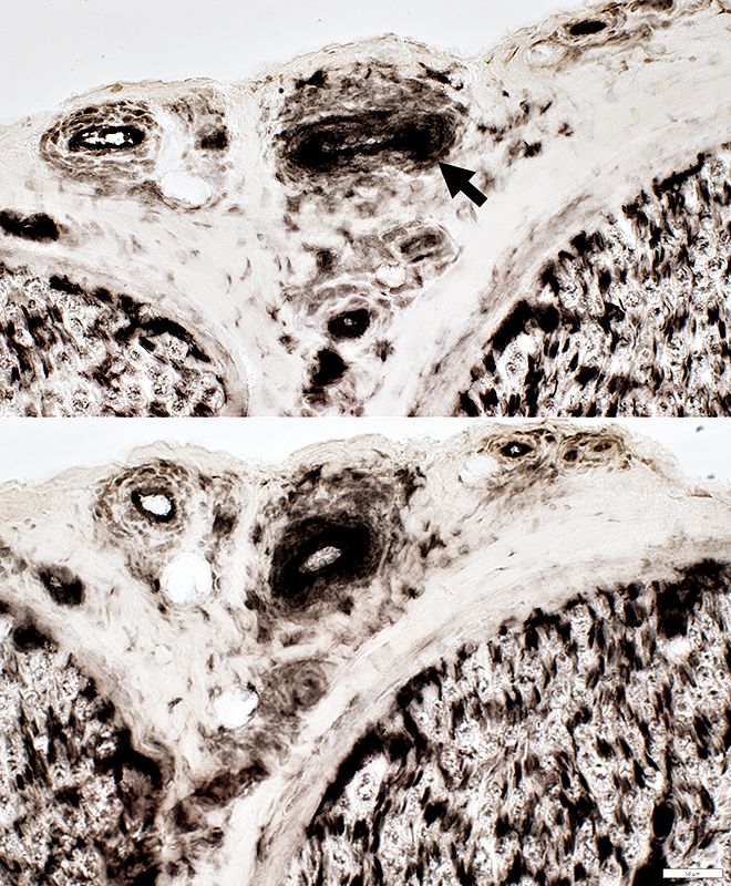
 VvG stain |
Increased numbers of smaller epineurial vessels
 UEA I stain |
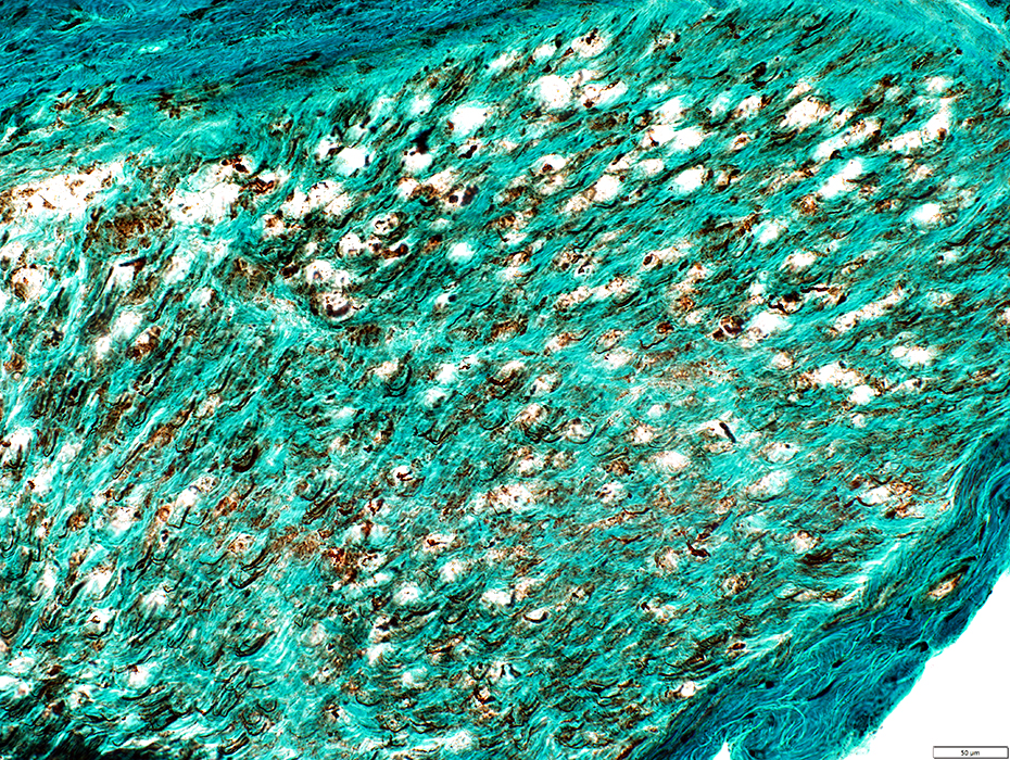
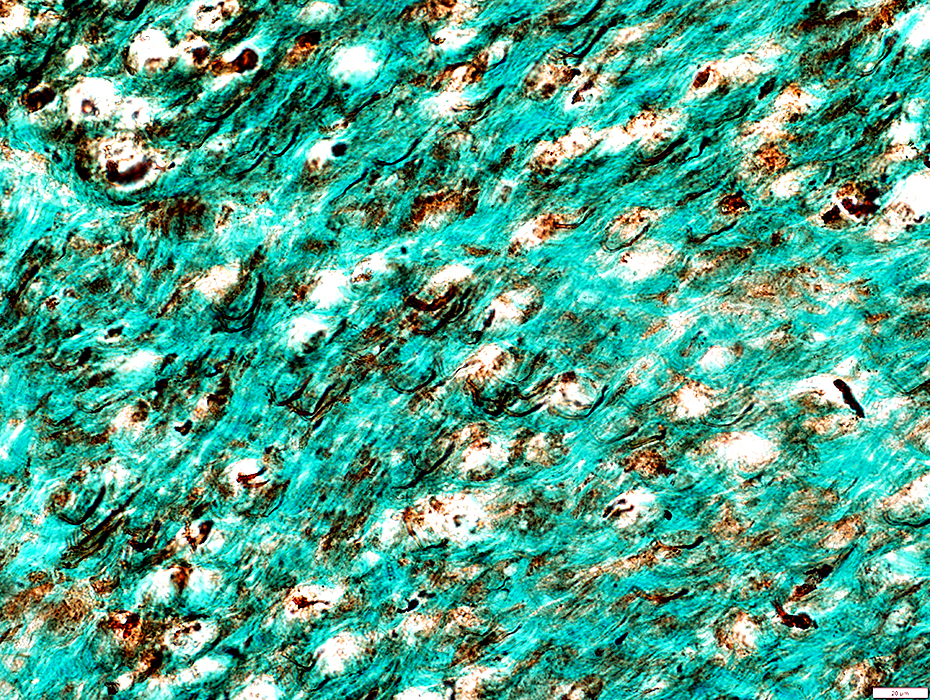
NSVN: Endoneurium
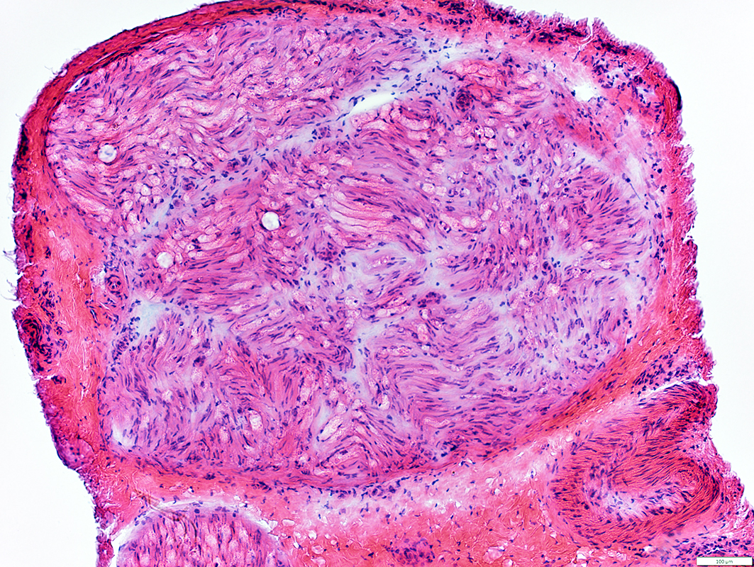 H&E stain |
Present in subperineurial & endoneurial regions between fascicles
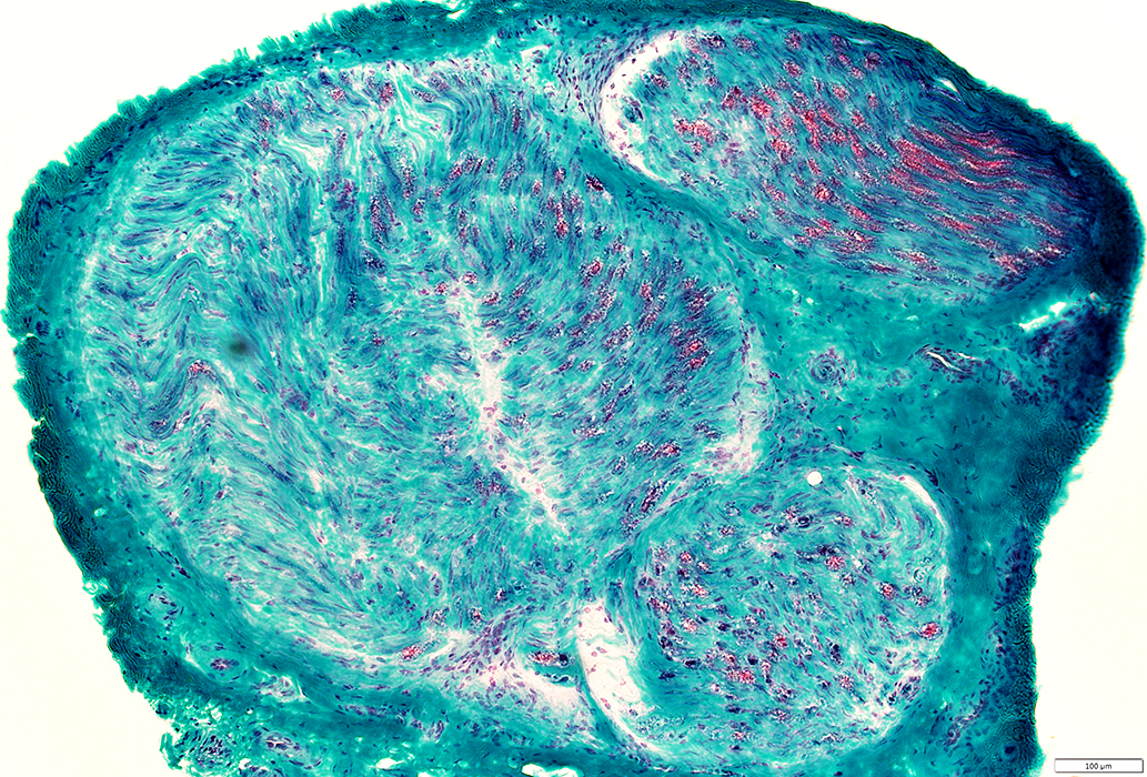 Gomori trichrome stain |
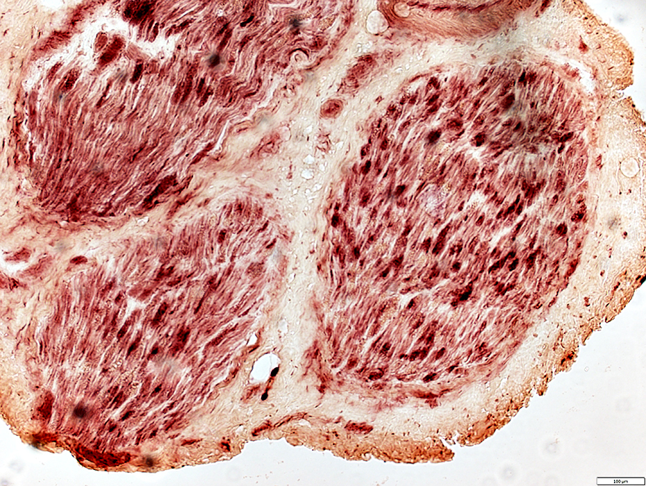
NSVN: Axons
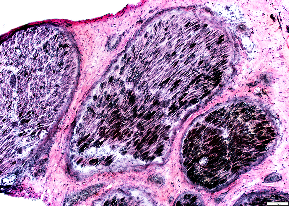 VvG stain |
Varied Loss (Above) & Degeneration (Above & Below) of myelin & axons within & among fascicles
VvG (Above) shows: Irregular staining of damaged myelin
Acid phosphatase (Below) stains: Damaged myelin sheaths
Epineurium
Damged structure in some regions
 Acid Phosphatase stain |
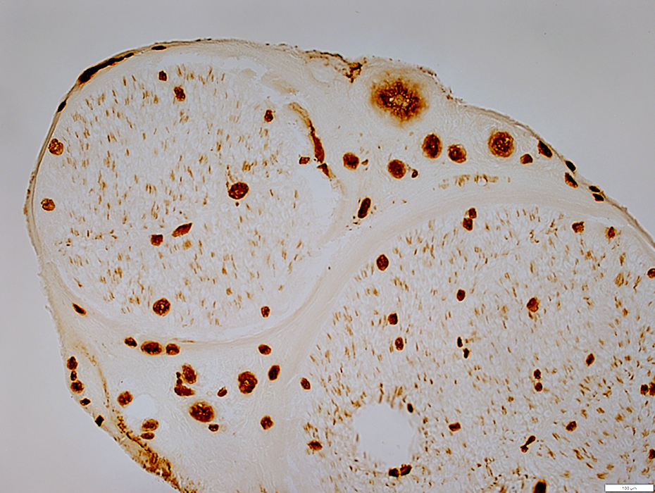
ATPase stain pH 4.3
Ischemia: Reduced staining in endoneurium
Vessels in endoneurium are normally stained
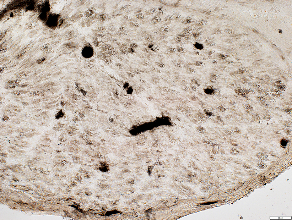 ATPase pH 4.3 stain |
NSVN: Wallerian degeneration
Axons: Degeneration & Loss
Myelin: Pathology, Phagocytosis & Loss
 Neurofilament stain |
Granular, punctate staining in areas that previously contained large myelinated axons
 Neurofilament stain |
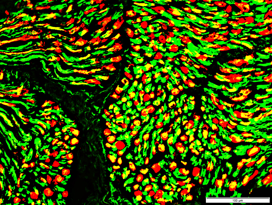
Axon loss: Large & Small
Differential intrafascicular loss of both large and small axons (bottom right of image).
The many empty non-myelinating Schwann cell (NCAM; Red) sheaths show loss of small axons.
Large axons (Green) are also lost in the damaged region compared to the more intact area (upper left)
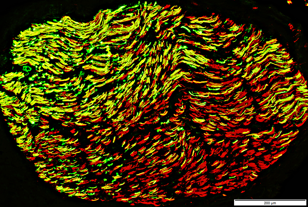 Neurofilament (Green) + NCAM (Red) stain |
Differential Intrafascicular Axon loss
More loss in lower regions
Involves both Large (Empty myelin sheaths (Red)) & Small (Green) axons
Ongoing axon loss: Punctate neurofilament staining within P0 myelin sheaths
Complete axon loss: Some P0 myelin sheaths have no neurofilament-stained axon
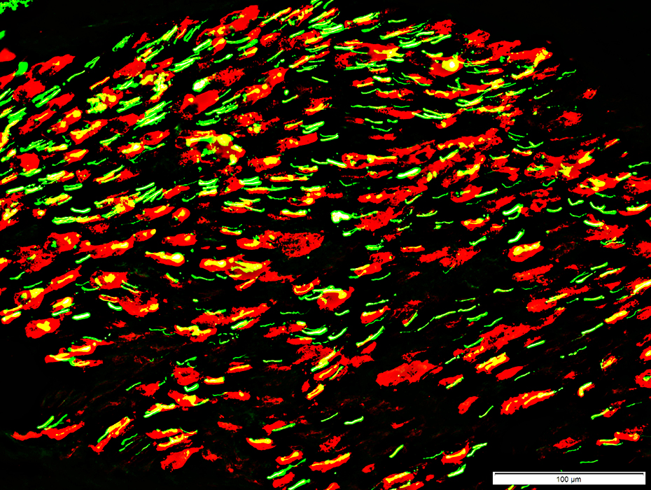 Neurofilament (Green) + P0 (Red) stain |
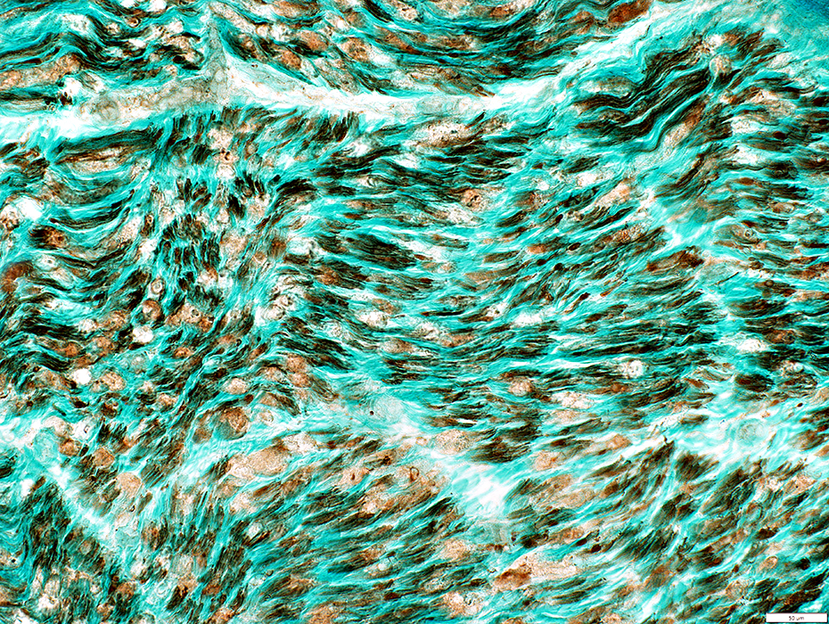
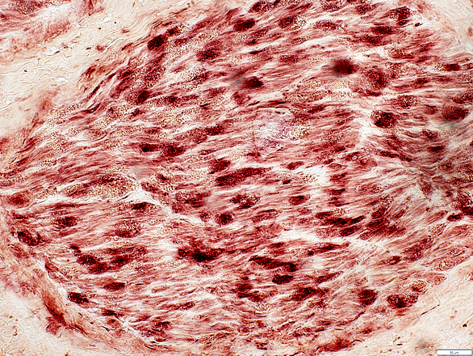
Wallerian degeneration, Early: Myelin pathology
Damaged myelin sheaths: Irregularly stained by VvG
 VvG stain |
Wallerian degeneration, Early: Myelin pathology
Schwann cells associated with abnormal myelin around degenerating myelinated axons abnormally express NCAM
 NCAM stain Wallerian degeneration, Early |
 NCAM (Green) + P0 (Red) stain Schwann cells associated with abnormal myelin around degenerating myelinated axons abnormally co-express NCAM (Green) & P0 (Red) |
Wallerian degeneration
Acid phosphatase stains myelin & some phagocytic endoneurial cells
 Acid Phosphatase stain |
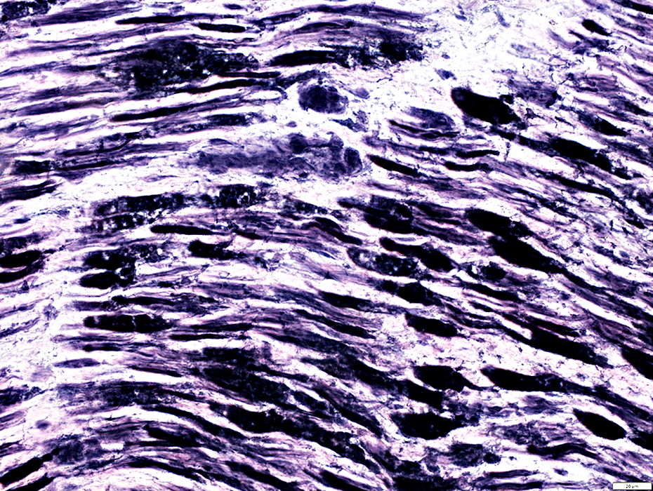
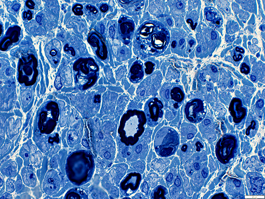 Toluidine blue stain |
Phagocytic endoneurial Schwann cells & Histiocytes contain myelin debris & lipid droplets
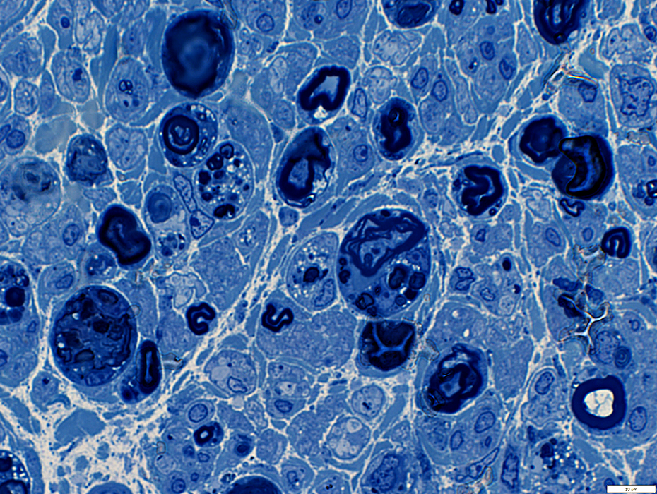 Toluidine blue stain |
 Toluidine blue stain |
|
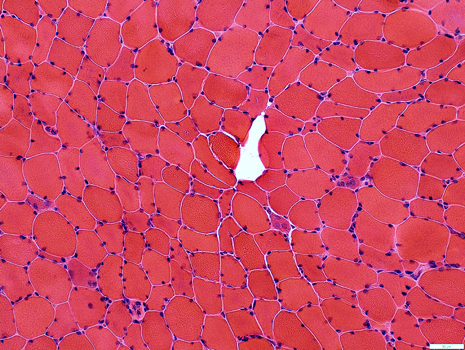
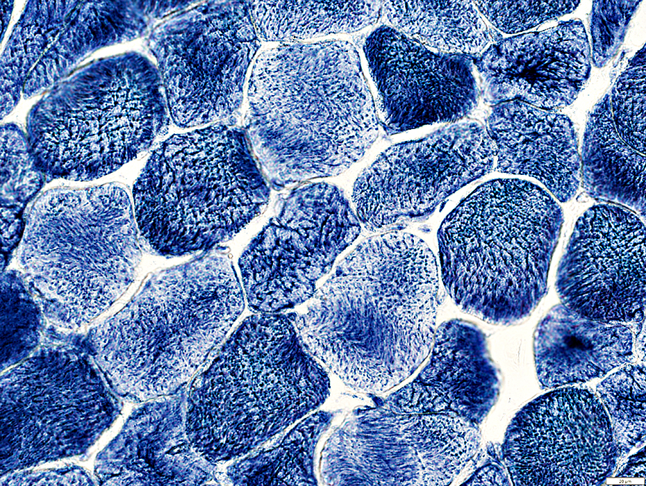
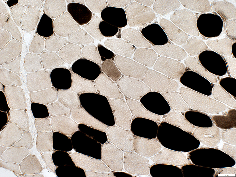
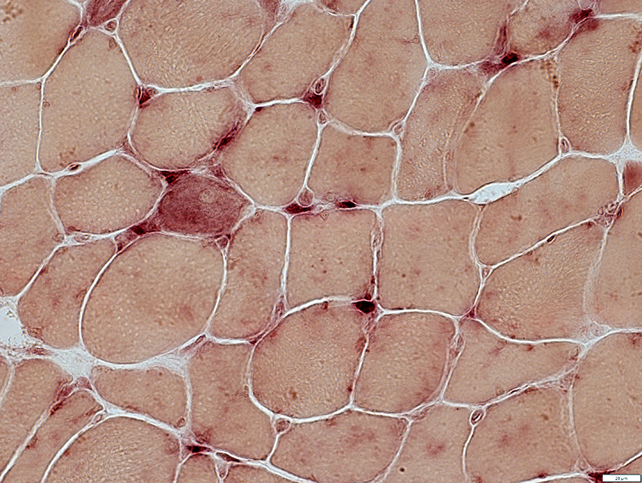
Muscle fibers: Midly small; Myonuclei are mildly large
|
|
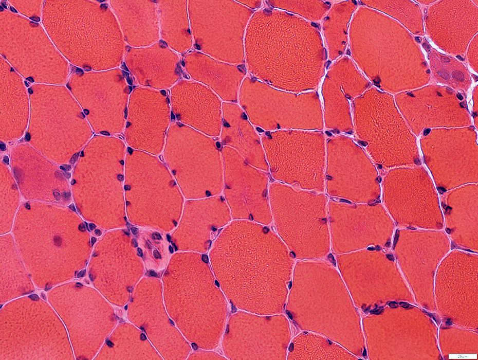
Muscle fibers: Irregular staining of cytoplasm
|
|
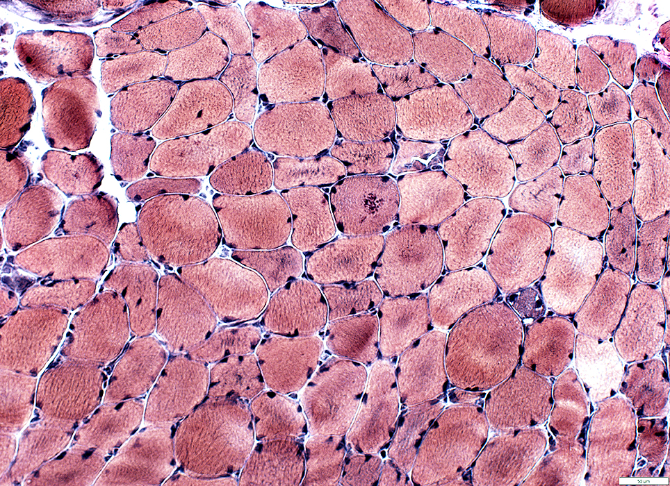
Muscle fibers: Irregular internal architecture
|
|
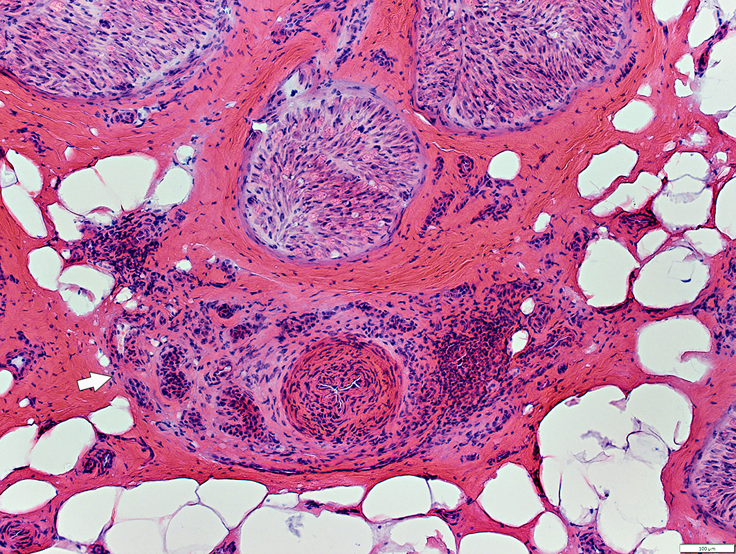
Large Vessel involvement
 H&E stain |
Epineurium & Vessel walls: May have neovascularization
 H&E stain |
NSVN: Neovascularization
 H&E stain |
 H&E stain |
NSVN: Large vessel damage
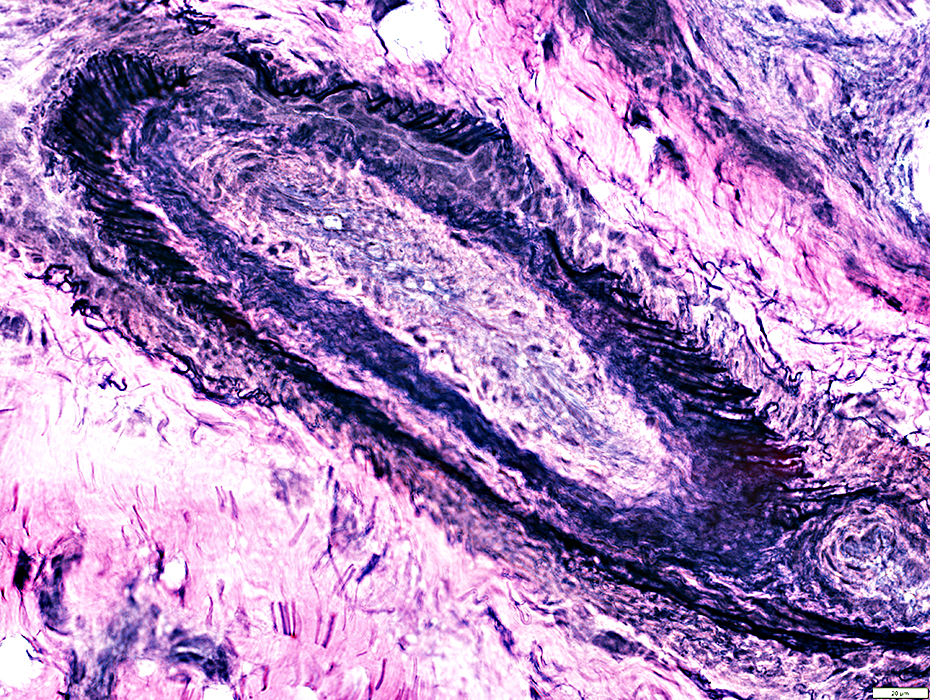 VvG/font> |
Return to Muscle biopsies
9/21/2024