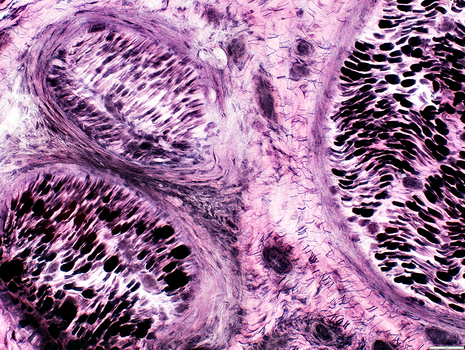
Neurolymphomatosis
|
Axon loss Cells |
Lymphoma Cells: Endoneurial & Epineurial (Perivascular)
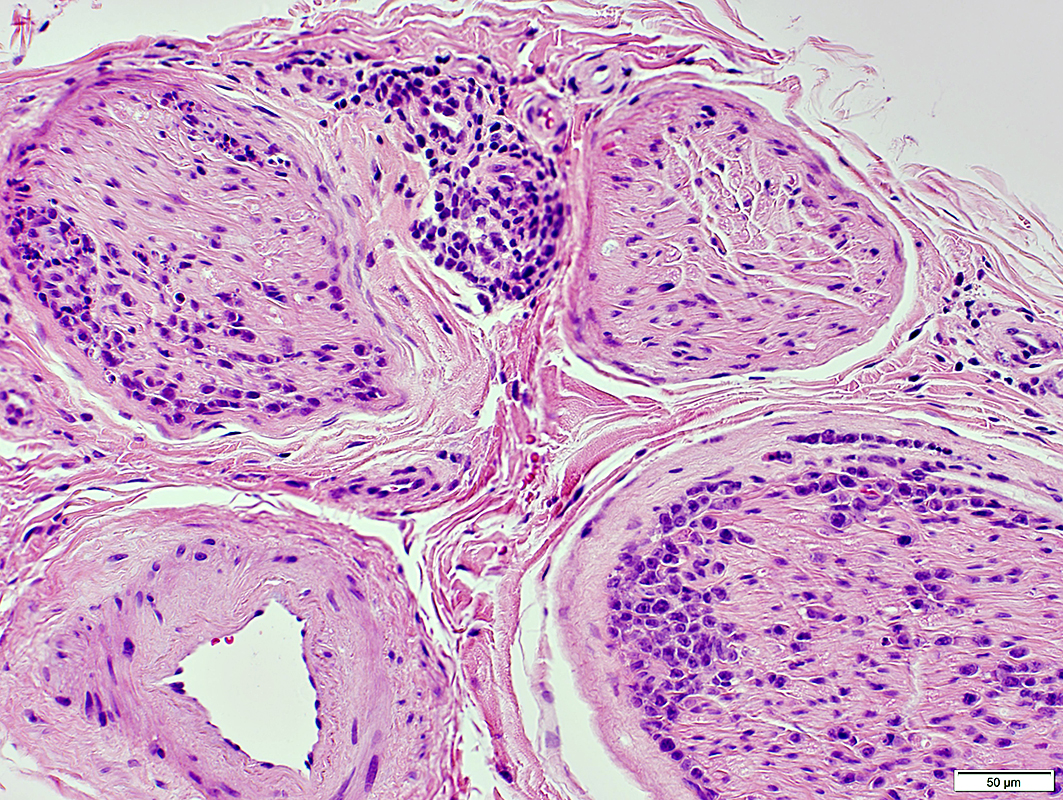 H&E stain |
Cell locations
Endoneurial: Subperineurial; Internal; Perivascular
Epineurial: Perivascular
 H&E stain |
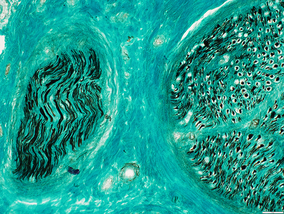
 H&E stain |

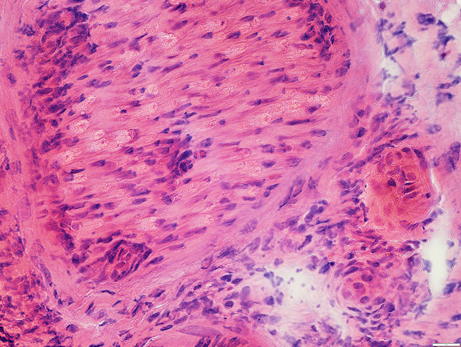 H&E stain |
 H&E stain |
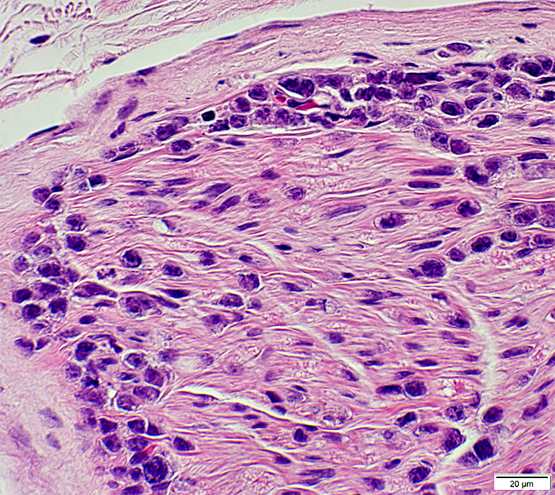
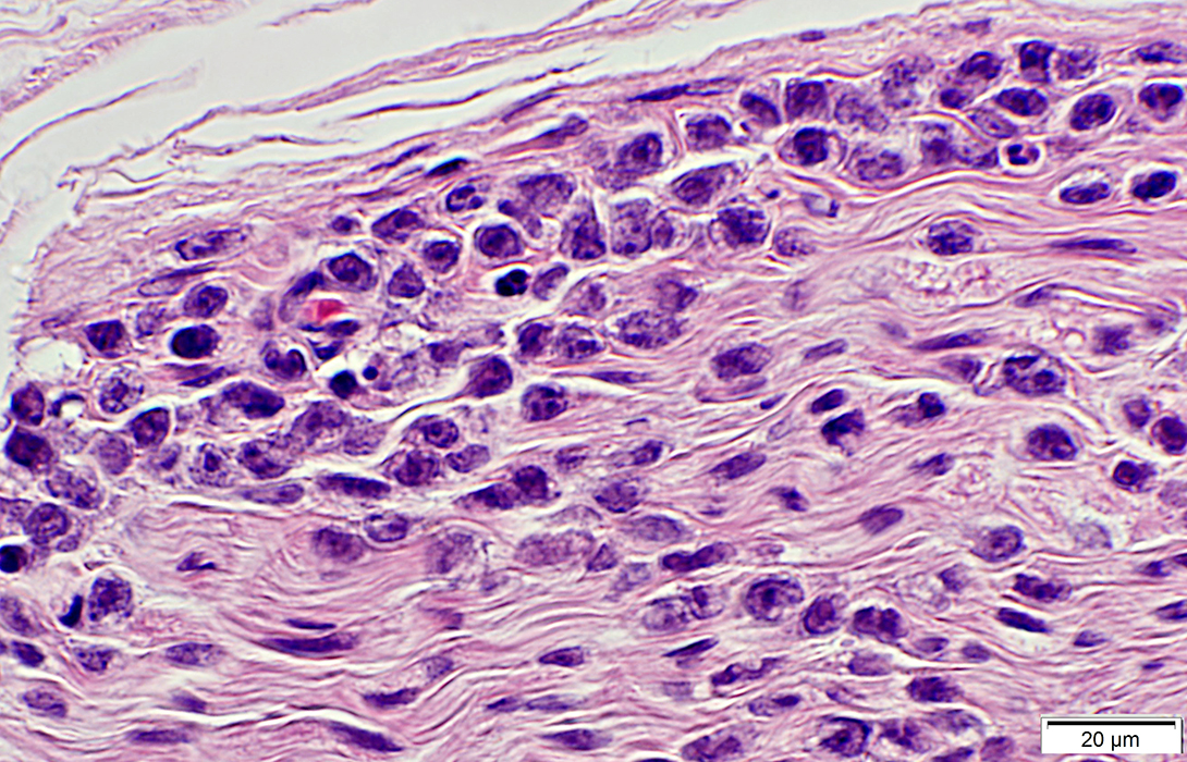
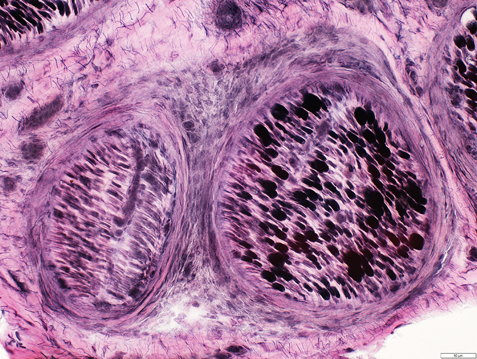
Locations: Subperineurial & Endoneurial
Cells
Nuclei: Large; Atypical; Nucleoli, One or more
Cytoplasm: Minimal
Mitotic activity
 H&E stain |
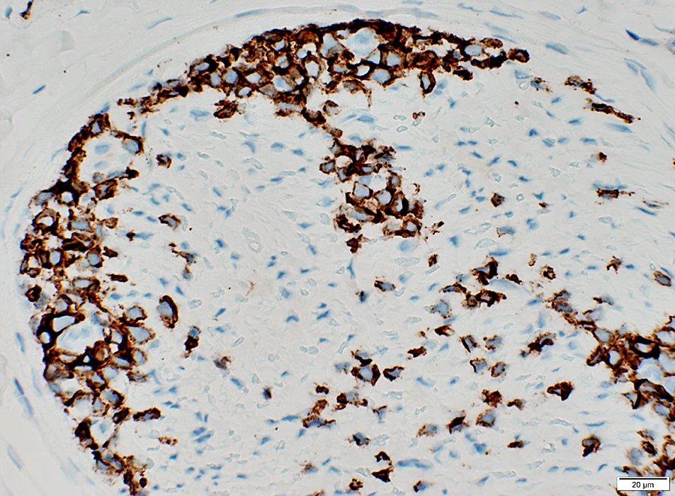
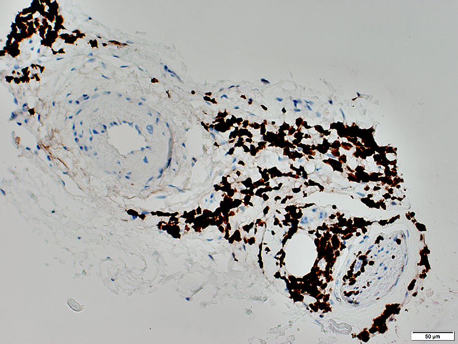
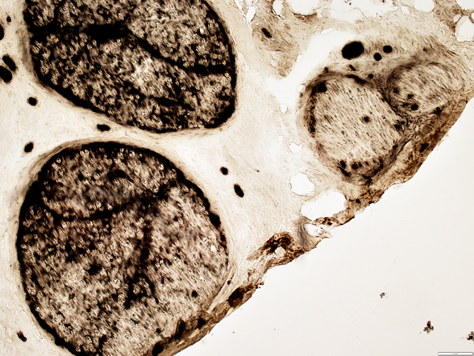
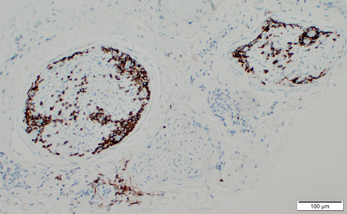 CD20 stain |
B-cells: CD20 stain
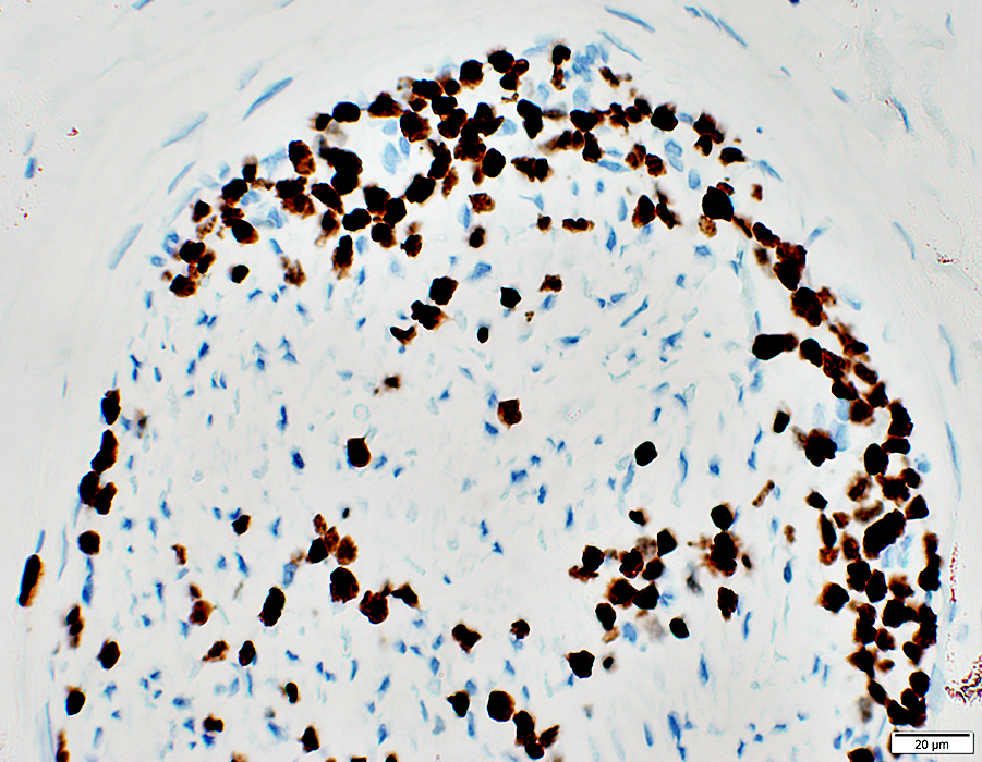
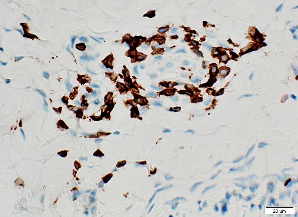
Proliferating: Ki67 stain
 Ki67 stain |
 CD20 stain |
B-cells: CD20 stain
Proliferating: Ki67 stain
 Ki67 stain |
 CD20 stain |
B-cells: CD20 stain
Proliferating: Ki67 stain
Ki67 stain |
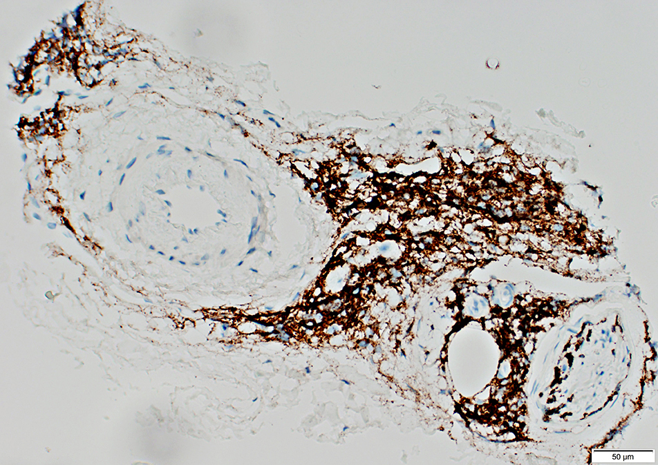
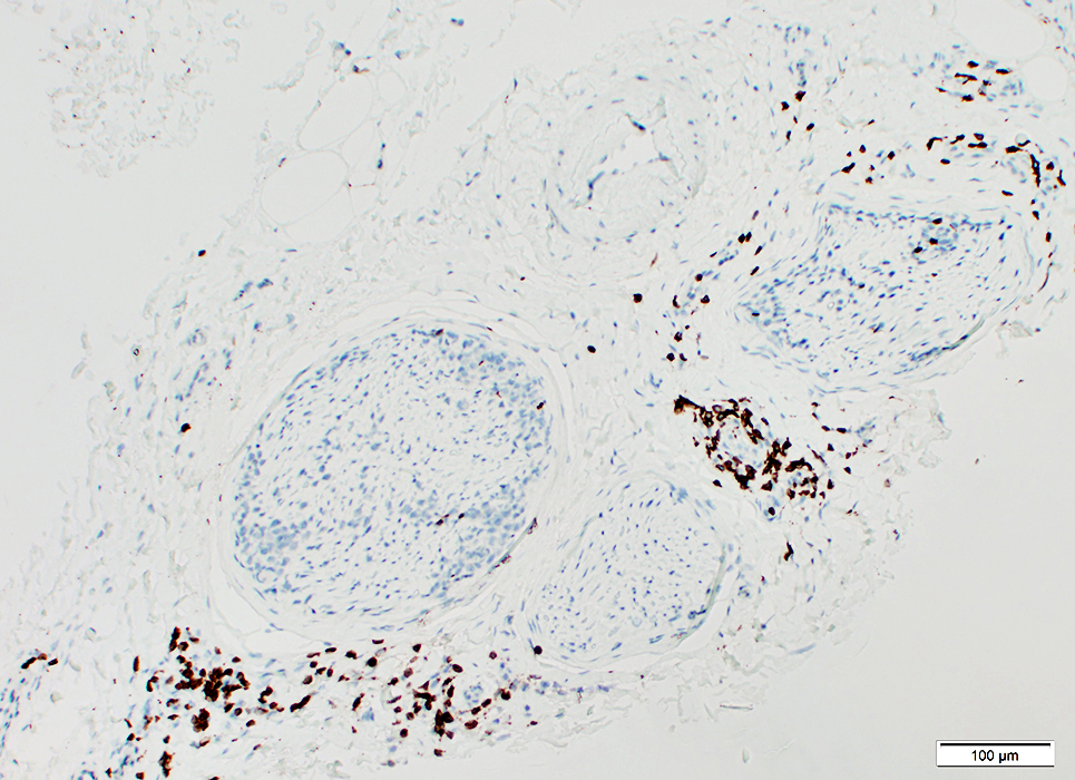 CD3 stain |
T-cells: CD3 stain
Not neoplastic
 CD3 stain |
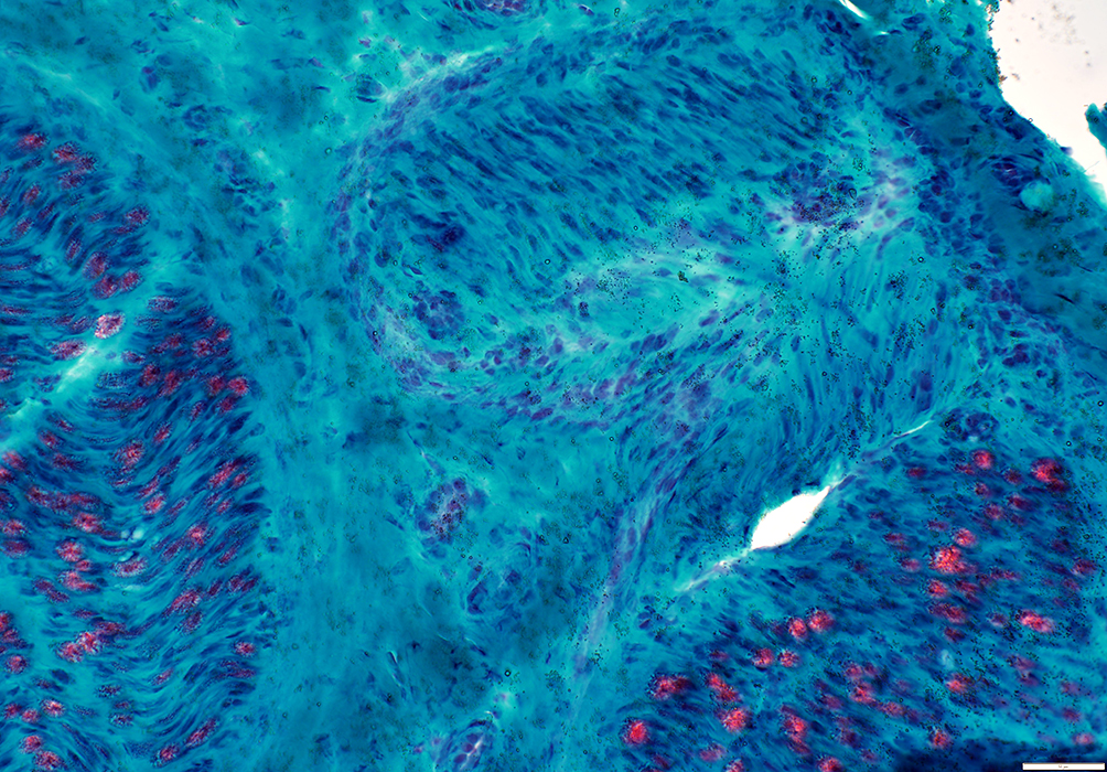
Axon Loss: Differential fascicular
Patchy: More in some fascicles than others
|
|
|
More in some fascicles than others
Large > Small
|
ATPase stain: Lost in some fascicles but not others
|
Axon loss, Chronic: Regions of P0 staining with no axons (Arrow)
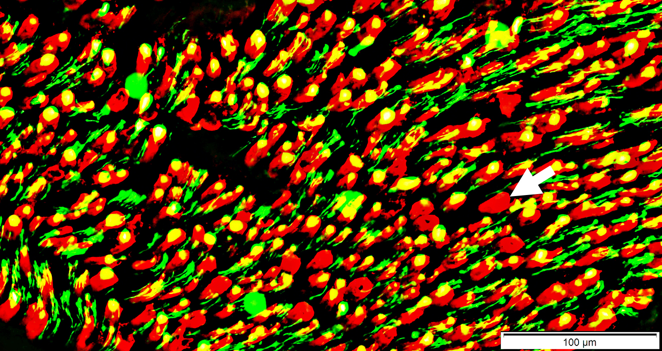 Neurofilament (Axons) = Green & Yellow; P0 = Red |
Return to: Lymphoma
Return to: Neuromuscular Home Page
References
1. Diagn Cytopathol 2010;38:208-212
3/12/2019