Mitochondrial disease pathology: Single, Large mtDNA Deletions (KSS & PEO)
|
Kearns-Sayre PEO Ultrastructure Also see Mitochondrial disease ultrastructure |
mtDNA deletion: Very large
Size & Location: 5 kB; ~8,300 to 14,300; ATPase6 to ND5
Clinical: Kearns-Sayre Syndrome
|
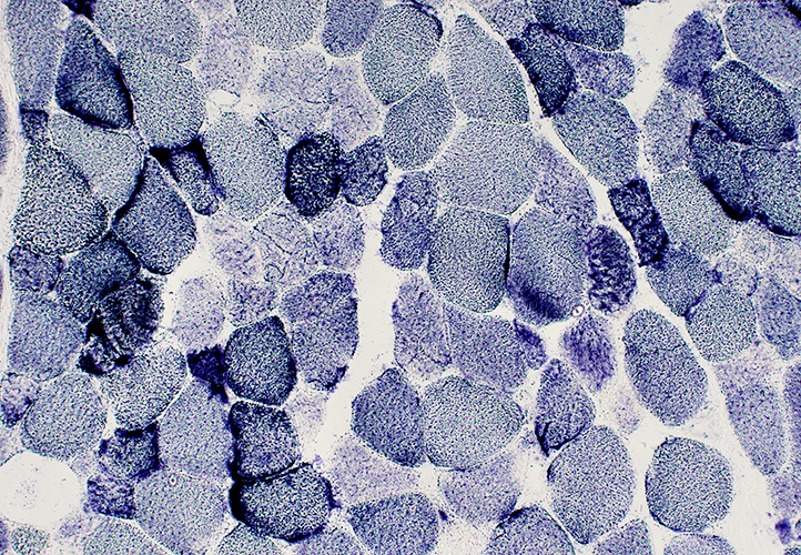
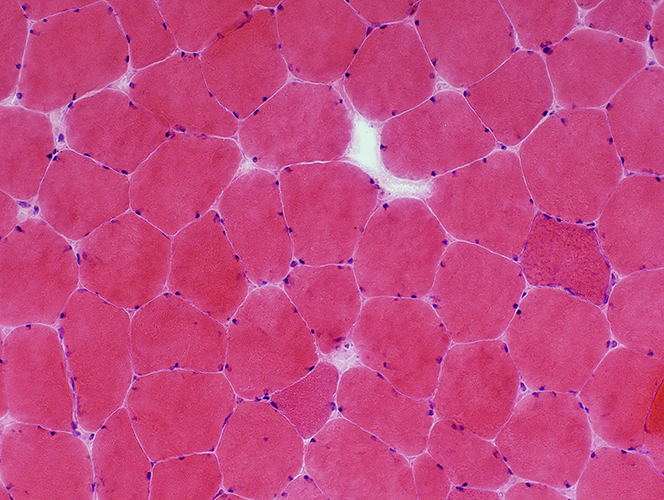
Muscle Fiber Morphology Scattered muscle fibers with Varied size Irregular internal architecture Large nuclei Basophilic cytoplasm Endomysial connective tissue: Normal 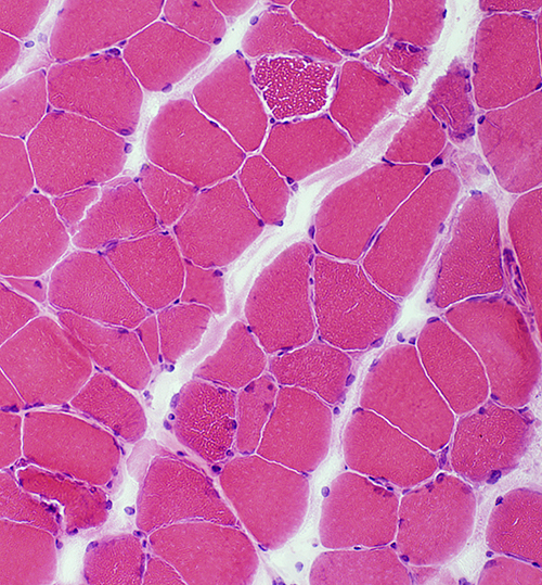 H&E stain |
 H&E stain |
|
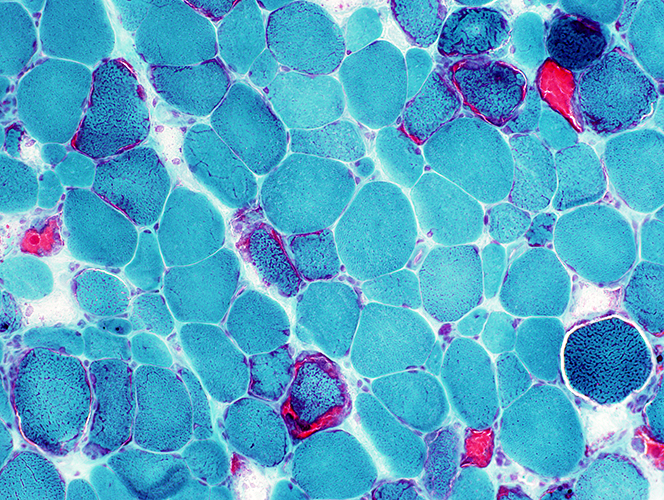
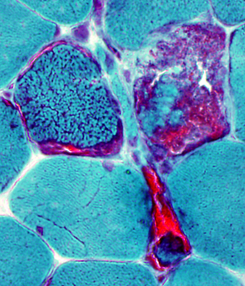
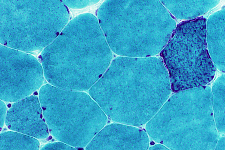
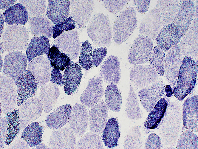
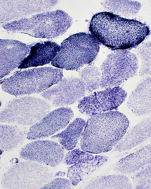
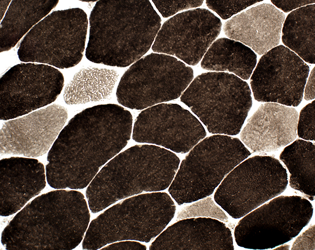
| Ragged red muscle fibers Mitochondrial proliferation: Varied Degrees Muscle fiber cytoplasm Entirely replaced by mitochondria (Red) in some segments Coarse internal architecture in other segments |
 Gomori trichrome |
 Gomori trichrome |
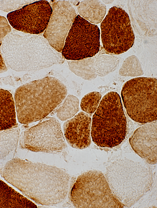
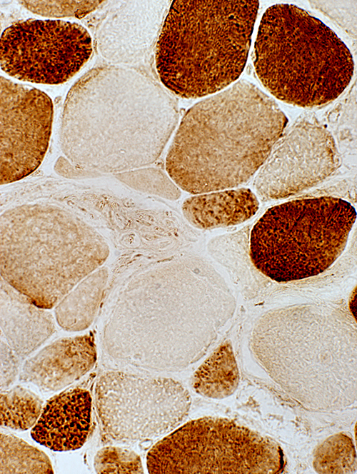
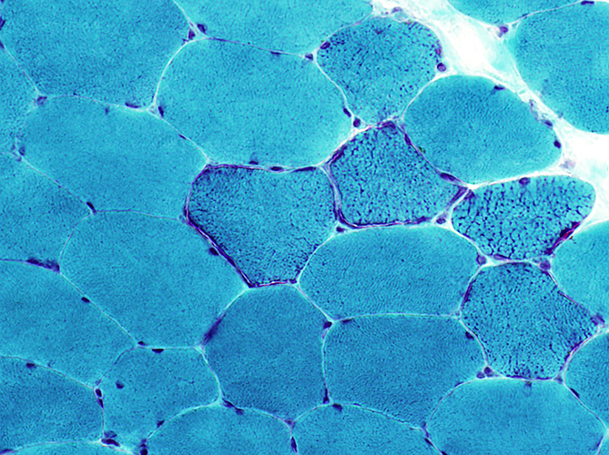
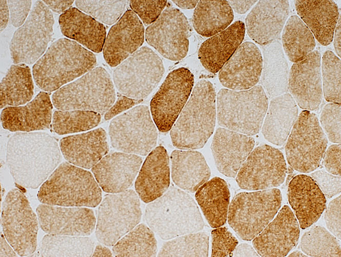
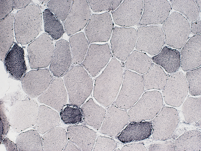
 SDH stain Mitochondrial proliferation: SDH+ muscle fibers with variably increased degrees of staining 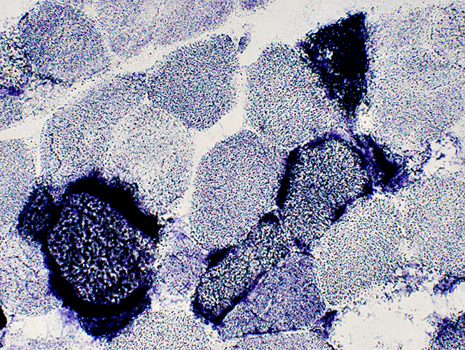 SDH stain |
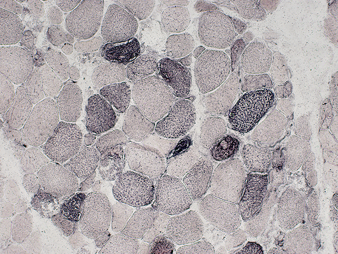
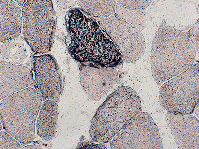
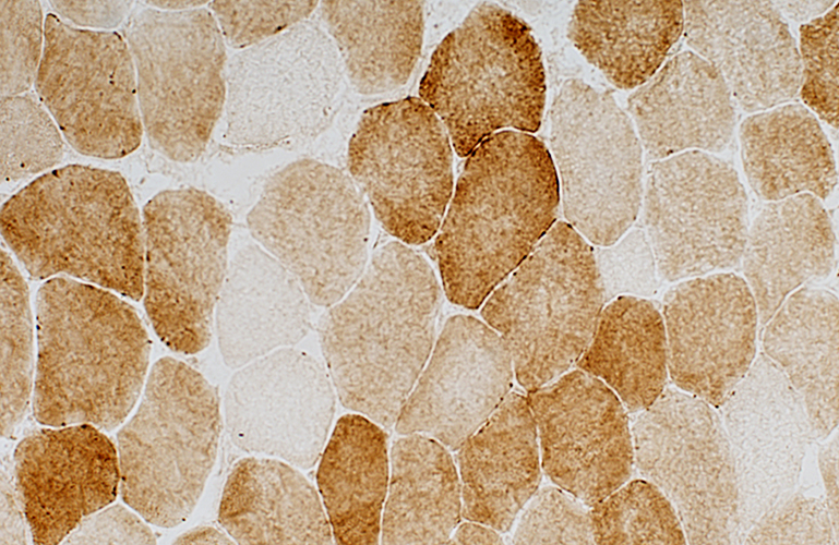
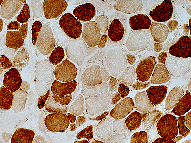 COX stain Cytochrome oxidase: Scattered muscle fibers with reduced or increased staining |
|
 COX stain |
 COX stain |
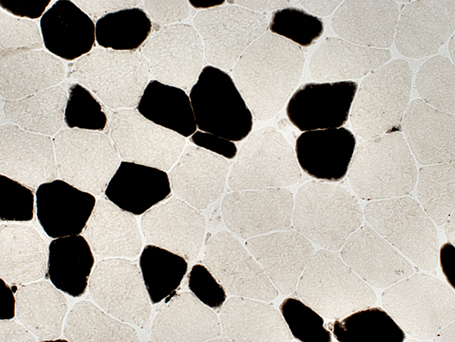
 Sudan black |
| Lipid: Increased size of lipid droplets in scattered muscle fibers |
 Sudan black |
mtDNA deletion: Moderately large
Size & Location: ND4 to ND5
Clinical: PEO
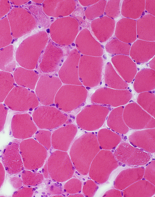
 H&E |
|
Muscle fiber size: Mild variation Basophilic mucle fibers: Occasional; Intermediate sized |
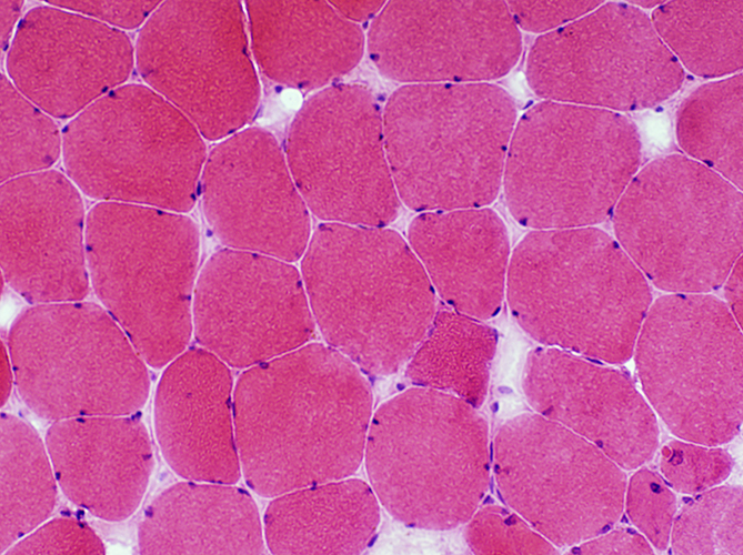 H&E |
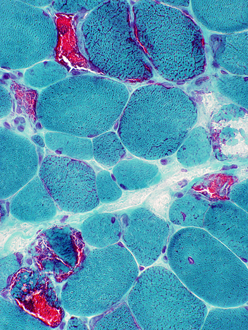
 Gomori trichrome |
| Ragged red muscle fibers Mitochondrial proliferation: Mild Muscle fiber cytoplasm Coarse internal architecture in some segments |
 Gomori trichrome |
 SDH stain | |
|
Mitochondrial proliferation SDH+ muscle fibers with variably increased degrees of staining 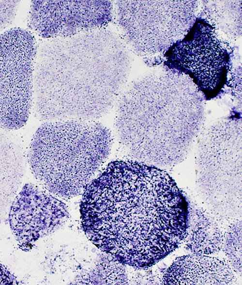 SDH stain |
 SDH stain |
 COX |
|
Cytochrome oxidase: Scattered muscle fibers with reduced staining |
 COX |
 Sudan |
|
Scattered muscle fibers with increased staining |
 ATPase pH 9.4 |
|
Fiber types: Type 2 predominance; Smallest fibers are type 1; No 2 C fibers |
 ATPase pH 4.3 |
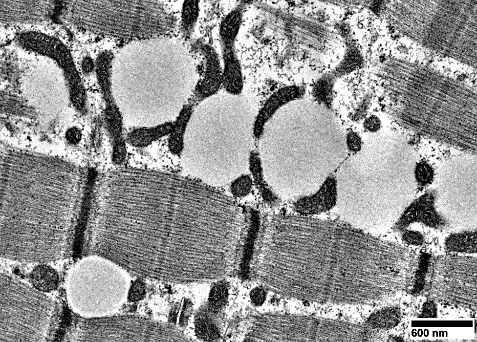
PEO: Muscle Ultrastructure
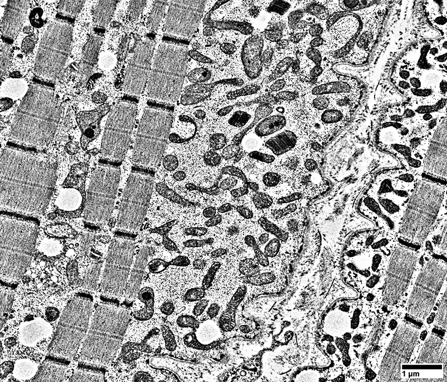 From: Robert Schmidt |
Proliferation of mitochondria in subsarcolemmal regions
Mitochonria have: Varied sizes & Irregular shapes
Lipid droplets are associated with some mitochondria
Mitochondria are separated by glycogen granules
 From: Robert Schmidt |
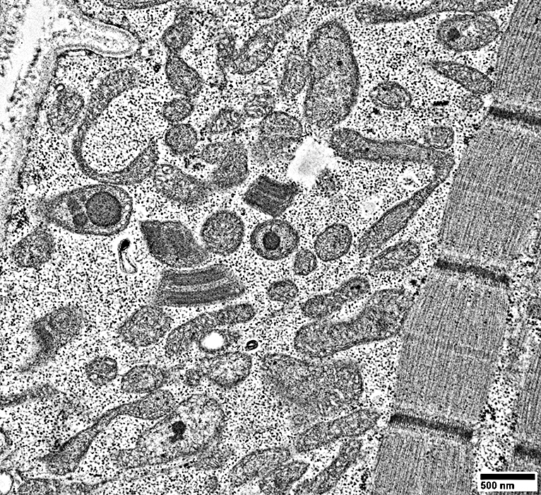
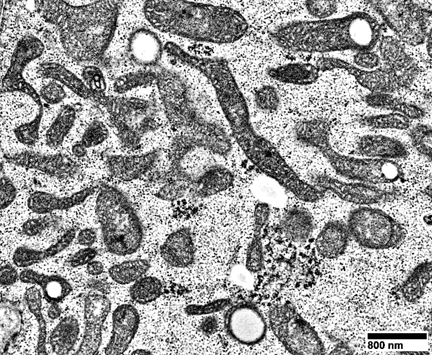
PEO: Mitochondria
Shape: A mitochondrion has a long process
 From: Robert Schmidt |
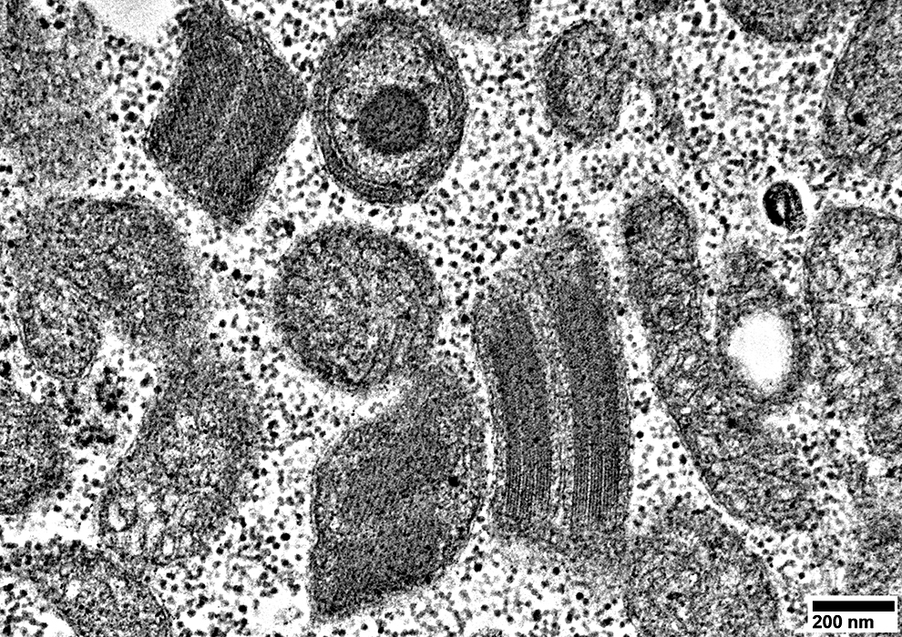
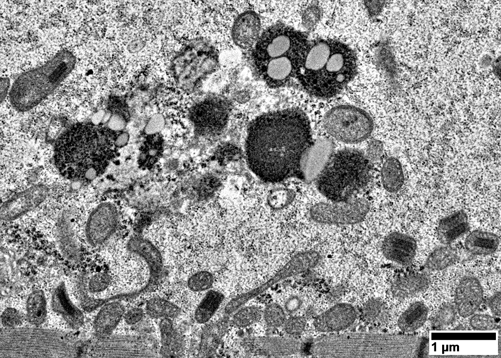
PEO: Crystalloid Inclusions (Parking lot bodies)
|
 From: Robert Schmidt |
Mitochondria may cluster around lipid droplets (Above)
Lipid droplets appear to be within mitochondria (Below)
 From: Robert Schmidt |
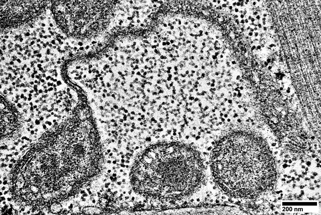
PEO: Lipopigment agggregates
 From: Robert Schmidt |
Return to Mitochondrial pathology
Return to Mitochondrial syndromes
Return to Muscle biopsies
12/25/2022