Macrophagic Myofasciitis (MMF)
|
Adult Histochemistry Child Aluminum Ultrastructure |
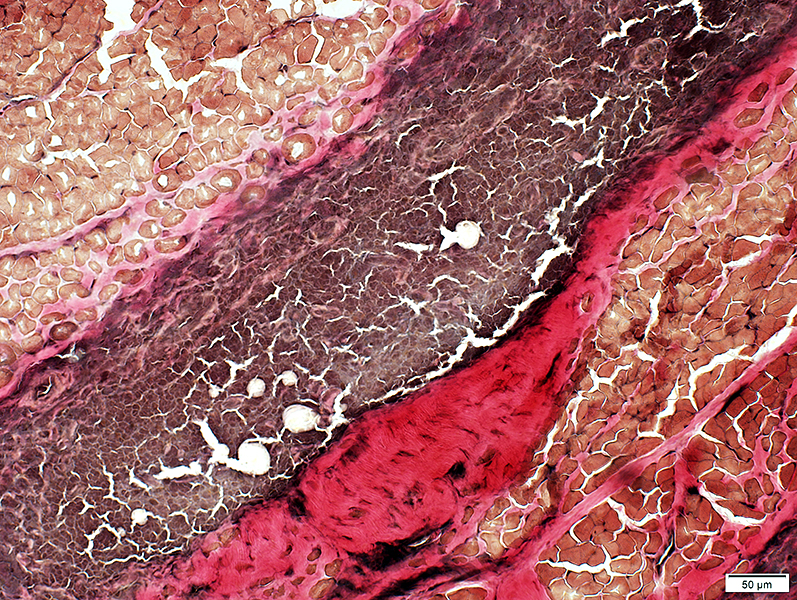
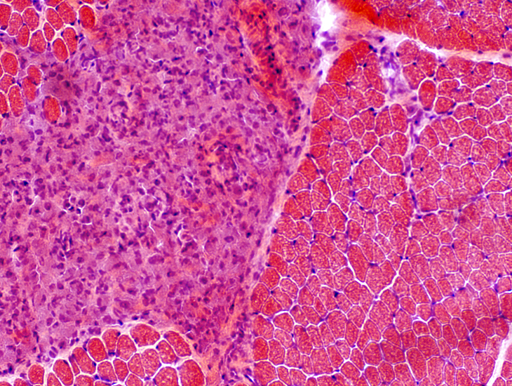
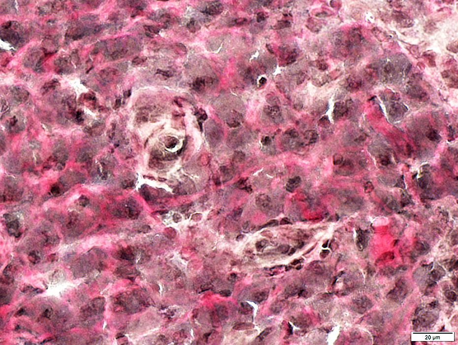
MMF: Adult
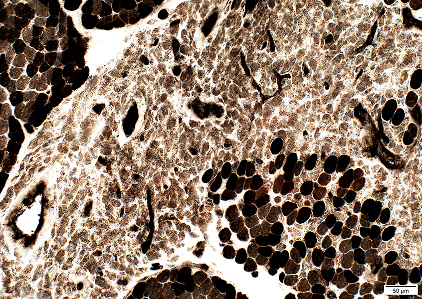
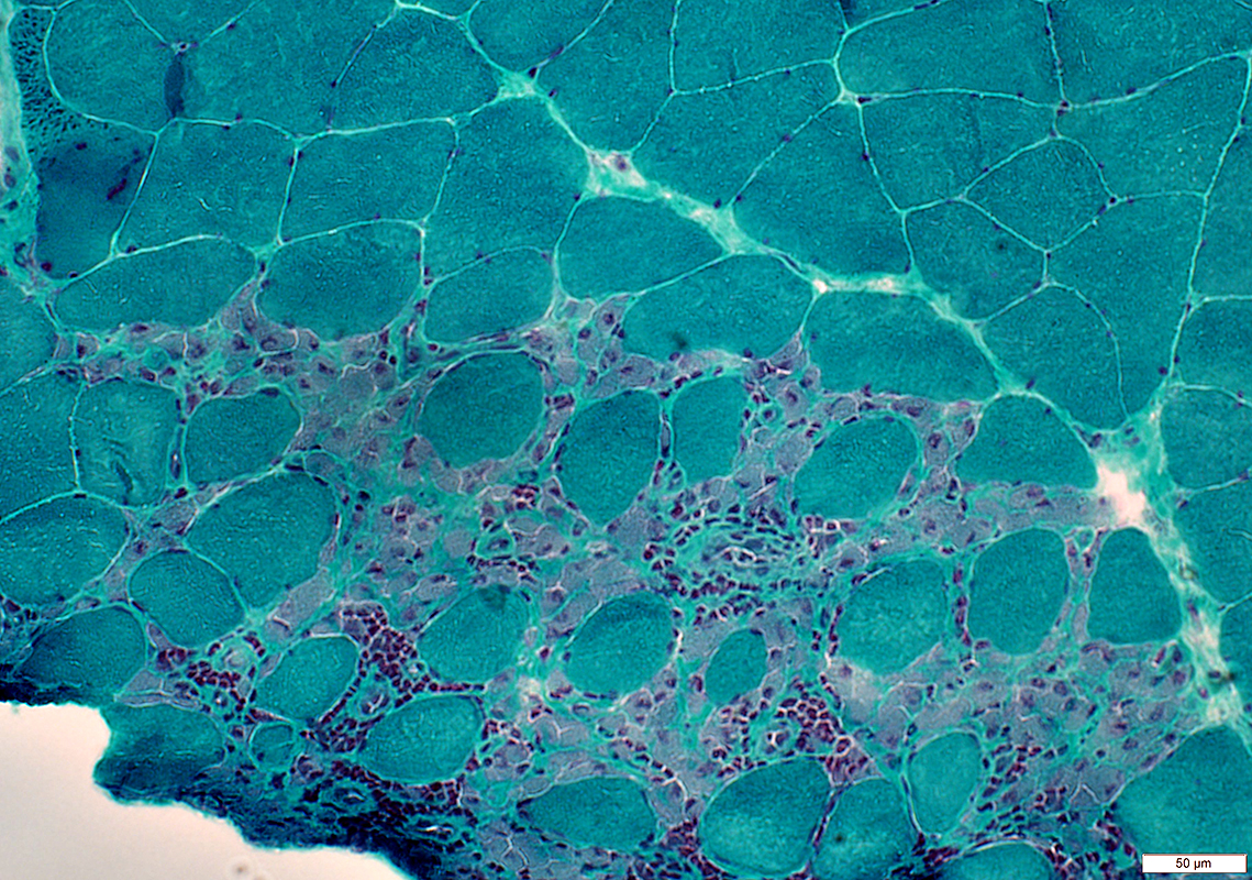 Gomori trichromestain |
Location: Endomysium
Cell features
Large
Abundant cytoplasm
Contain: Aluminum (Morin stain); Inclusions
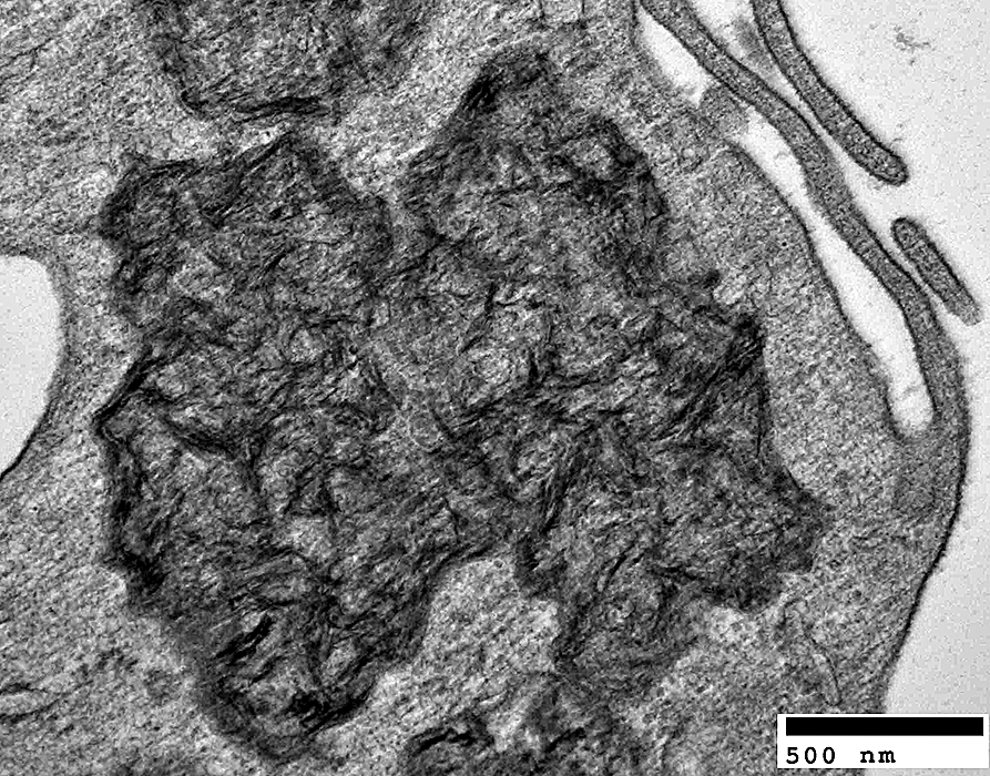
Ultrastructure
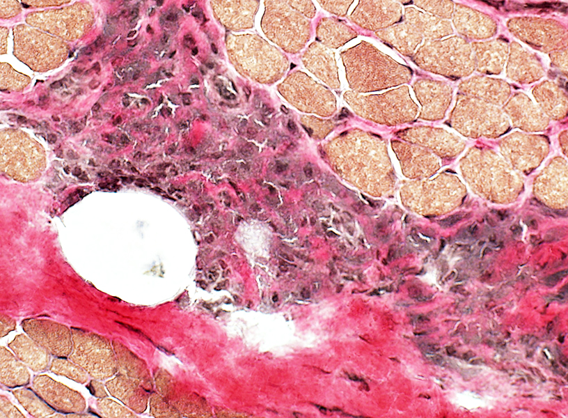
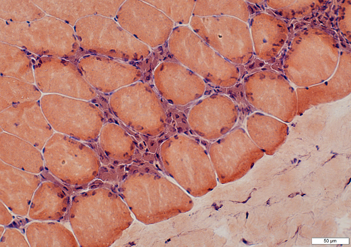 VvG trichromestain |
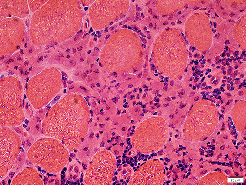 H&E stain |
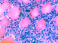 H&E stain 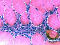 H&E stain |
|
 H&E stain |
Nuclei: Large; Irregular shapes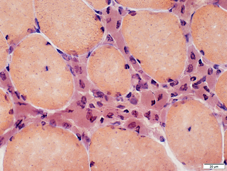 Congo red stain |
|
Cells in infiltrates
Mostly histiocytic Stain strongly with: Esterase & Acid phosphatase. |
|
 Esterase stain |
 Esterase stain 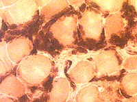 Esterase stain |
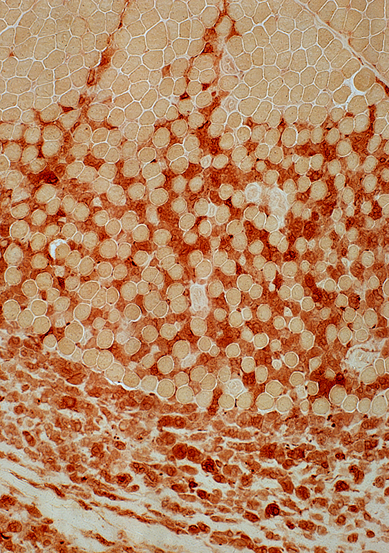
 Acid phosphatase stain |
|
MMF cells: Confluent, strong acid phosphatase staining; Individual cell outlines in foci not visible |
 Acid phosphatase stain |
 VvG stain |
|
MMF cells contain cytoplasmic lipid: Stains with VvG and Sudan |
 Sudan stain |
PAS stain |
|
| Cells also stain moderately with PAS | |
MMF: Children
 H&E stain |
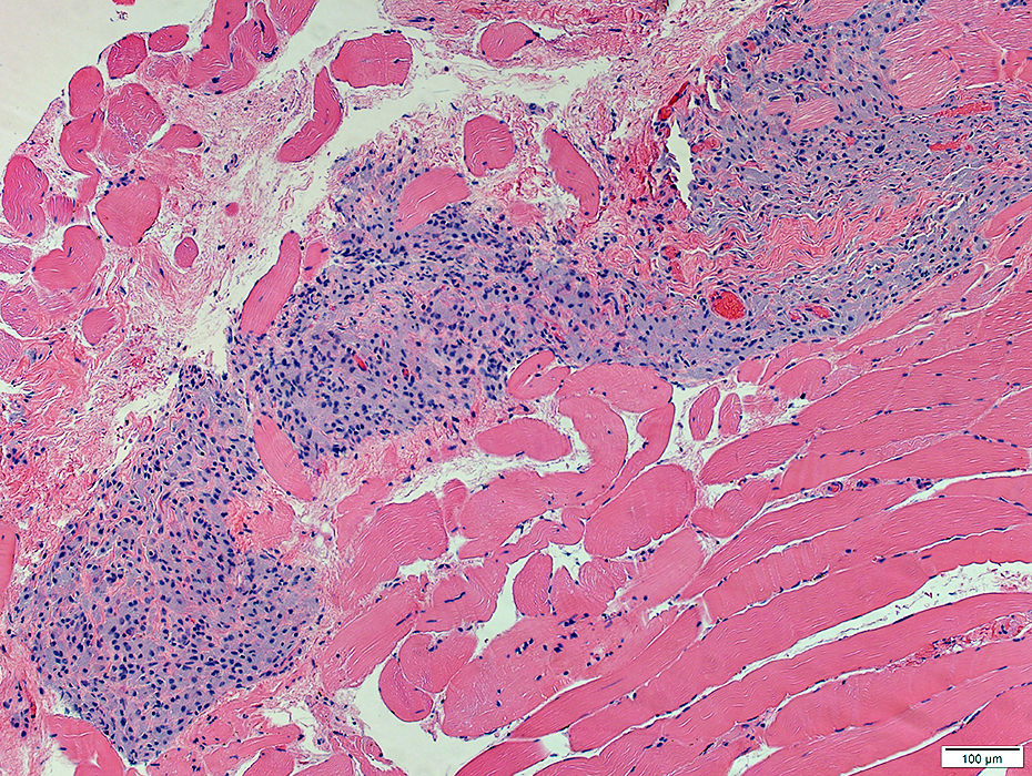
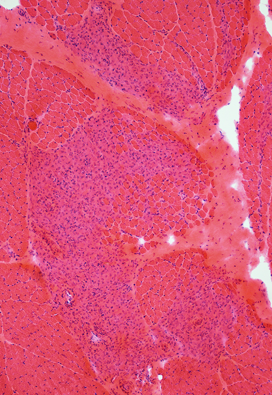
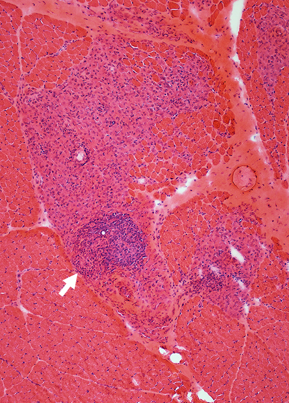
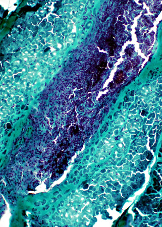
MMF: Lesions
Locations: Perimysium & EndomysiumCells: Mostly large & Histiocytic; May contain sub-foci of non-histiocytic cells (Arrow)
 H&E stain |
 H&E stain |
 H&E stain |
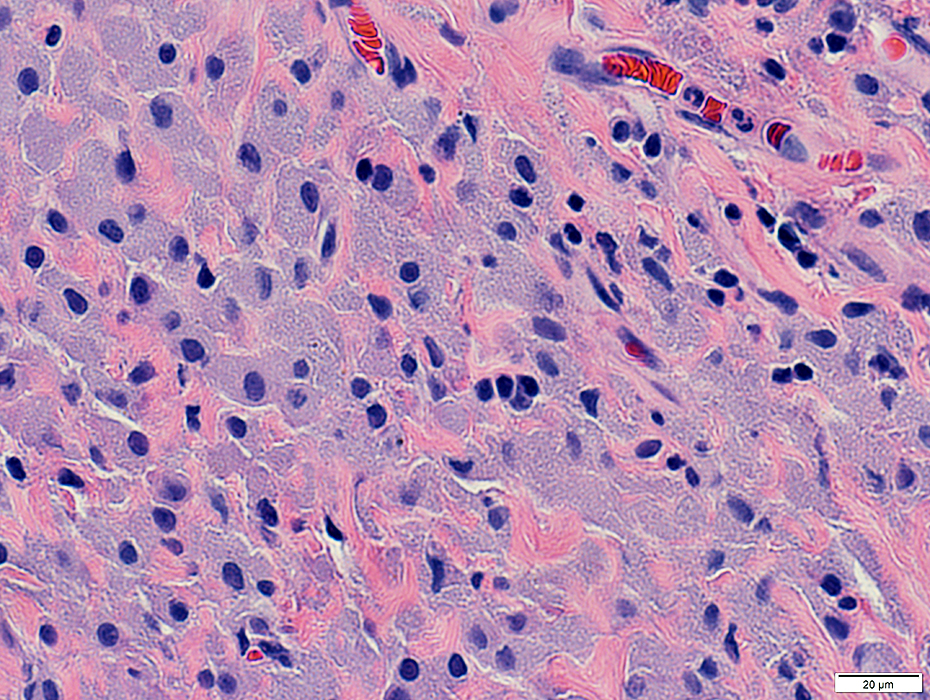
MMF: Clusters of Histiocytic cells
 VvG stain |
MMF: Smaller lesion confined to perimysium
 VvG stain |
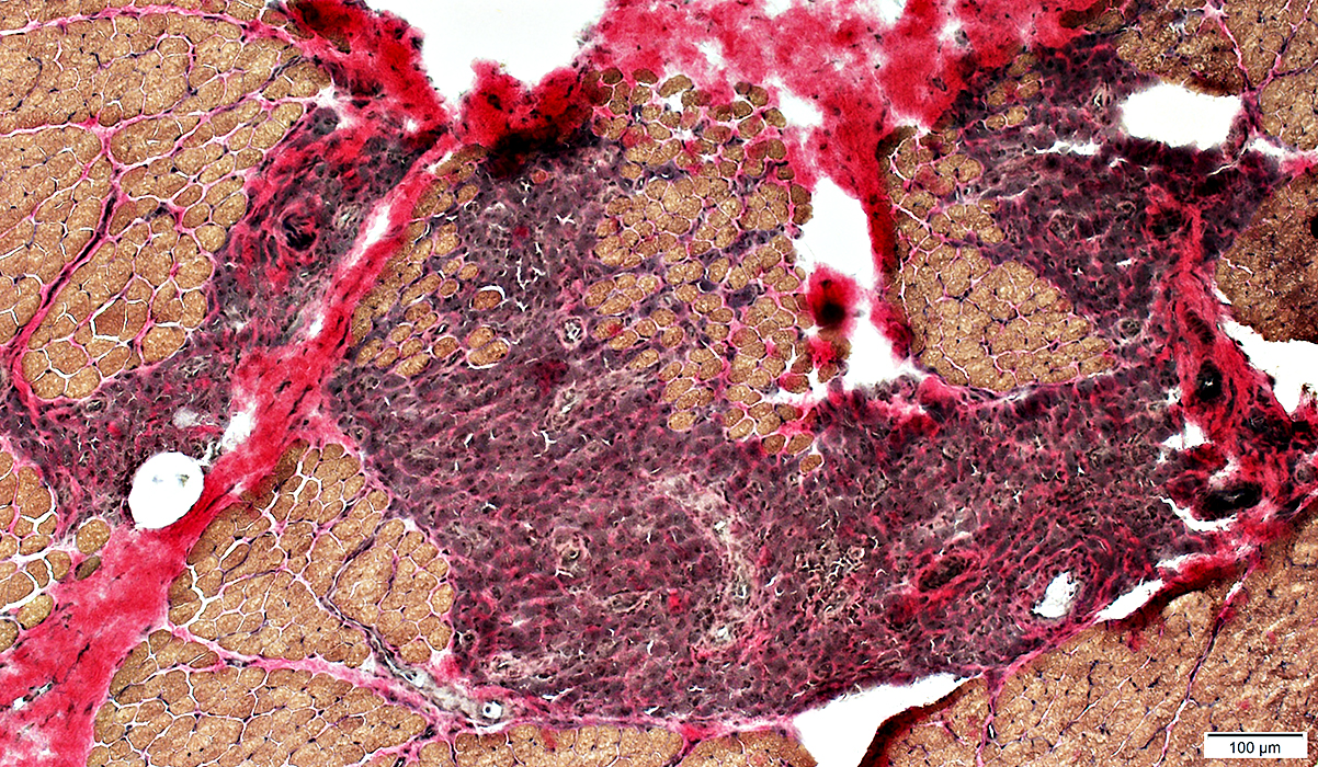
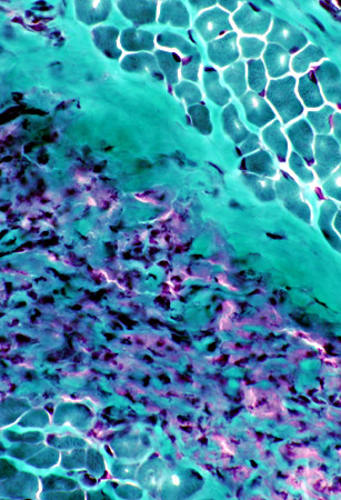
MMF: Vascularization
Cell foci often contain smaller vessels, some with thick basal lamina
 VvG stain |
Vessels in perimysial cell foci stain for Ulex
 UEAI stain |
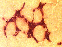
 Alkaline phosphatase stain |
 ATPase pH 9.4 stain |
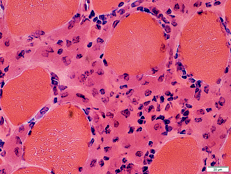
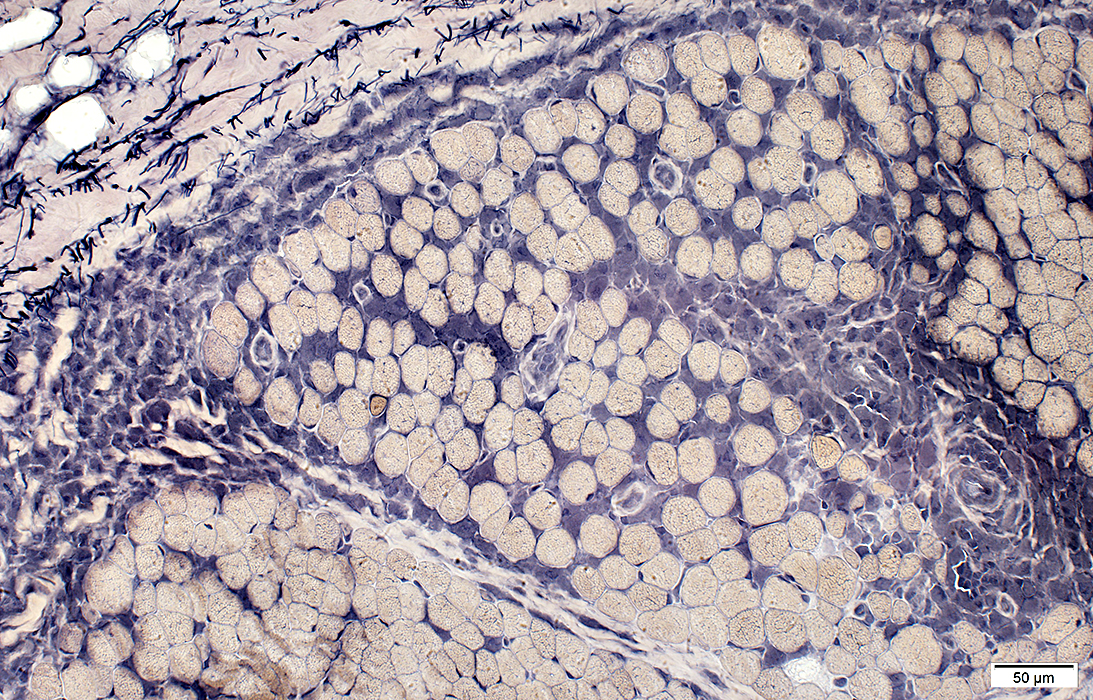
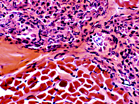
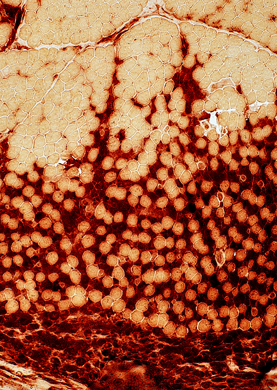
MMF: Cell FociLarge cells with abundant cytoplasmLocation: Perimysium > Endomysium 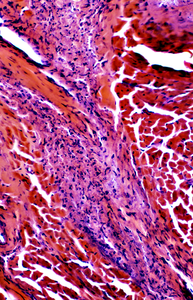 H&E stain |
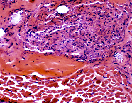  H&E stain |
 H&E stain MMF Cell Foci Large cells with abundant cytoplasm Location: Perimysium > Endomysium |
 Gomori trichrome stain |
 Congo red stain |
Cells: Large
Nuclei: Large; Irregular shapes
 VvG stain |
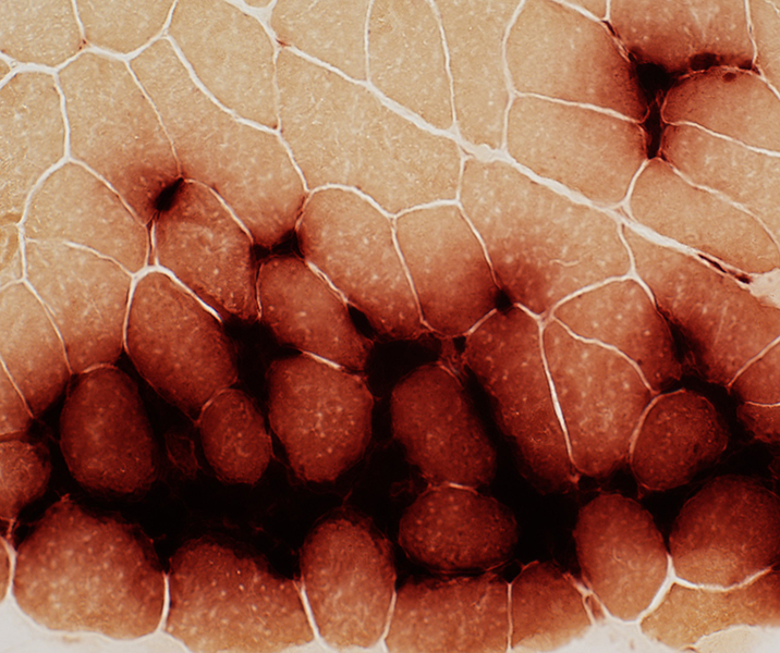
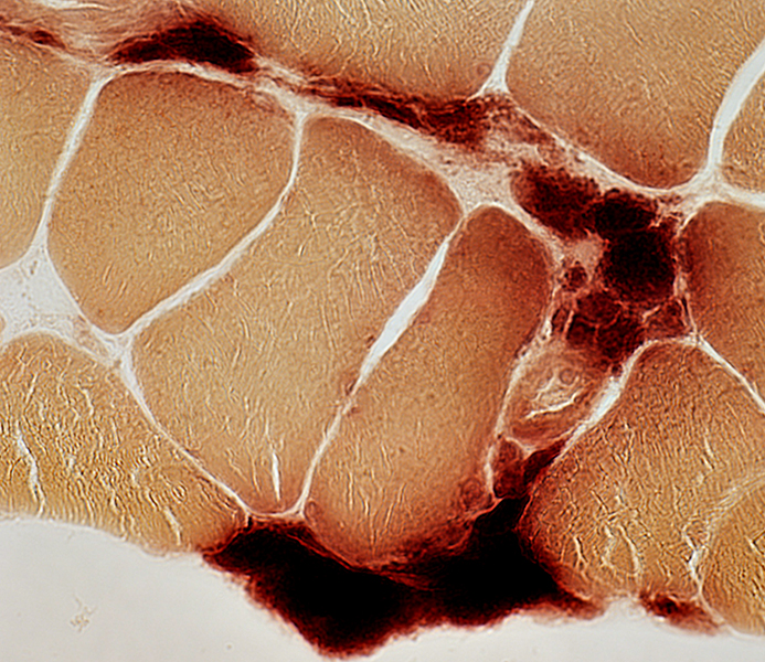
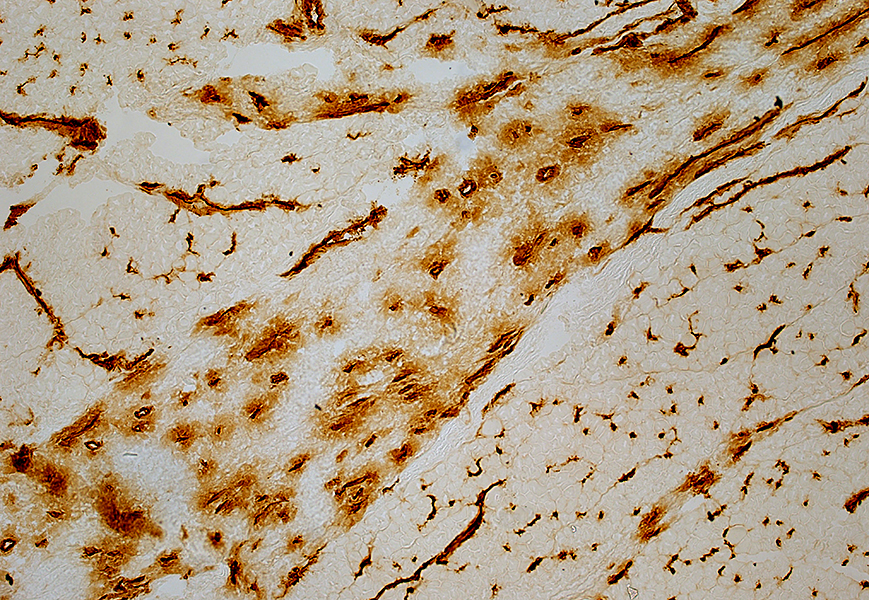
Histiocytic cellsAcid phosphatase positiveConfluent staining 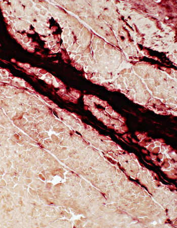 Acid phosphatase stain |
|
Histiocytic cells:
May extend from perimysium into endomysium
 Acid phosphatase stain |
 Esterase stain |
|
MMF cells in perimysium: Contain lipid Dark on Sudan Gray on VvG 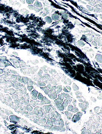 Sudan black |
 VvG |
MMF perimysial cells: May be PAS positive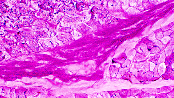 PAS stain |
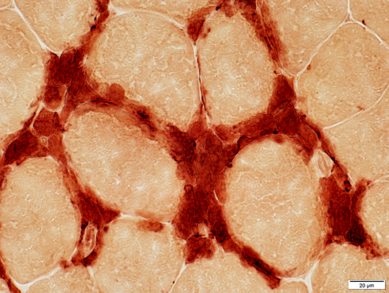
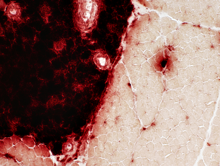
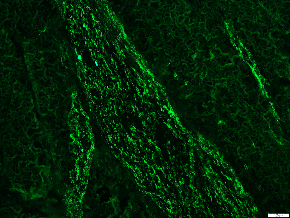
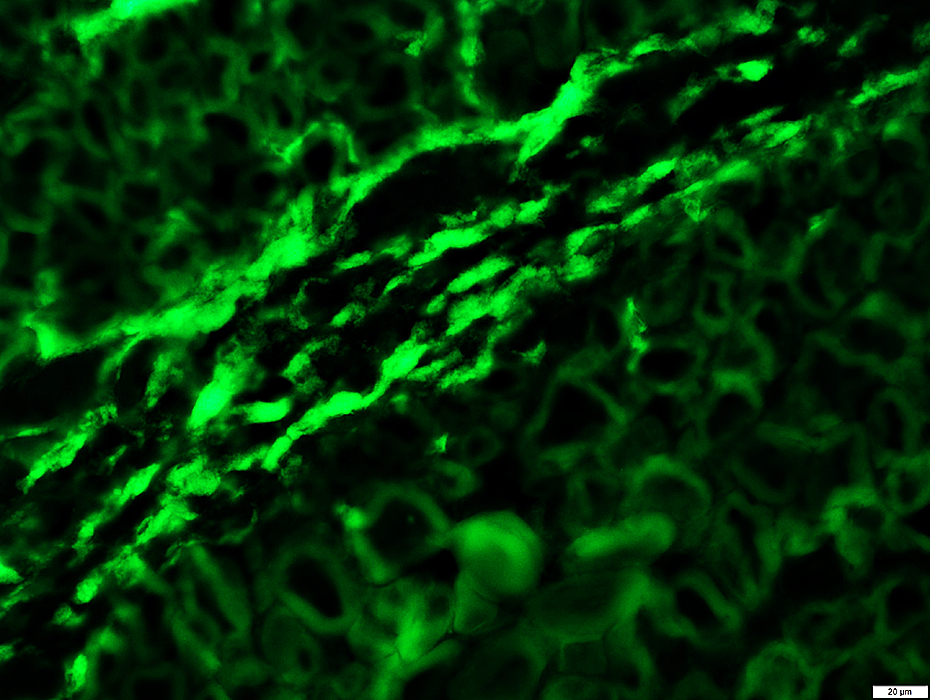
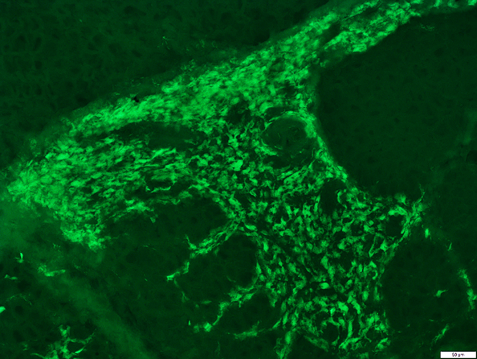
MMF: MHC Class I
Cells in foci: Strong stainingMuscle fibers: Upregulation by morphologically normal fibers
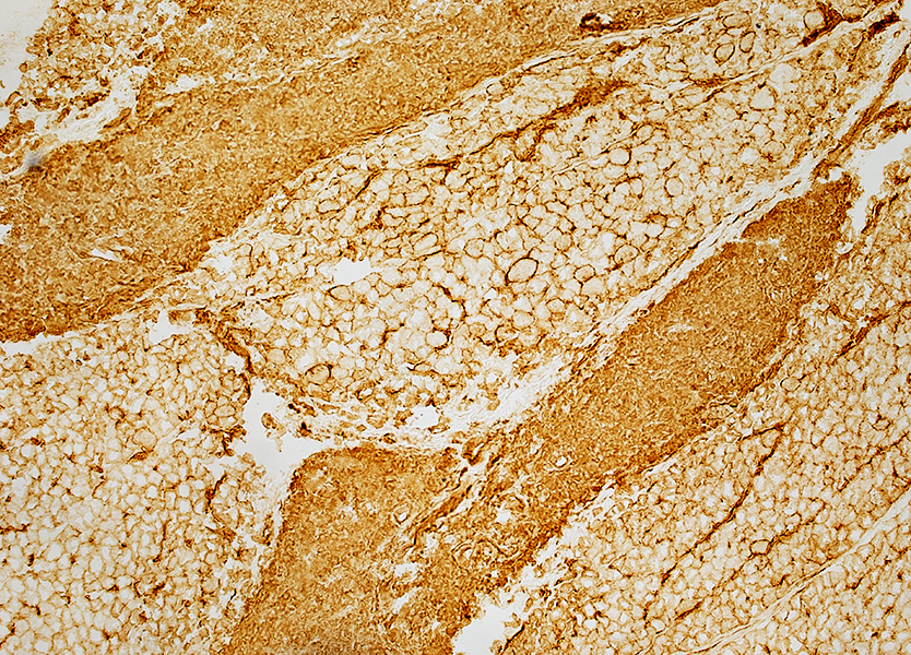 MHC Class I stain |
MMF: Cell Types
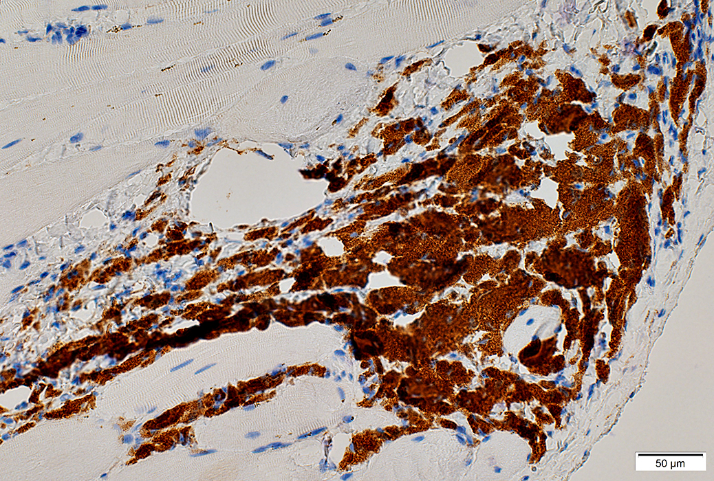 CD68 stain |
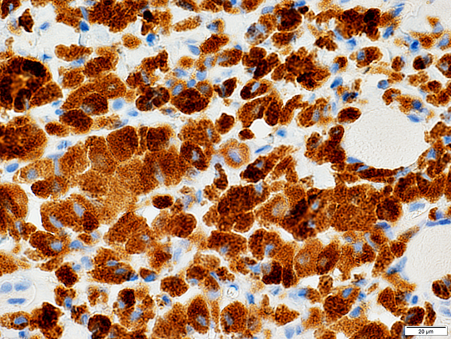 CD68 stain |
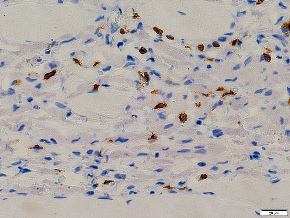 CD3 stain |
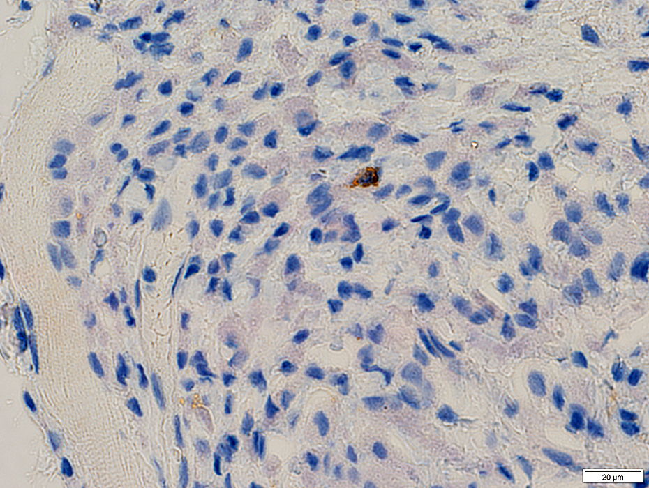 CD20 stain |
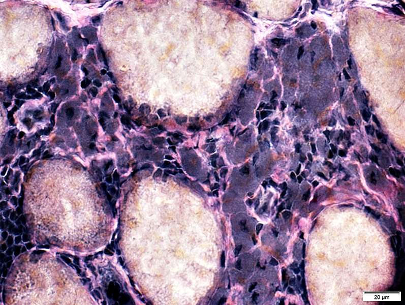
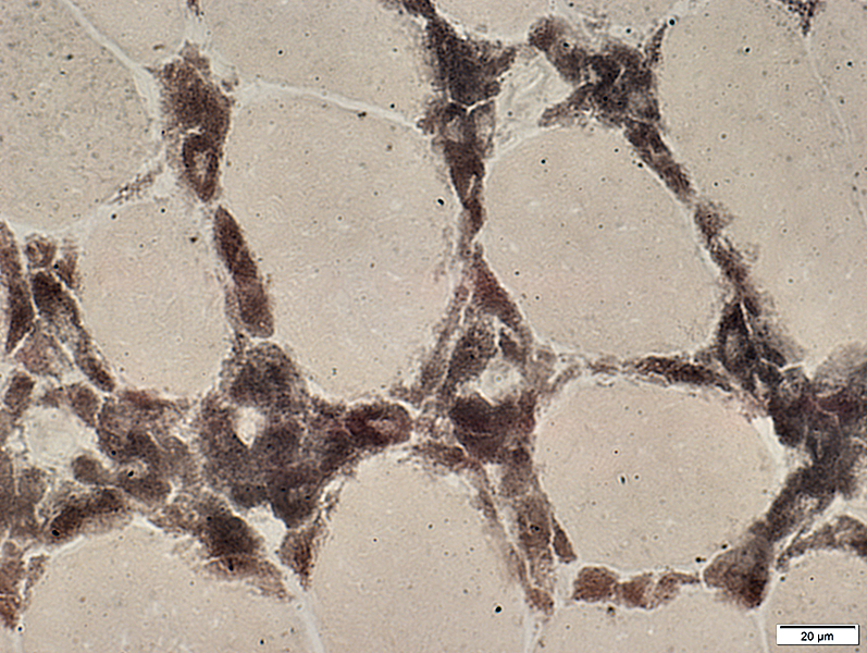
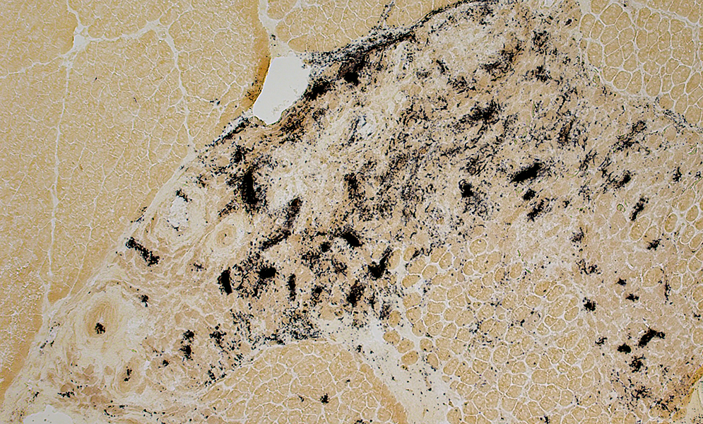
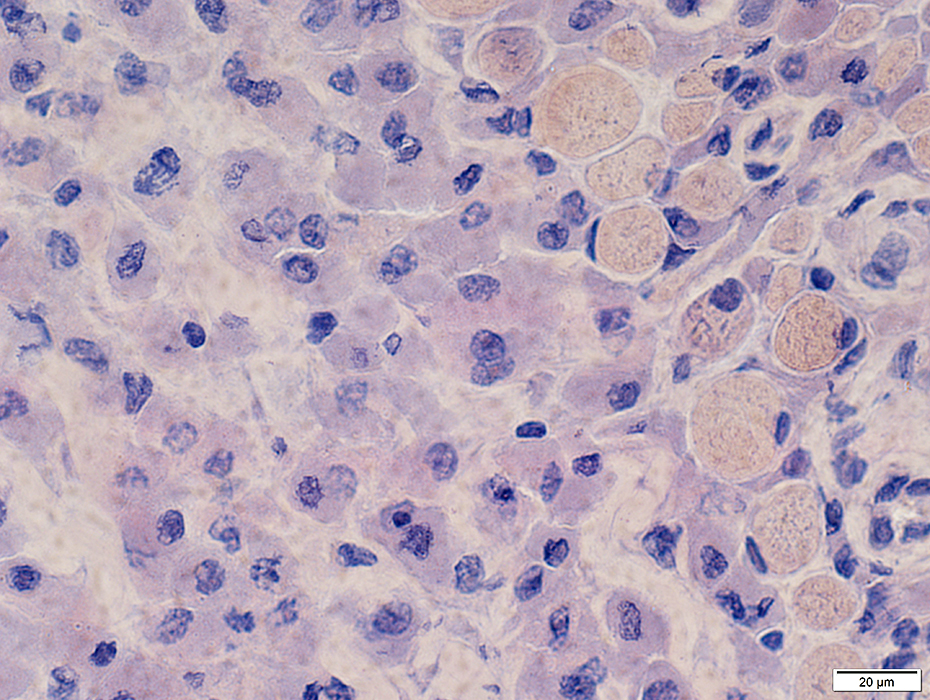
MMF: Aluminum stains
 Morin stain |
 Morin stain |
 Quercetin stain |
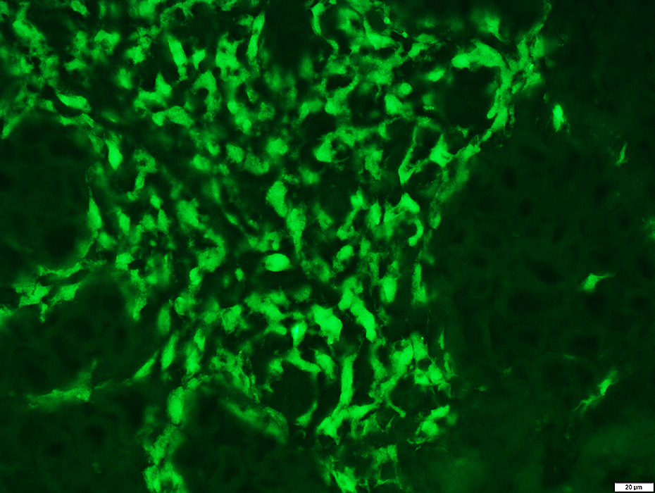 Quercetin stain |
Macrophagic Myofasciitis: Aluminum-containing Histiocytes
Also see: IMAM
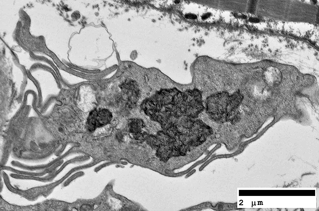 From: C Cai |
Contain inclusions: Spiculated; Electron dense (Typical of aluminum crystalloid)
 From: C Cai |
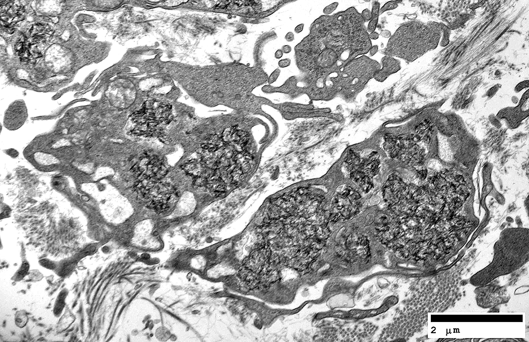
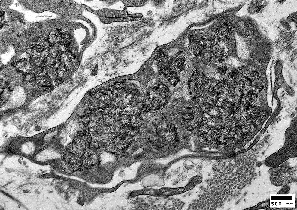 From: C Cai |
Contain inclusions: Spiculated; Electron dense (Typical of aluminum crystalloid)
 From: C Cai |
Return to Neuromuscular Home Page
Return to Inflammation
Return to Inflammatory myopathies
11/30/2021