Inclusion Body Myopathy with Dementia & Paget disease of Bone
|
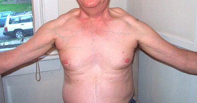
Patient pictures Distal weakness Scapular winging Muscle pathology |
|
|
|
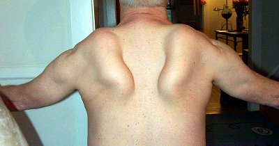
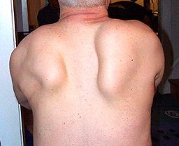
Scapular winging in patient with IBM + Paget's |
|
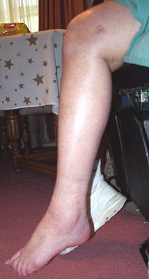
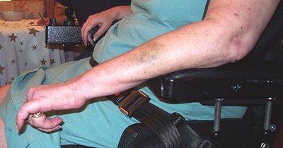
Distal weakness (Severe) in patient with IBM + Paget's
|
|
|
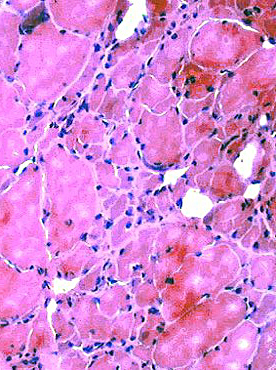
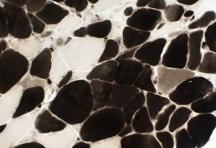
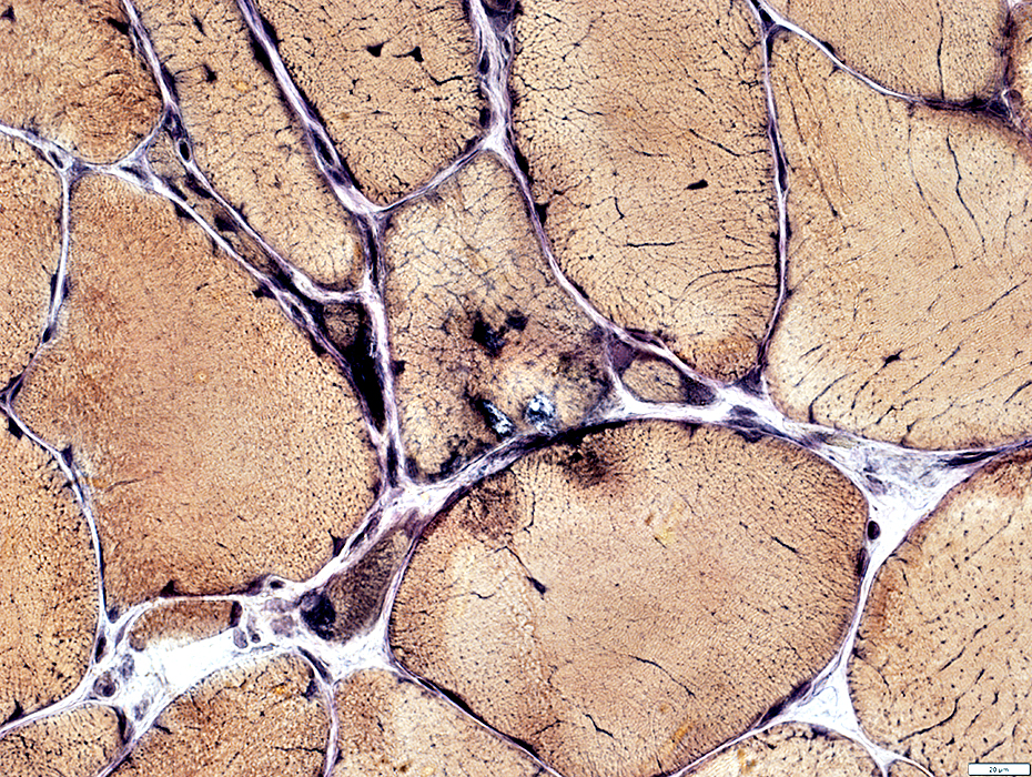
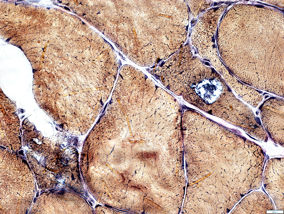
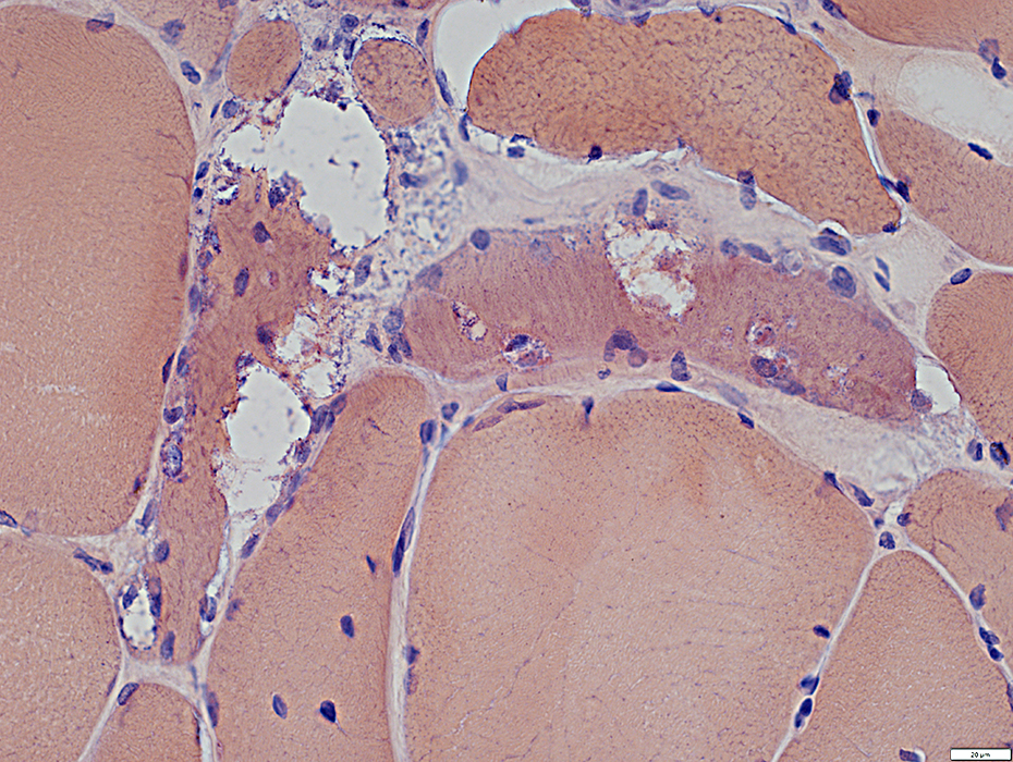
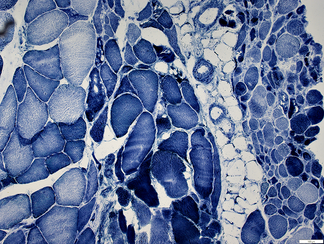
Muscle Pathology: Inclusion Body Myopathy with Dementia & Paget disease (MSP1)
|
Myopathy Vacuoles Aggregates |
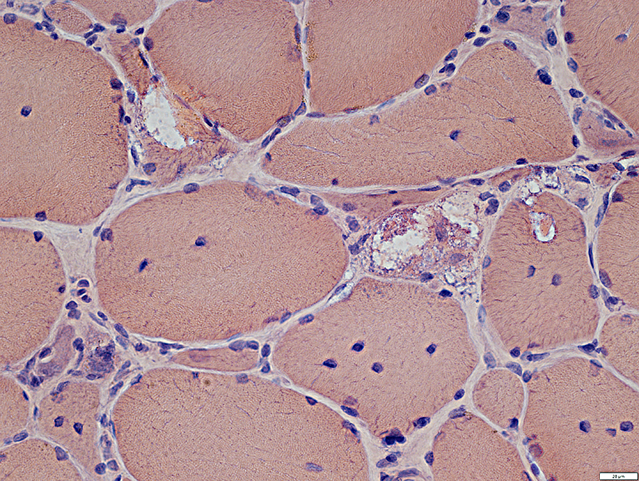
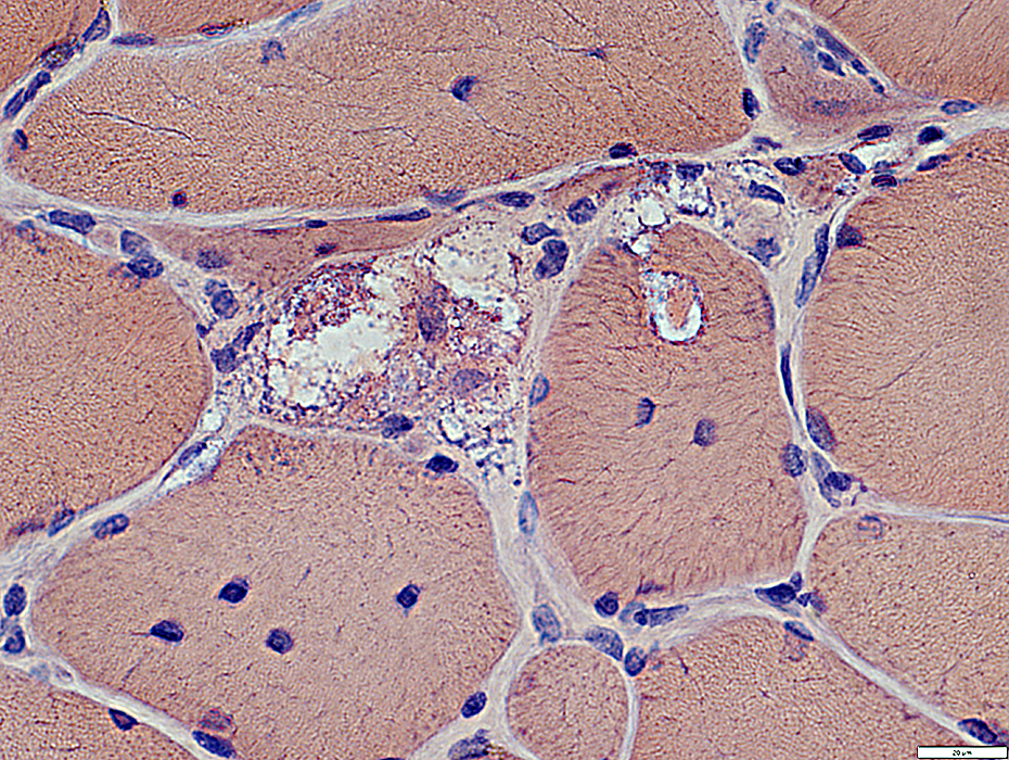
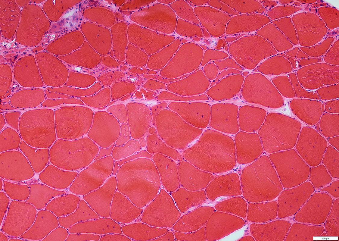 H&E stain |
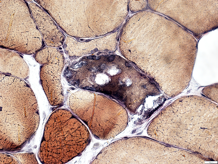
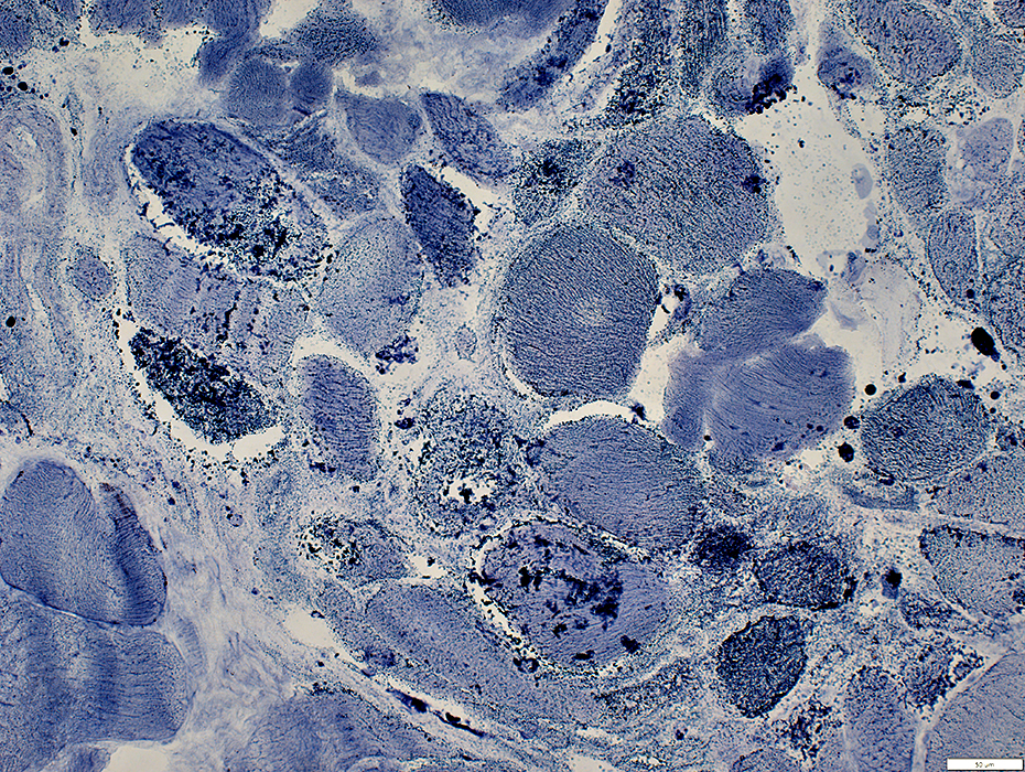
Muscle fibers
Atrophy: Often in clusters
Hypertrophy
Nuclei: Internal
Vacuoles: Irregular shapes; May contain granular debris
Aggregates
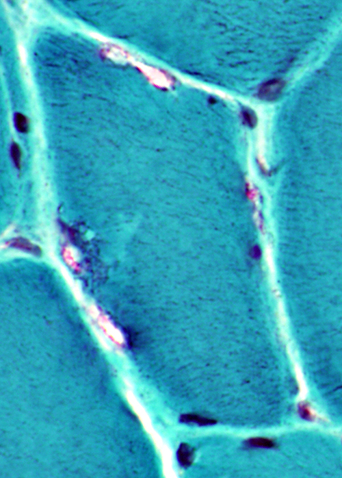
Endomysial connective tissue: Mildly increased
 H&E stain |
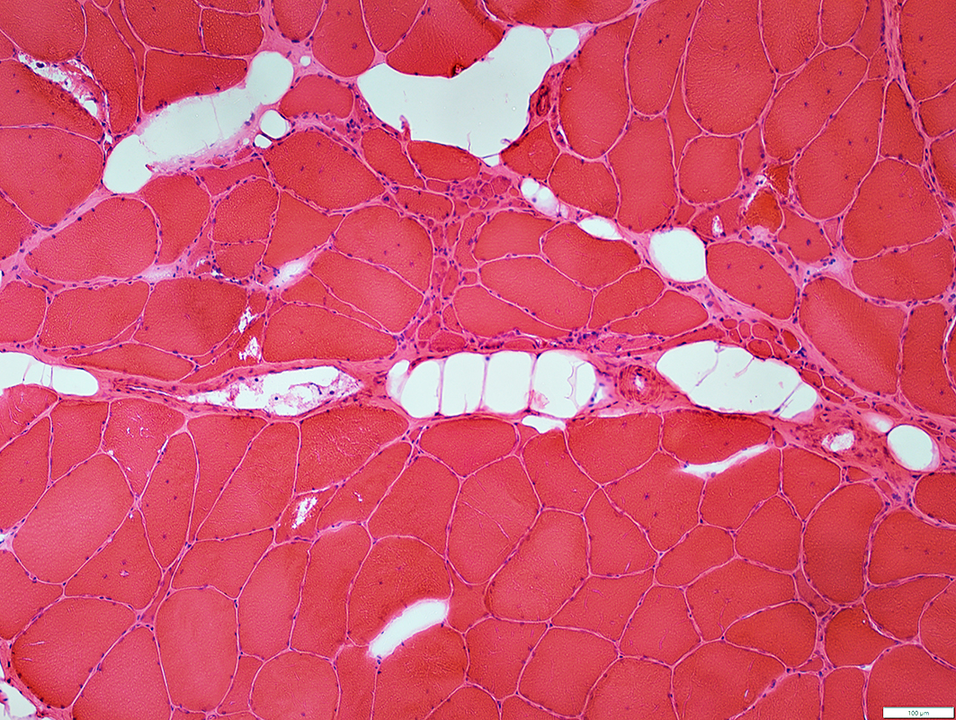
 H&E stain |
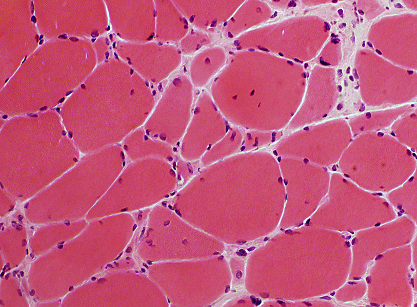
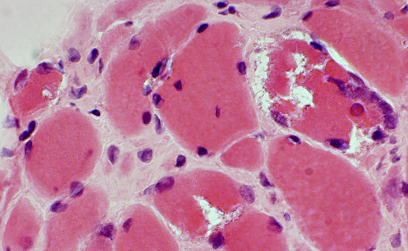
Grouped atrophy: Myopathic
|
 H&E stain |
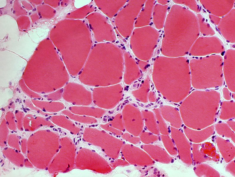
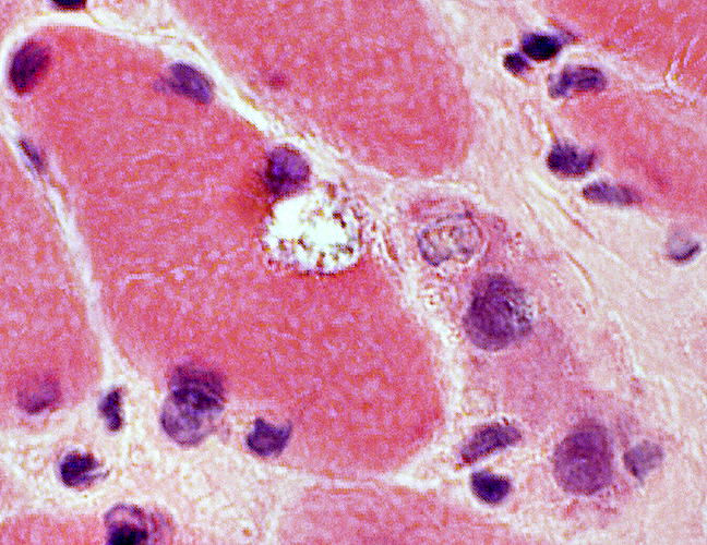
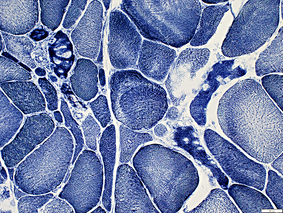
Myopathy
Fiber sizes: Varied
Small fibers
Shapes: Round or Angular
Cytoplasm: Mildly basophilic
Myonuclei: Large
 H&E stain |
Fiber Type Features
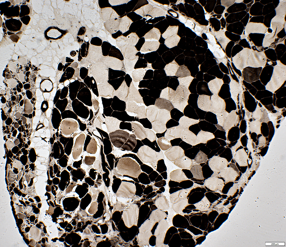
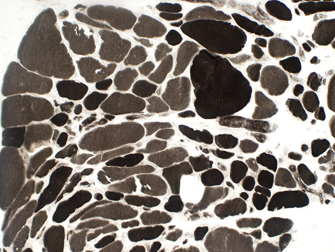 ATPase pH 9.4 stain Type 1 muscle fiber predominance Small & Large fibers are both types No fiber type grouping. |
 ATPase pH 4.3 stain |
 ATPase pH 4.3 stain Type 1 muscle fiber predominance Scattered type 2C (Intermediate-staining) muscle fibers |
|
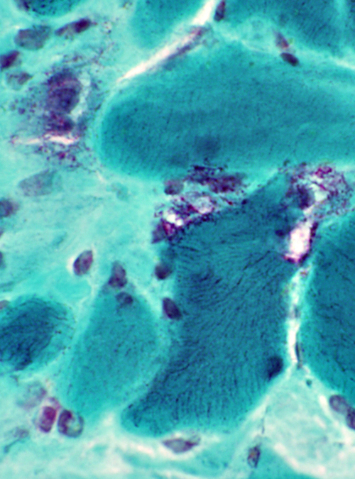
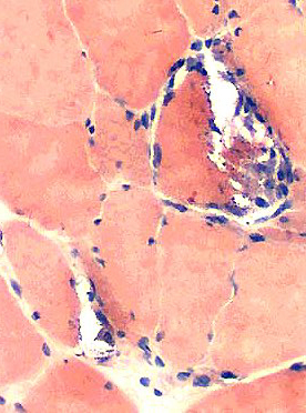
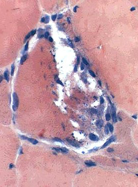
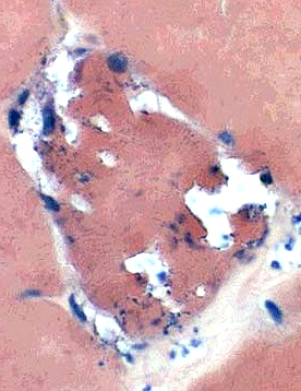
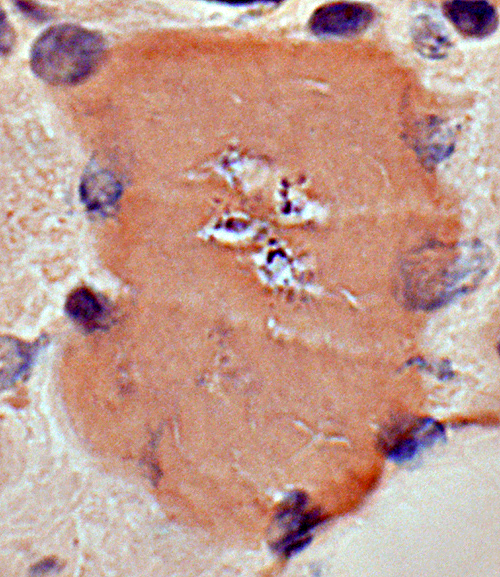
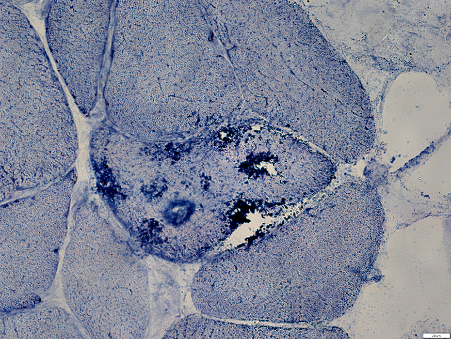
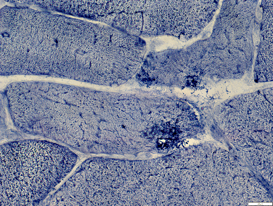
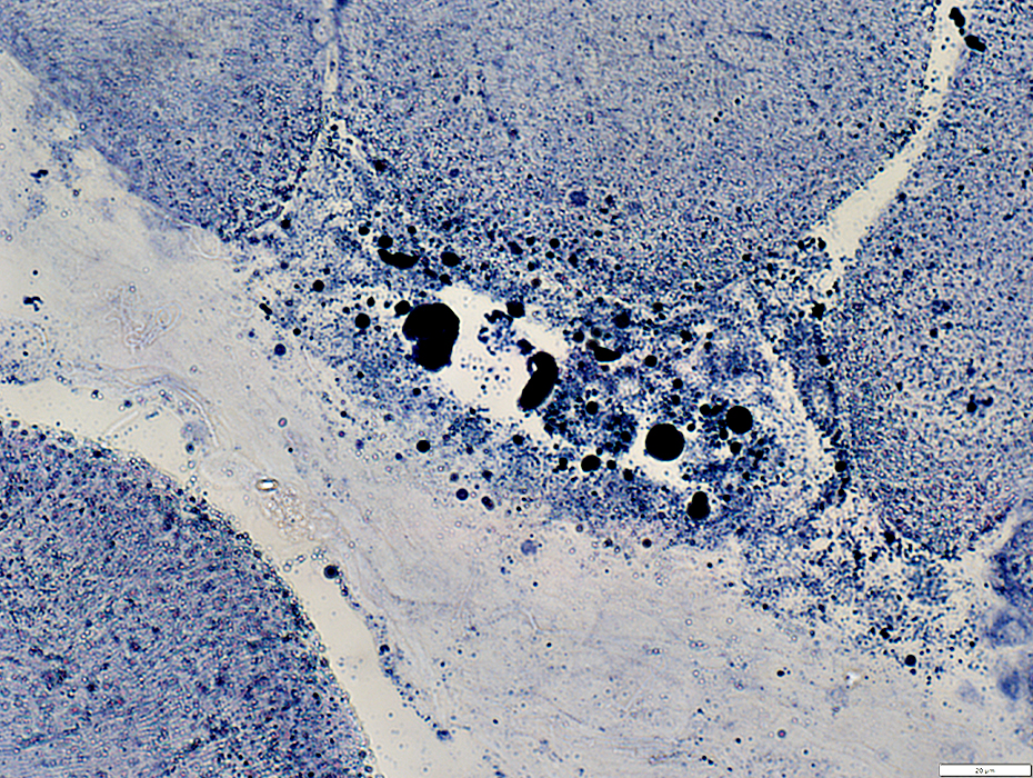
Vacuoles Rimmed Shapes: Irregular Contents: Granular debris 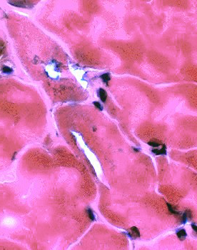 H&E stain |
 H&E stain |
 H&E stain |
|
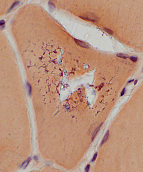
 Gomori trichrome stain |
 Gomori trichrome stain |
|
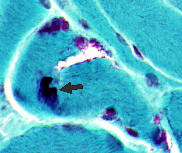
Vacuoles Red-rimmed Aggregates Cytoplasmic bodies (Below, Arrow) | |
 Gomori trichrome stain | |
VCP Myopathy: Vacuoles
 VvG stain |
Contain: Fragmented debris
Aggregates
Irregular, dark-stained regions of sarcoplasm
 VvG stain |
Contain: Fragmented debris
Aggregates
Irregular, dark-stained regions of sarcoplasm
 VvG stain |
VCP Myopathy: Vacuoles
 Congo red stain |
Contain: Fragmented debris
Shapes: Irregular
Sizes: Varied; Small or Most of fiber area
Number: One, or several, in individual muscle fibers
 Congo red stain |
 Congo red stain |
Contain: Fragmented debris
Shapes: Irregular
Sizes: Varied; Small or Most of fiber area
Number: One, or several, in individual muscle fibers
|
Shapes: Irregular
Contents & Neighboring areas
Debris: Granular, Basophilic
Myonuclei in vacuolar fibers
Size: Large
Mildly pale staining
 Congo red stain |
 Congo red stain |
 NADH stain |
Small fibers: Stain dark
Vacuoles: Pale staining
 NADH stain |
 Acid phosphatase stain |
 AMPDA stain |
 AMPDA stain |
 AMPDA stain |
 AMPDA stain |
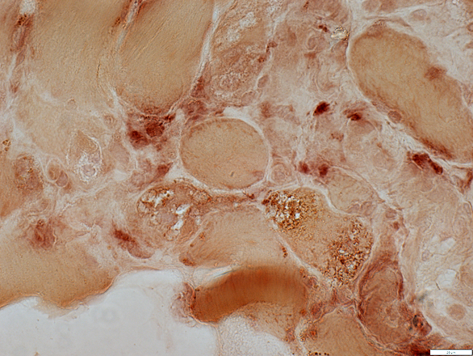
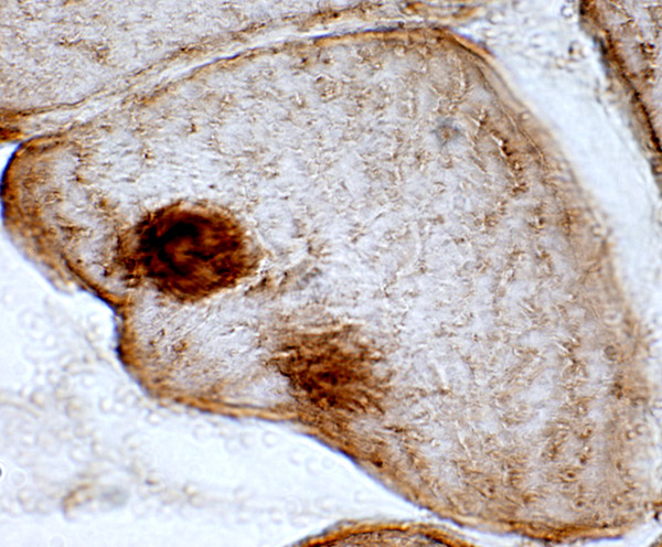
VCP stained inclusions
Normal & Mutant p97/VCP in vitro: Distribution of VCP & Ubiquitinated proteins
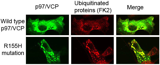
- Normal VCP in cells (Top)
- VCP staining: Diffusely distributed in cytoplasm
- Ubiquitinated proteins (FK2 staining): Present in small clusters in cytoplasm
- Mutant VCP in cells (Bottom)
- VCP staining: Aggregates in cytoplasm
- Ubiquitinated proteins (FK2 staining): Present in aggregates in cytoplasm
- VCP & Ubiquitinated proteins: May or not be present in same aggregates
Return to IBM with Paget's & Dementia
Return to Distal myopathy
Return to Muscle biopsies
Return to Biopsy illustrations
Return to Neuromuscular home page
Return to Polyneuropathy Index
4/2/2023