Inclusion Body Myositis (IM-VAMP pathology): 46 yo female with HIV
|
Inflammation Focal invasion CD4 & CD8 lymphocytes Myopathy MHC Class I upregulation by myofibers Vacuoles Aggregates Mitochondrial pathology Endomysial capillaries Also see: IM-VAMP pathology |
IM-VAMP during HIV
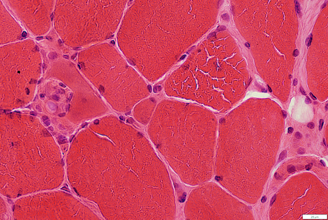
Fiber size: Varied
Small muscle fibers: Angular or Round
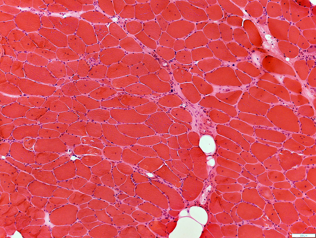 H&E stain |
 H&E stain |
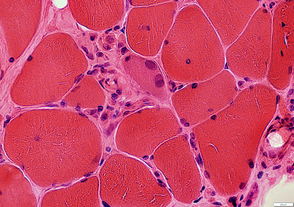
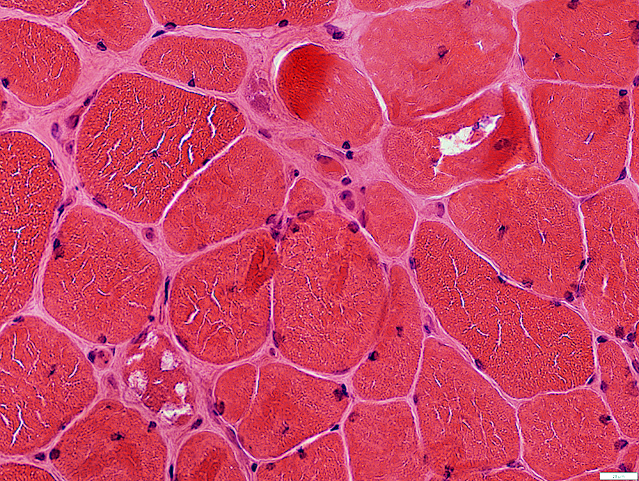
Muscle fibers
Size: Varied
Small muscle fibers: Angular or Round
Internal nuclei
Immaturity in some fibers: Cytoplasm basophilic; Nuclei large
Endomysial connective tissue: Increased
Endomysial capillaries: Large
 H&E stain |
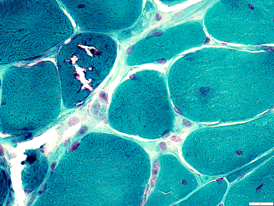
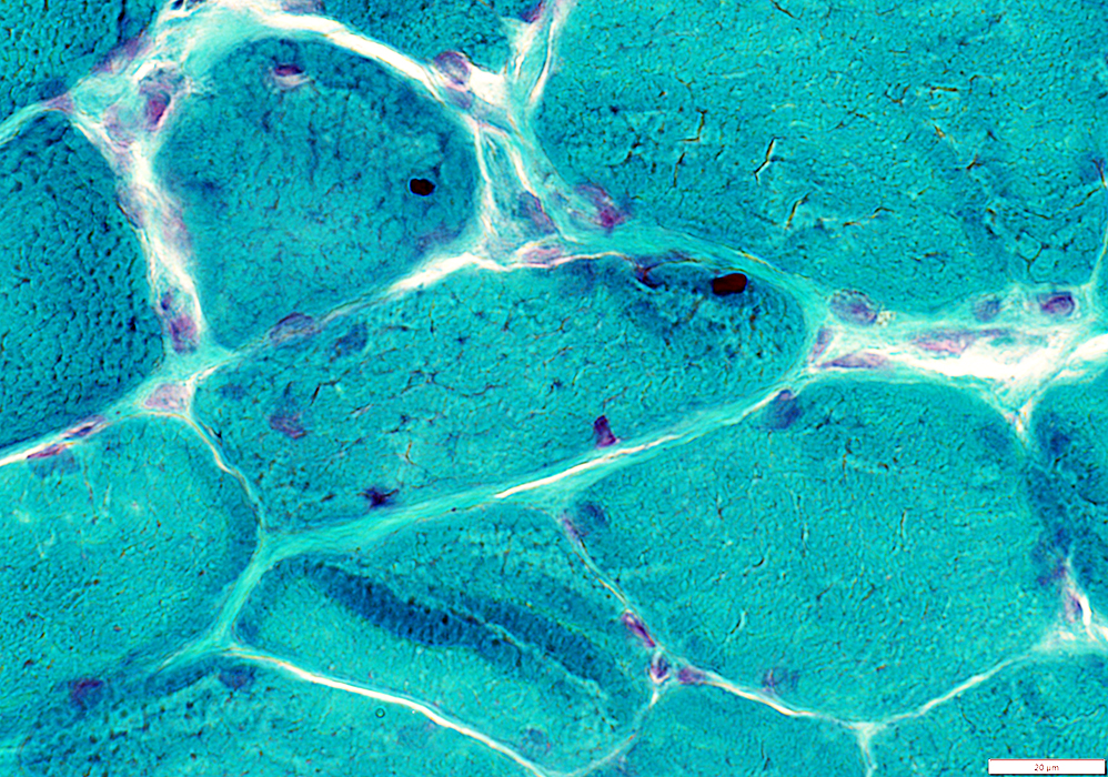
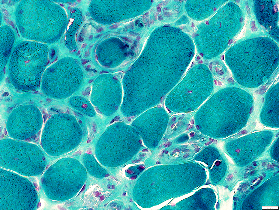 Gomori trichrome stain |
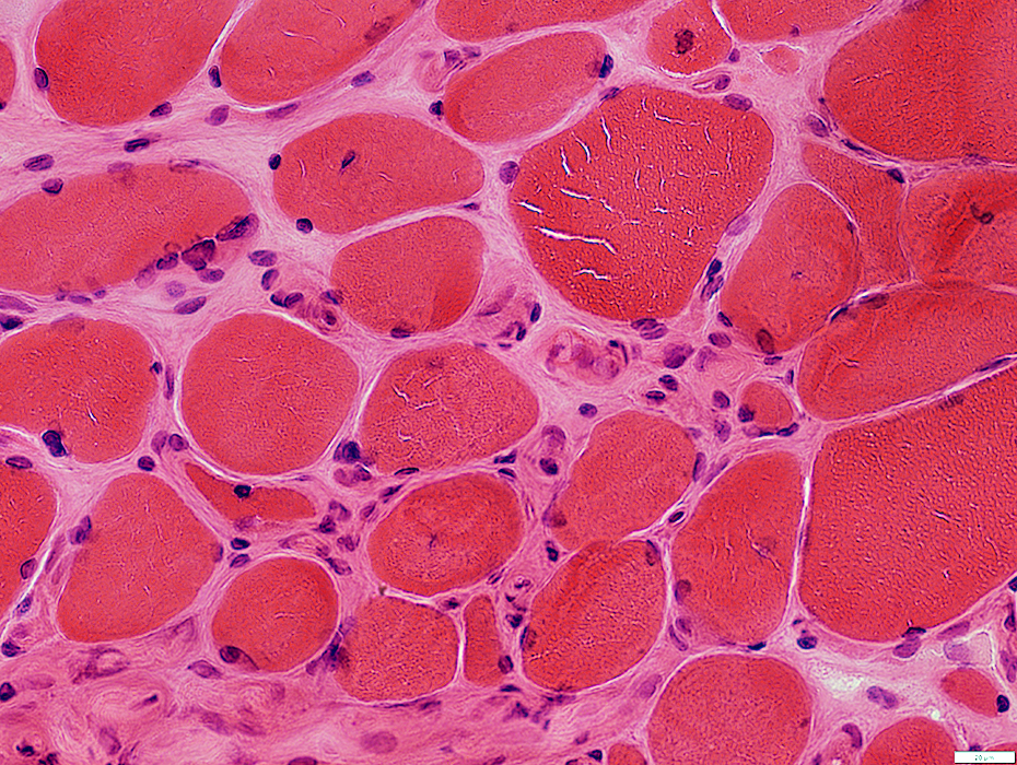
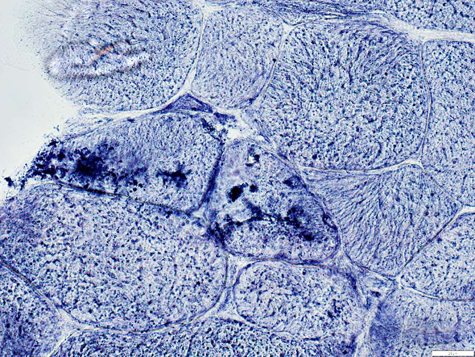
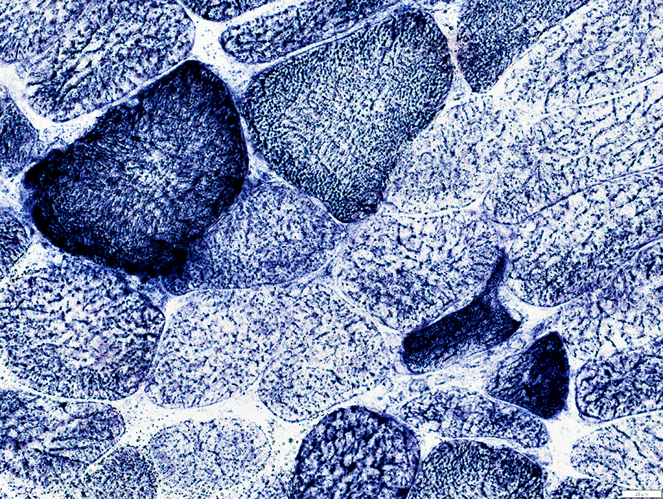
IM-VAMP during HIV
Internal architecture: Coarse in many muscle fibers
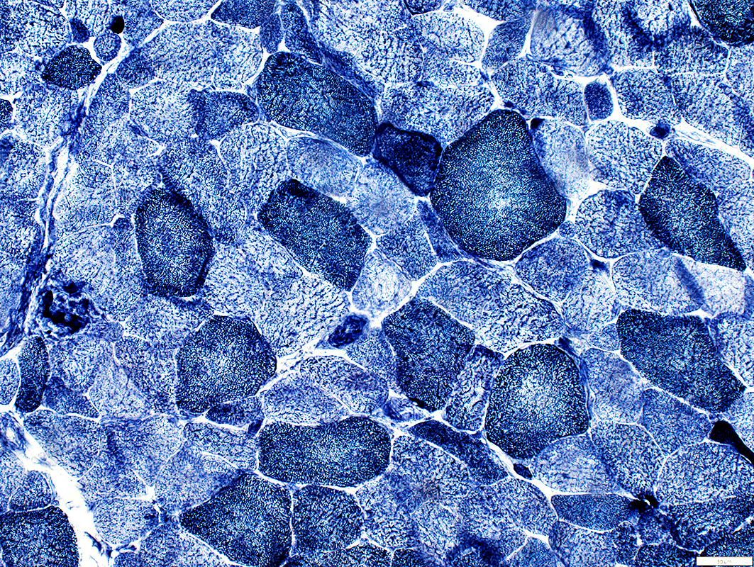 NADH stain |
 Congo red stain |
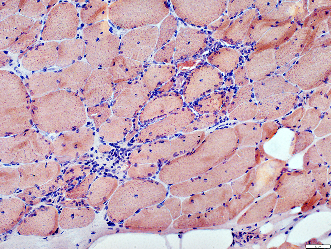
Inflammation: Endomysial
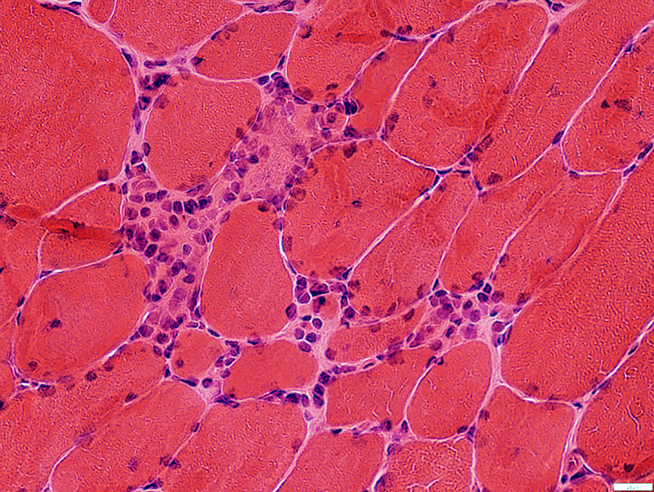 H&E stain |
 H&E stain |
Inflammation
Endomysial
Focal invasion of muscle fibers
 Congo red stain |
 Congo red stain |
Inflammation
Focal invasion of muscle fibers
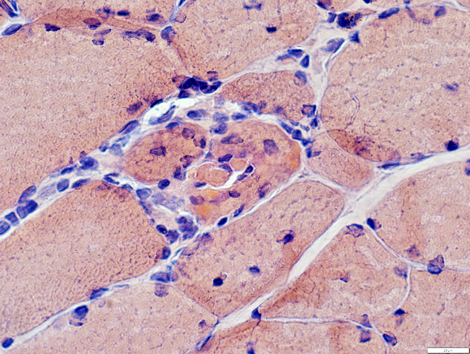
 Acid phosphatase stain |
 CD4 stain |
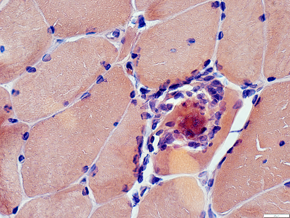
Inflammation, endomysial
Composed of CD4 & CD8 lymphocytes
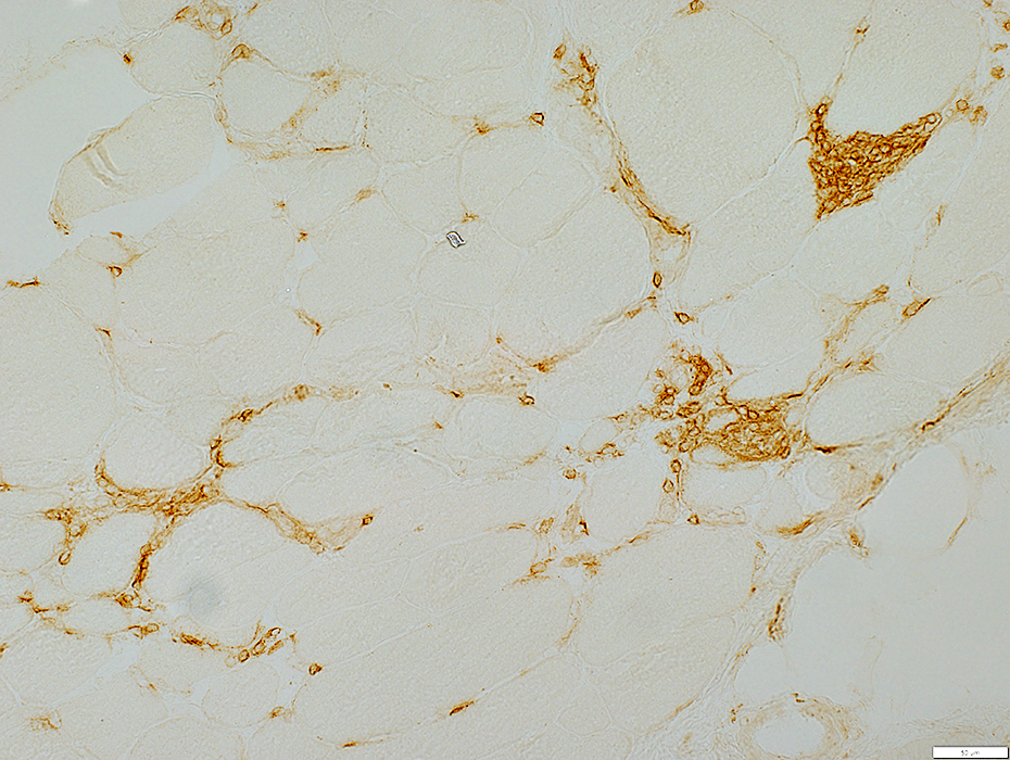 CD8 stain |
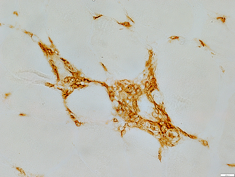 CD8 stain |
IM-VAMP during HIV
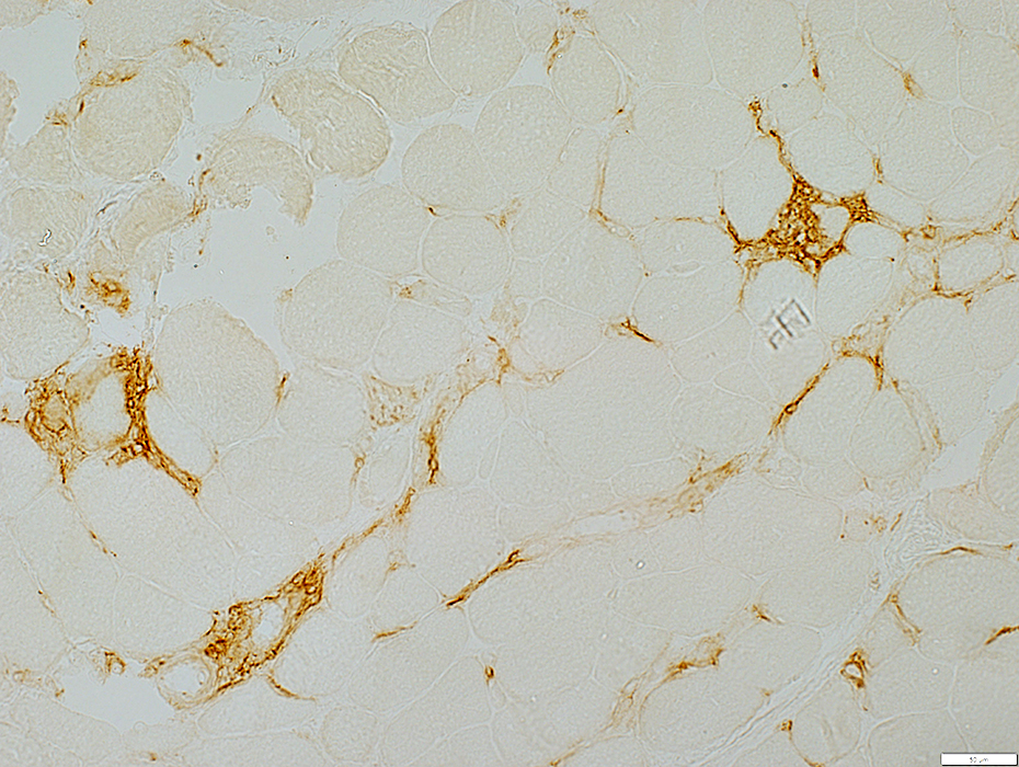
MHC Class I upregulation on surface of muscle fibers
MHC Class I is also upregulatied in the cytoplasm of some muscle fibers
Endomysial lymphocytes also express MHC Class I
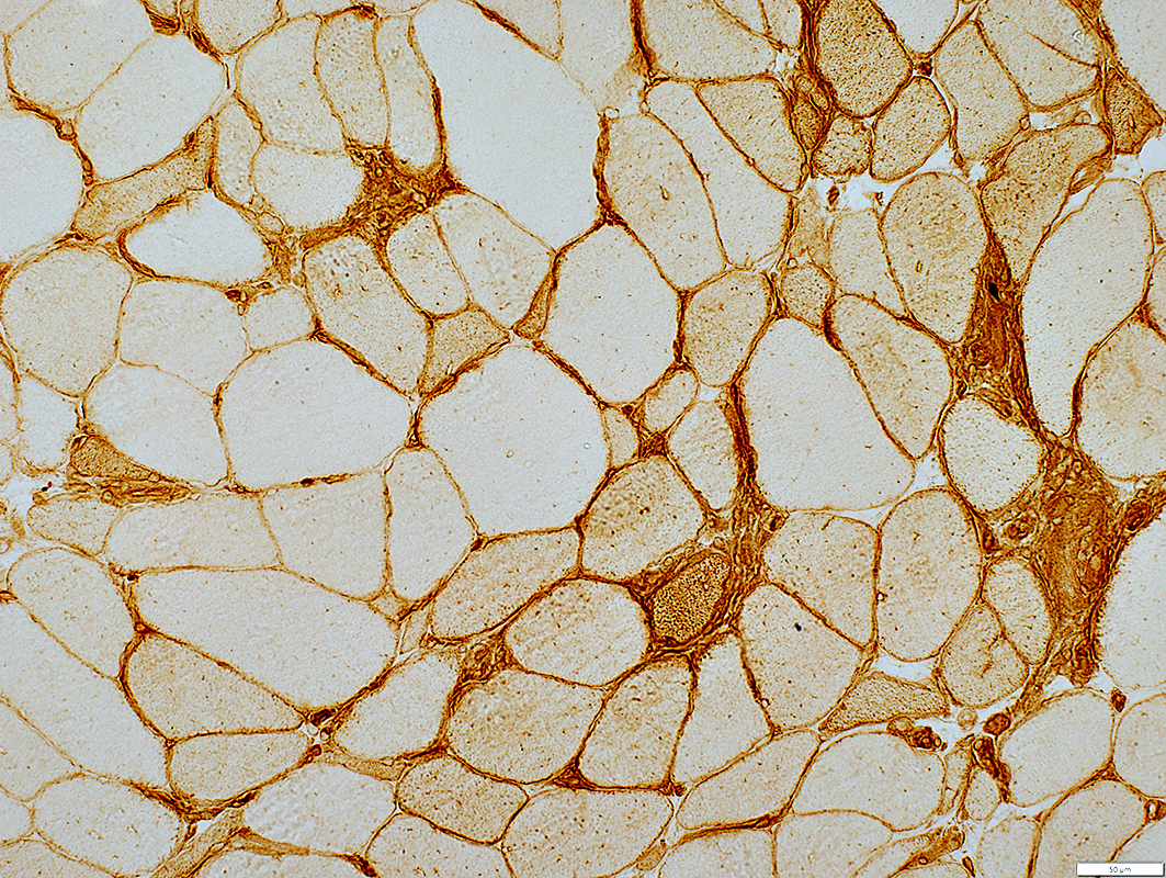 MHC Class I stain |
 H&E stain |
Irregular vacuoles in some muscle fibers
Some vacuoles have reddish border on Gomori trichrome stain
 Gomori trichrome stain |
IM-VAMP during HIV: Aggregates
Cytoplasmic bodies in muscle fibers
 Gomori trichrome stain |
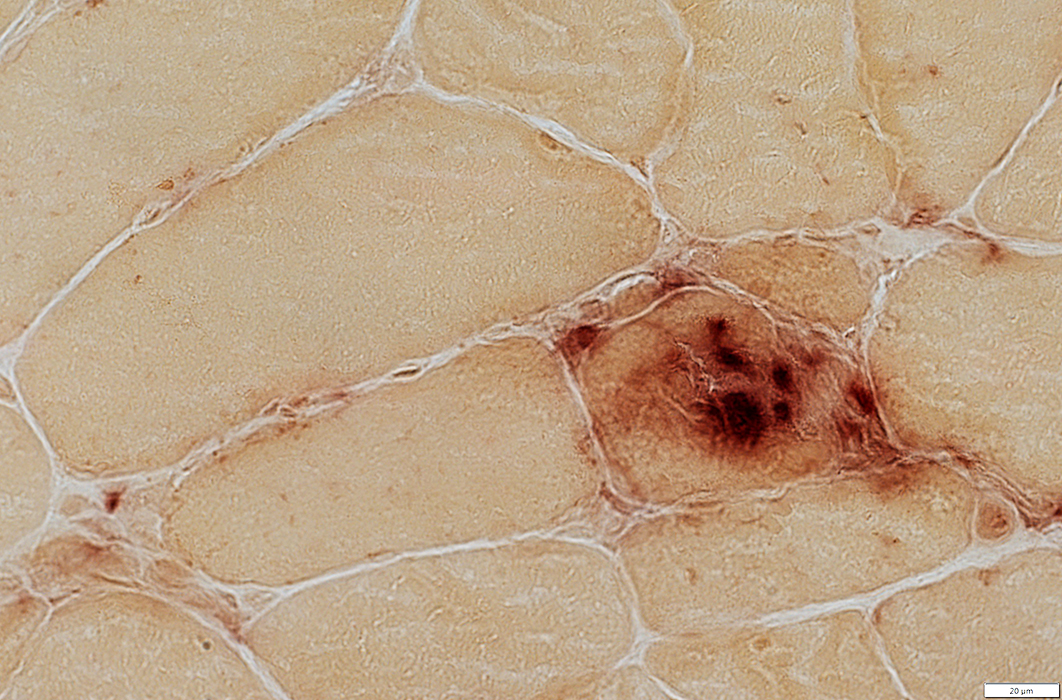
IM-VAMP during HIV
AMPDA aggregates in some muscle fibers
 AMPDA stain |
IM-VAMP during HIV
LC3 aggregates in some muscle fibers
 LC3 stain |
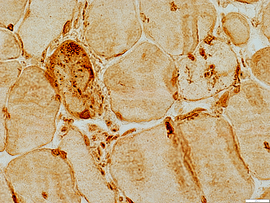
IM-VAMP during HIV: Mitochondrial pathology
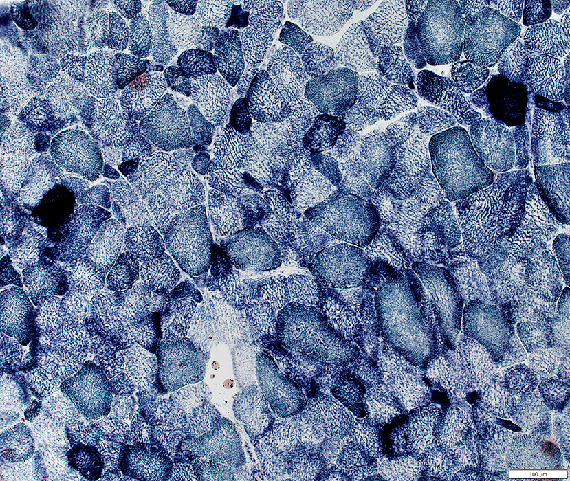 Succinate Dehydrogenase (SDH) stain |
 Succinate Dehydrogenase (SDH) stain |
COX negative muscle fibers: Scattered
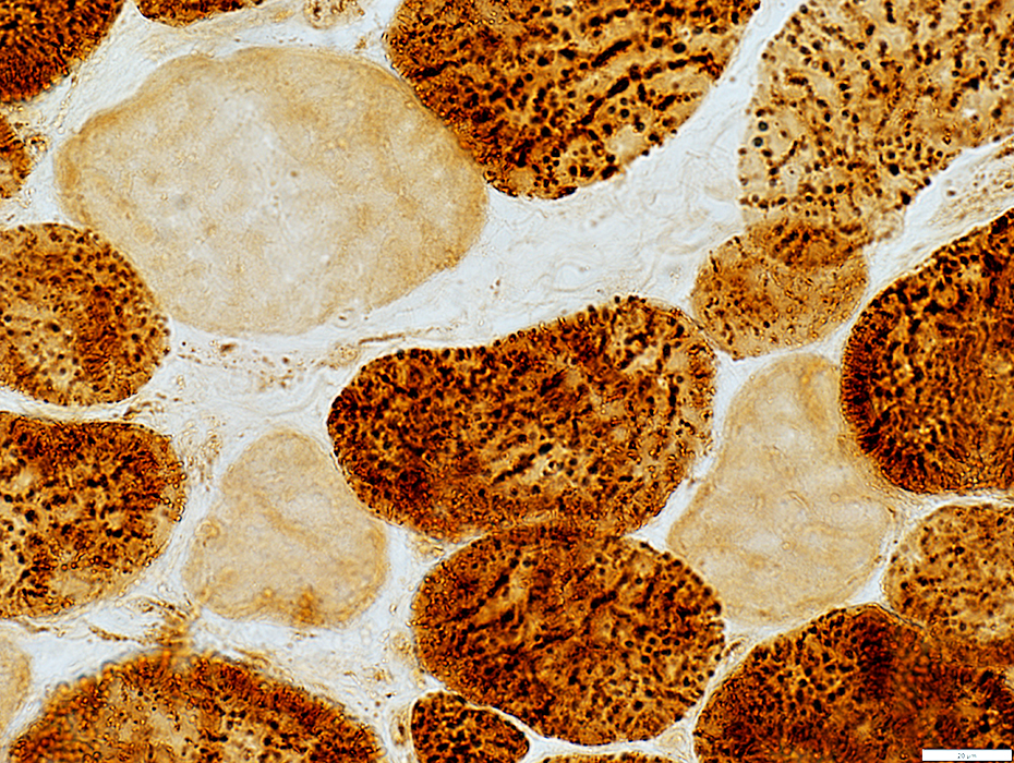 Cytochrome oxidase (COX) stain |
Return to: Neuromuscular Home Page
1/18/2021