Neuropathy with IgM antibody vs GD1b (CANOMAD)
No inflammation
Axon loss
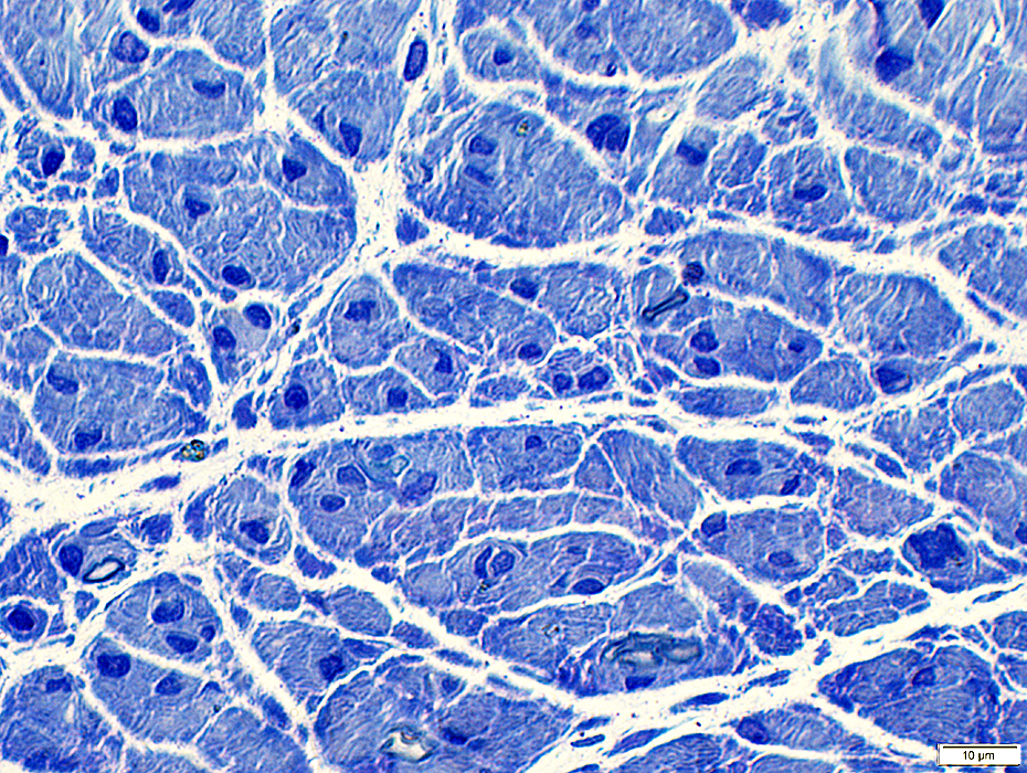
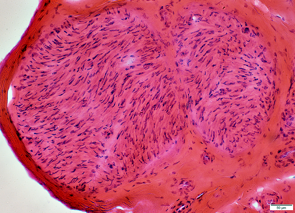 H&E stain |
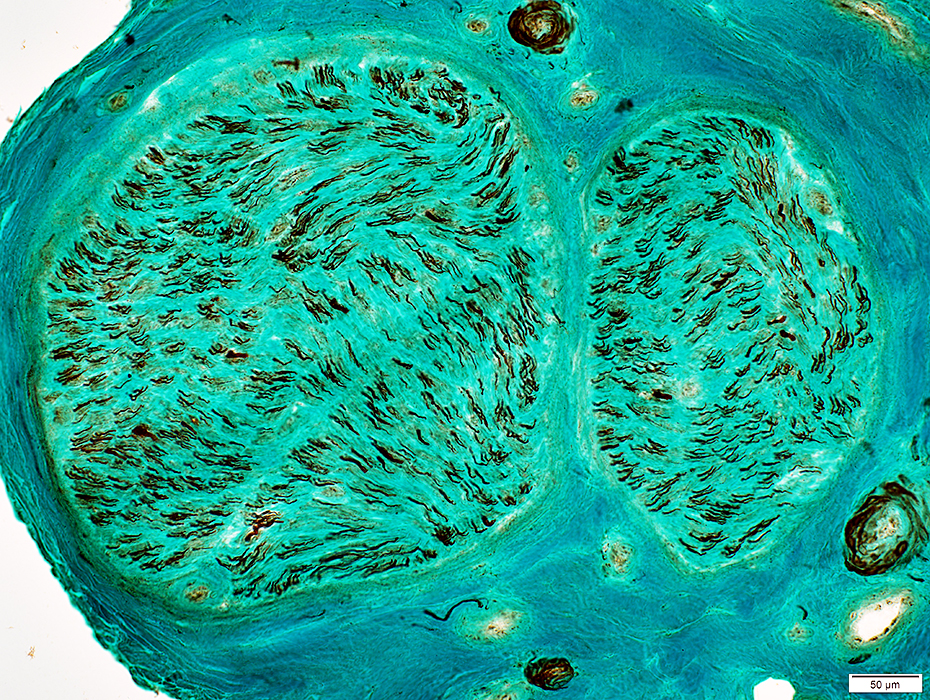
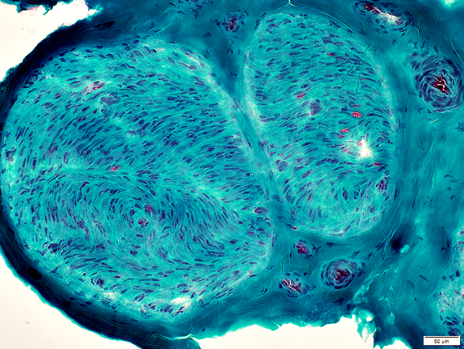 Gomori trichrome stain |
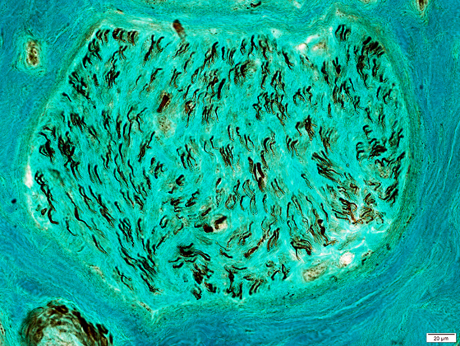
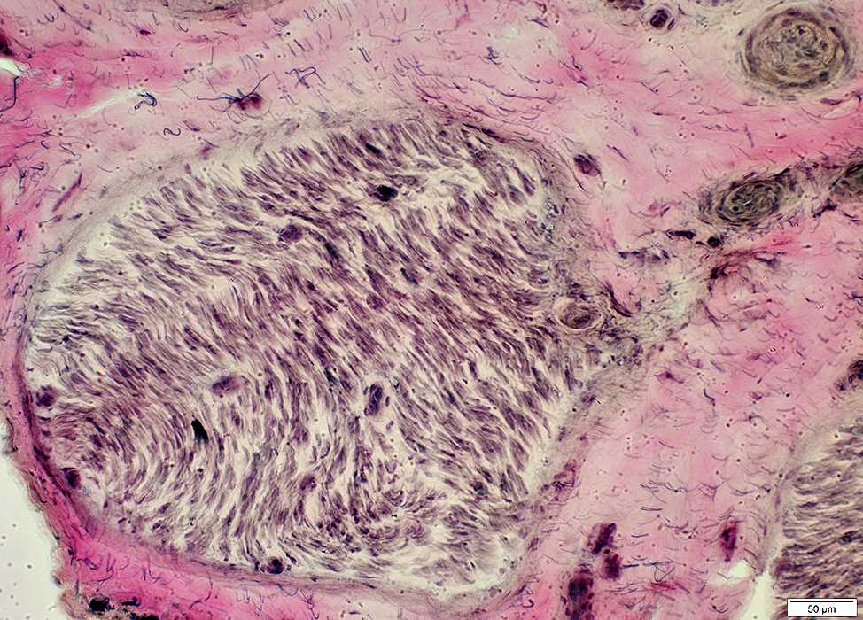 VvG stain |
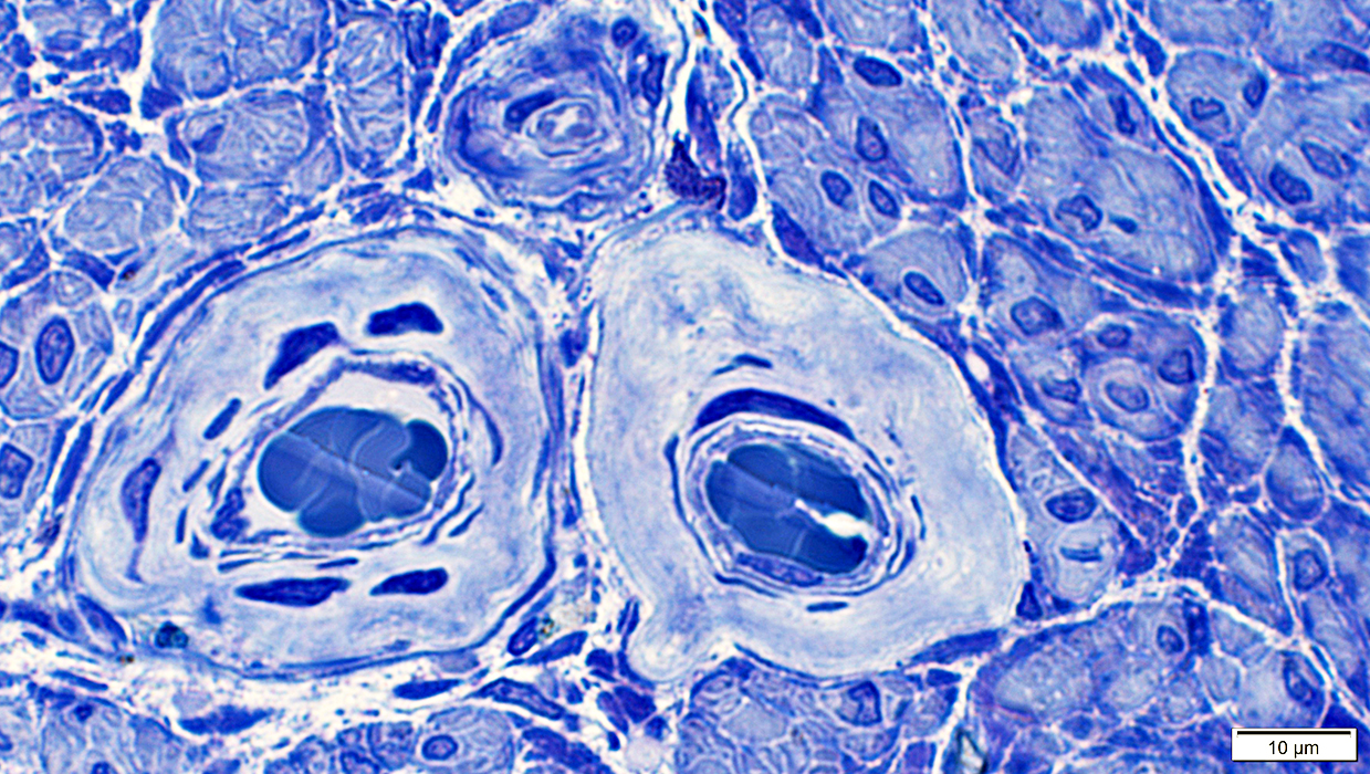
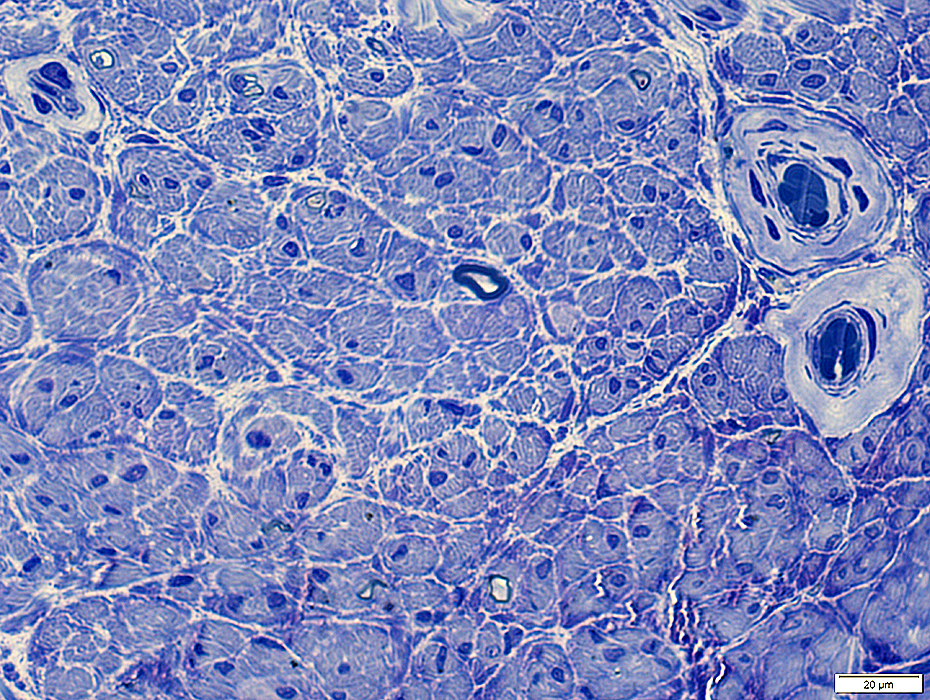 Toluidine blue stain |
Toluidine blue stain |
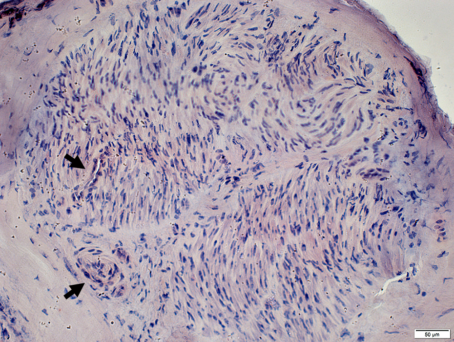
 Neurofilament stain |
Small axons: Relative preservation
See: Control nerve: Many large axons
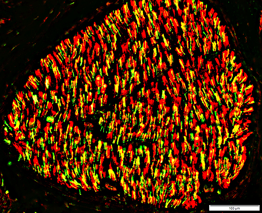
 Neurofilament stain |
-MBP(r).jpg) Neurofilament (Green) & Myelin basic protein (MBP) (Red) stains |
Severe loss
Only a few remaining axons have associated myelin basic protein
Normals: Largest myelinated axons commonly have myelin containing MBP
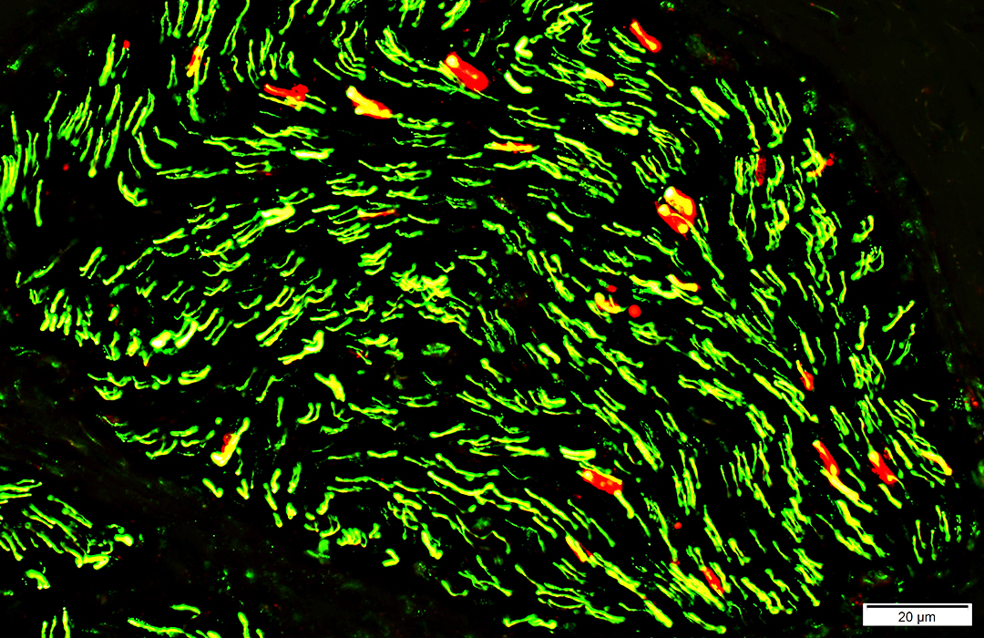 Neurofilament (Green) & Myelin basic protein (MBP) (Red) stains |
p0
In myelin around a few remaining larger axons
p0 is also present in Schwann cells with no associated axons (Büngner bands)
Normals
p0 is present in myelin on most myelinated axons
Few or no empty p0 cells without axons
 Neurofilament (Green) & P0 (Red) stains |
NCAM
Present around most small, unmyelinated axons
NCAM is also present in Schwann cells with no associated axons (Büngner bands)
Few large axon stained for neurofilaments
Normals
Few or no empty p0 cells without axons
Many large axons with no associated NCAM
-NCAM(r).jpg) Neurofilament (Green) & NCAM (Red) stains |
Büngner bands
p0 and NCAM co-localize in Schwann cells with no associated axons (Yellow)
Small NCAM positive cells (Green) are associuated with remaining small, unmyelinated axons
Normal: Many p0 positive myelin sheaths with no NCAM
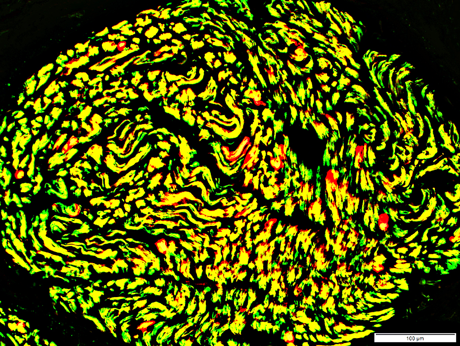 NCAM (Green) & p0 (Red) stains |
 Congo red stain |
 Toluidine blue stain |
Return to Neuromuscular Home Page
12/28/2018