Muscle: Fetal
|
Extramedullary hematopoesis Fascicles Muscle fibers Nerves: Intramuscular Organizarion Spindles Vessels |
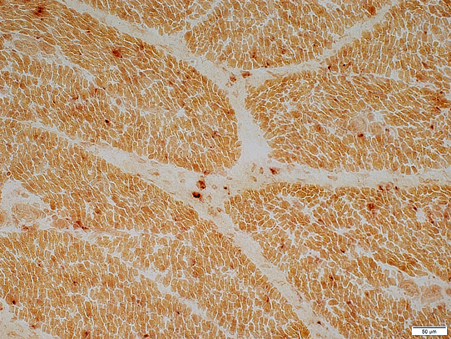
Organization
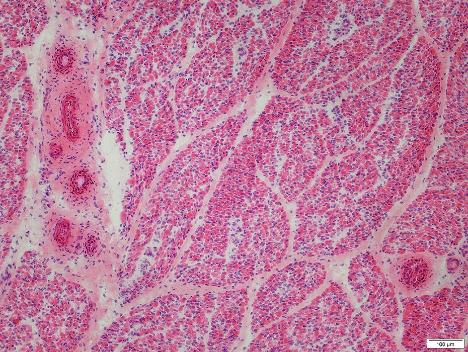 H & E stain |
Arteries & Veins: In bundles
Arterioles: In center of fascicles (in Vascular perimysium)
Muscle fibers
In fascicles
Surrounded by avascular perimysium
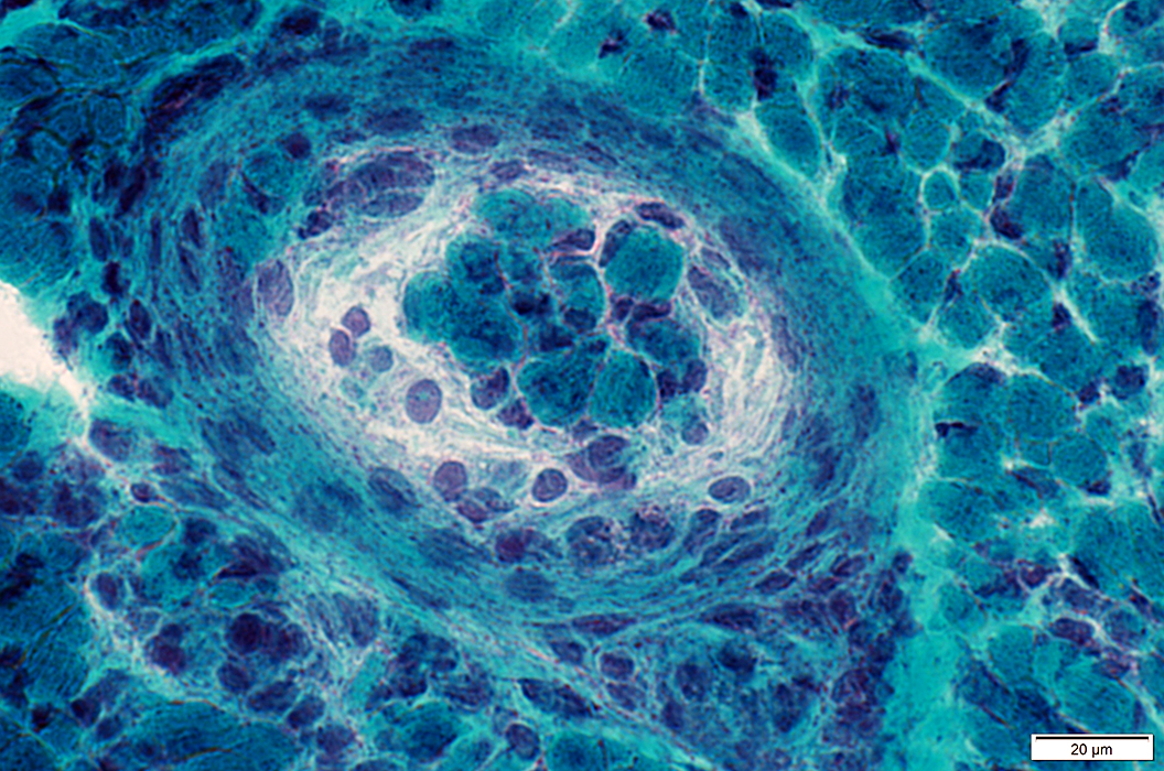
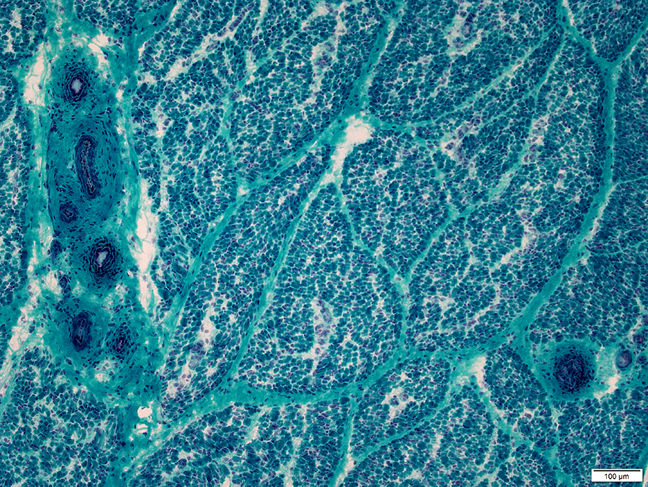 Gomori trichrome stain |
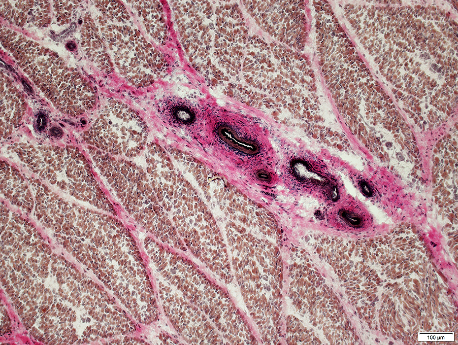 VvG stain |
Fascicles of Muscle fibers
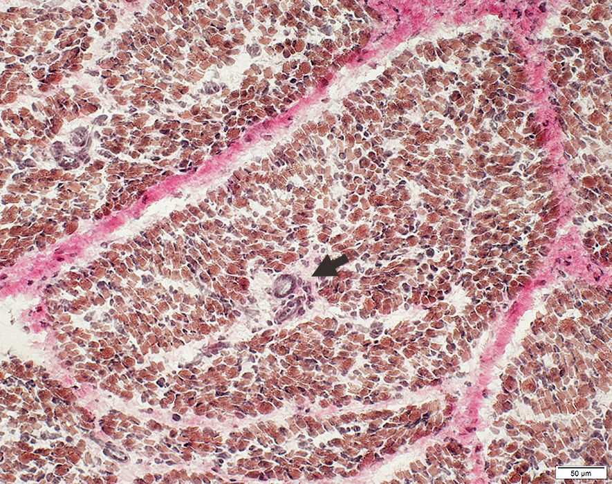 VvG stain |
Surrounded by avascular perimysium
Contain Arterioles & Veinules in center (Arrow)
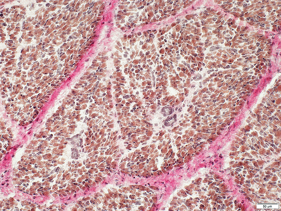 VvG stain |
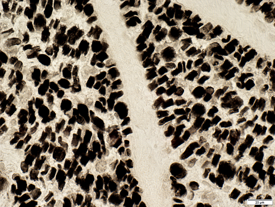
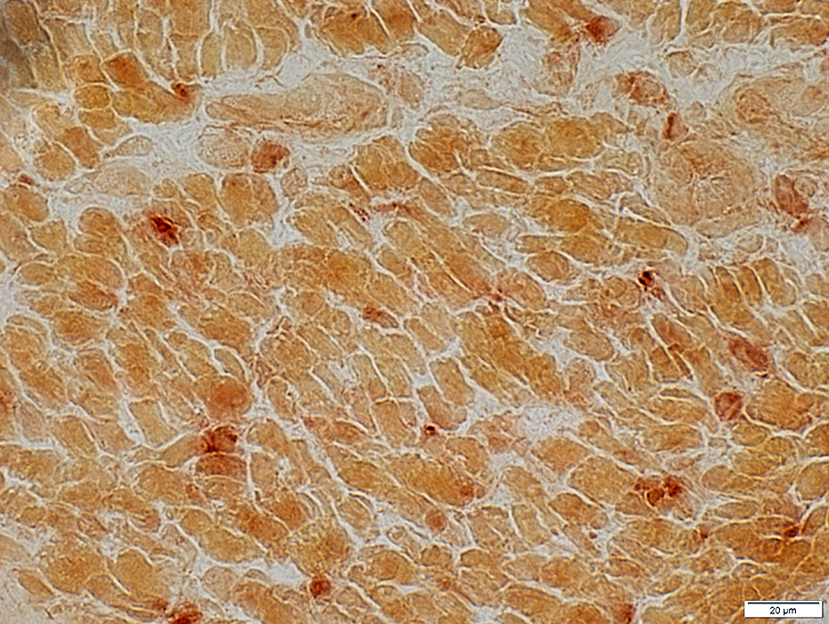
Muscle fibers
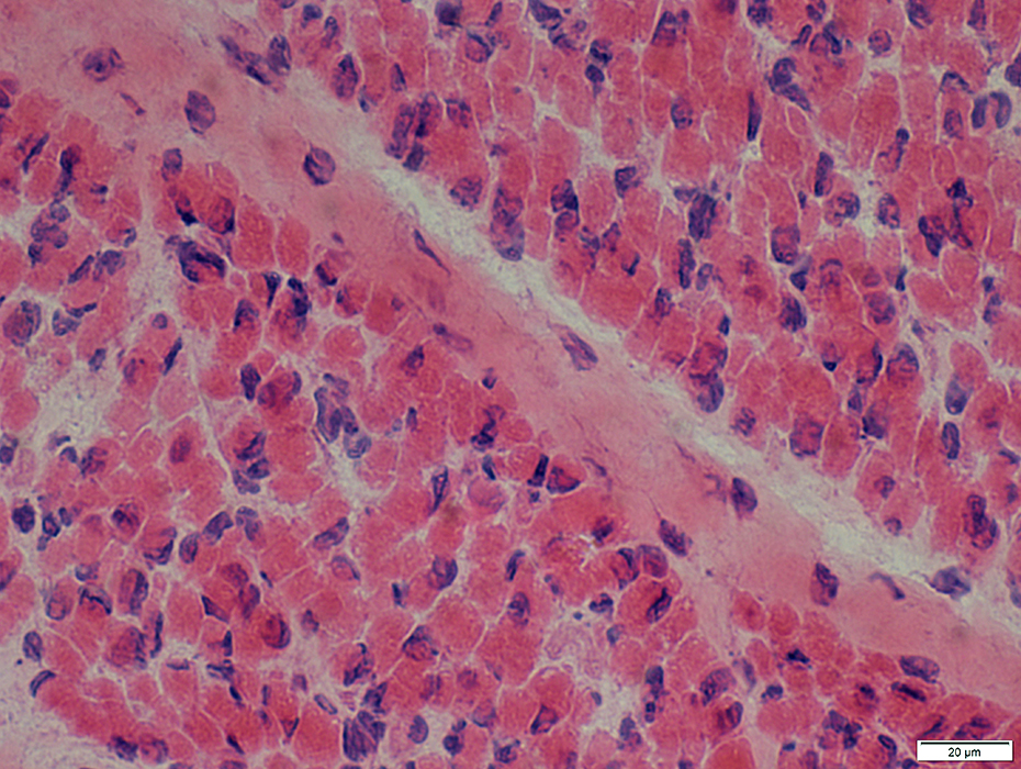 H&E stain |
Sizes: Small (3 to 10 µM diameter)
Nuclei: Large; Irregular shapes
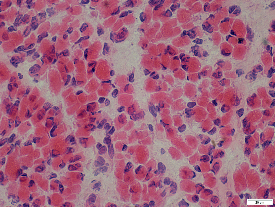 H&E stain |
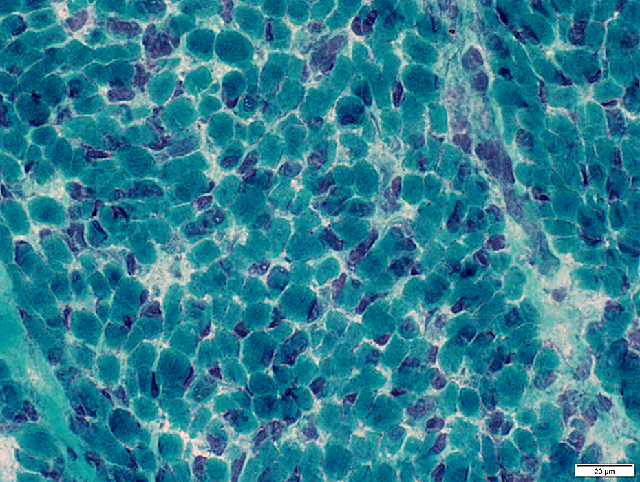 Gomori trichrome stain |
Fiber types: Checkerboard pattern of types 1 & 2
 ATPase pH 4.3 stain |
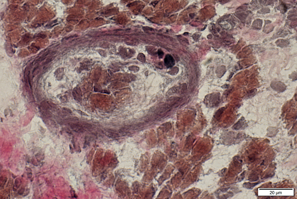
Vessels
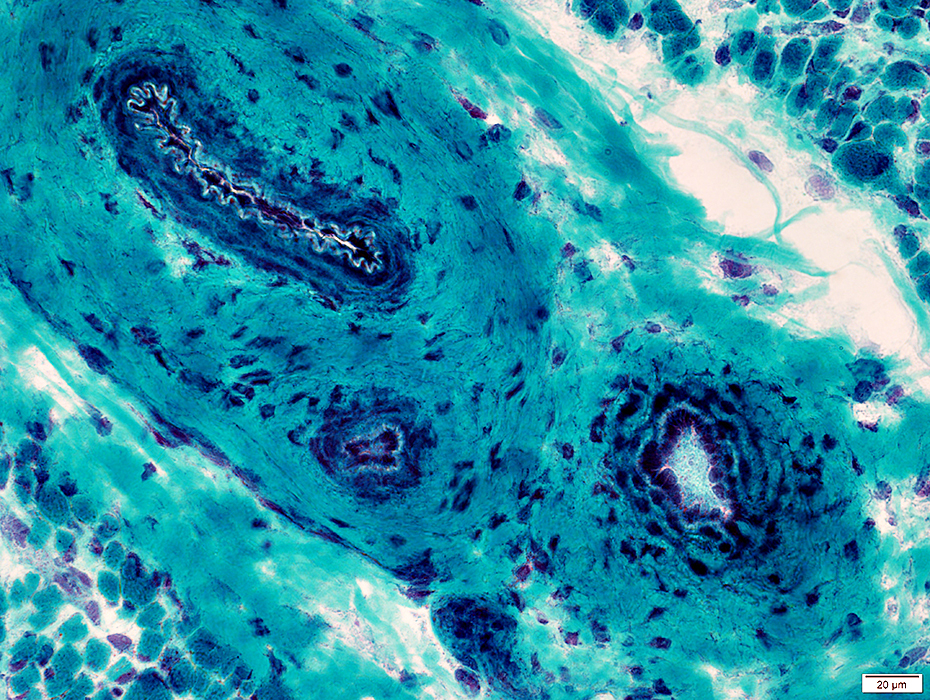 Gomori trichrome stain |
Arteries
Inner layer: Contains continuous elastin layer; Different from fibrillar structure in older people
Smooth muscle layer: Moderately thick
Surround: Connective tissue; More prominent than in older patients
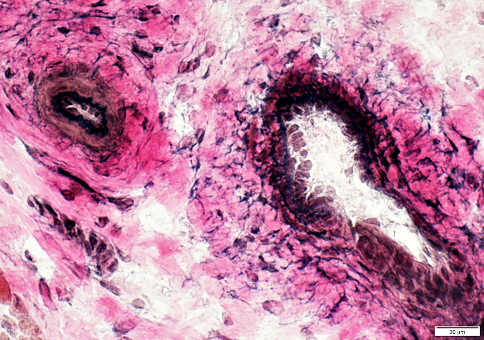 VvG stain |
Artery
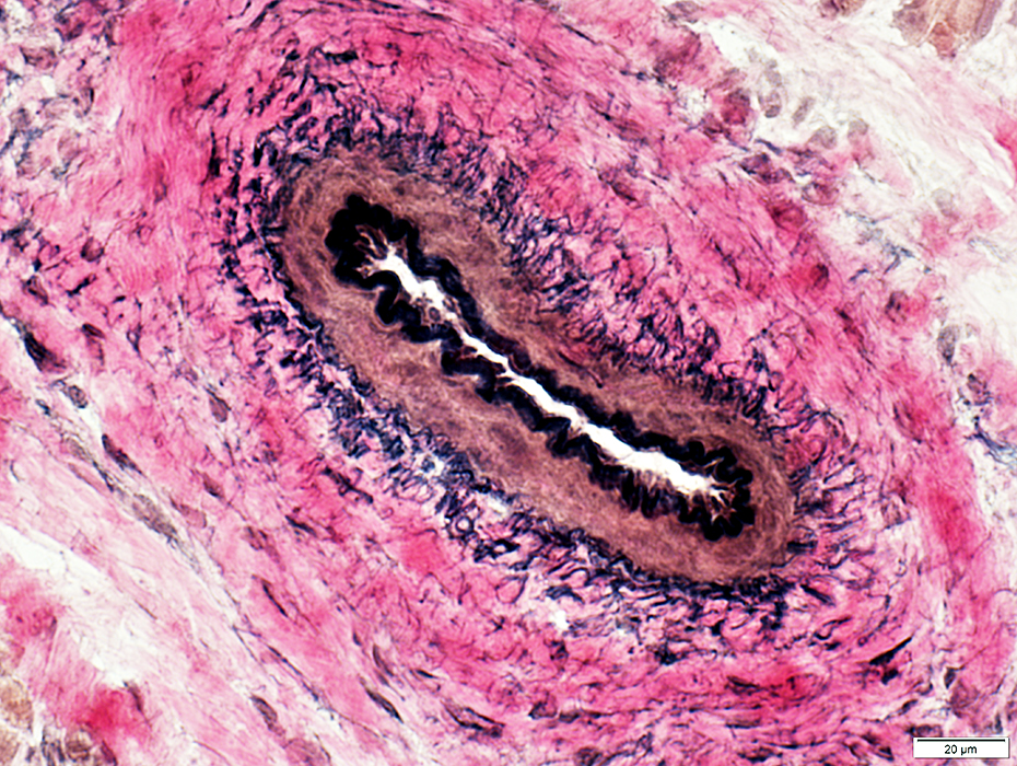 VvG stain |
Vein
Thinner wall
Contains irregular fibrils
 VvG stain |
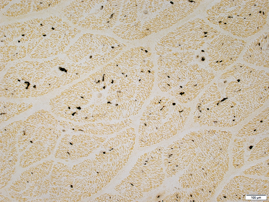 Alkaline phosphatase stain |
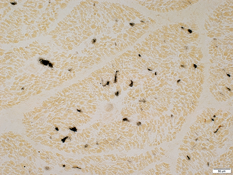 Alkaline phosphatase stain |
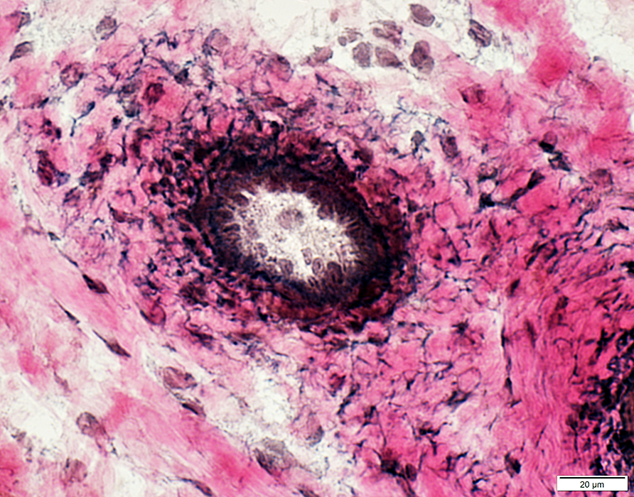
Spindles
 H & E stain |
 Gomori trichrome stain |
 VvG stain |
 Acid phosphatase stain |
 Acid phosphatase stain |
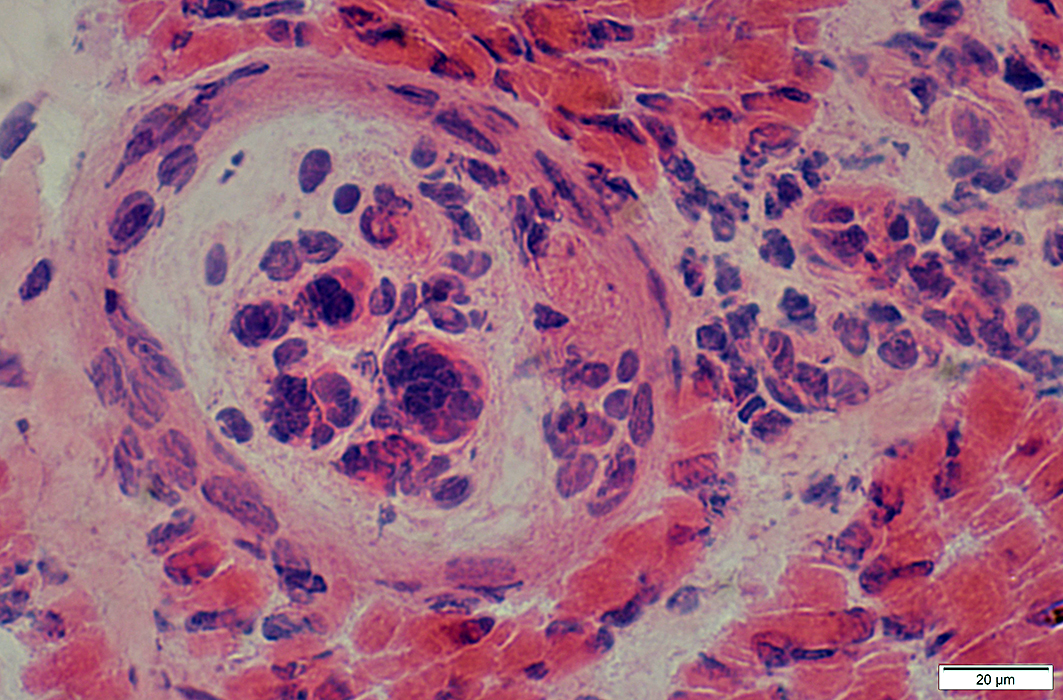
Fetal muscle: Extramedullary Hematopoesis
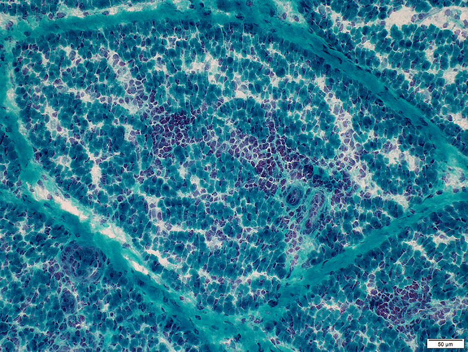 Gomori trichrome stain |
Cells surrounding smaller vessels in vascular perimysium
<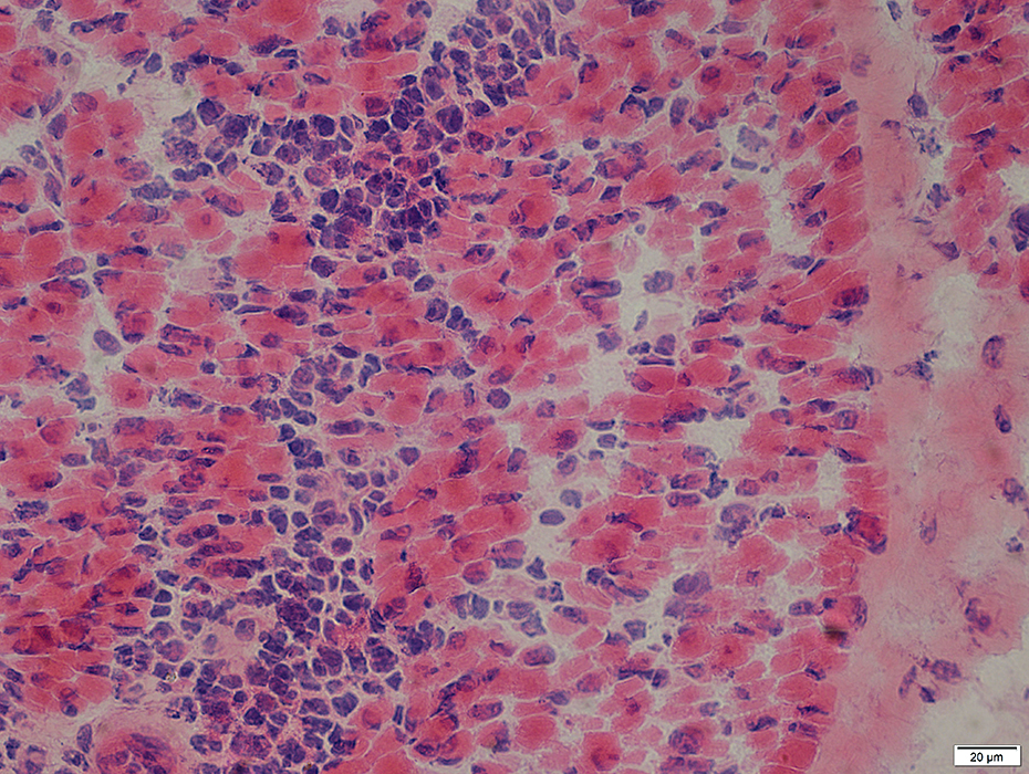 H&E stain |
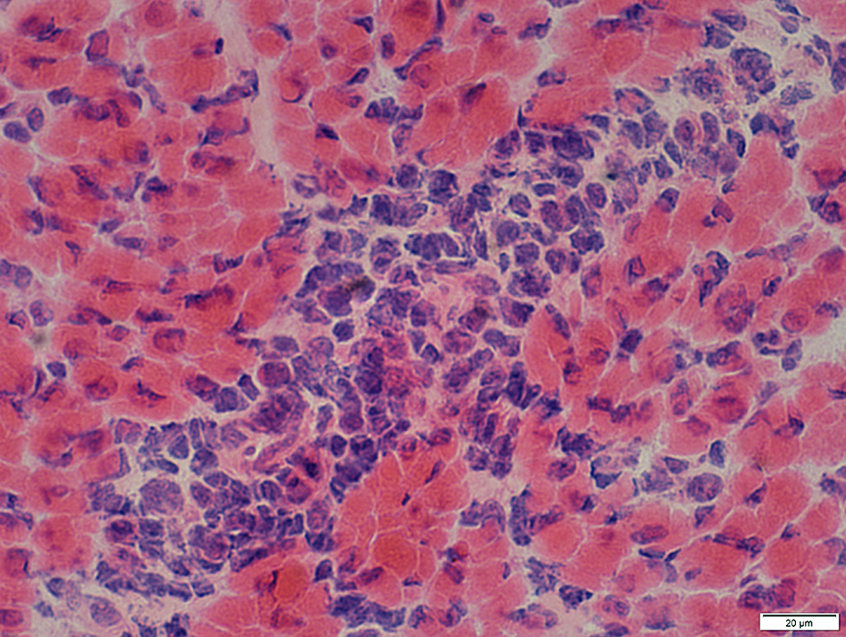 H&E stain |
Intramuscular Nerves
Axons surrounded by myelin
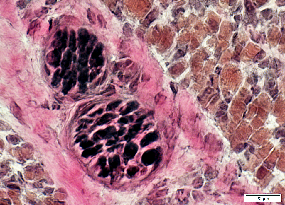 VvG stain |
Return to Neuromuscular Home Page
Return to Pathology index
6/11/2017