
Neuromuscular Junctions
See: Terminal Motor Axons
|
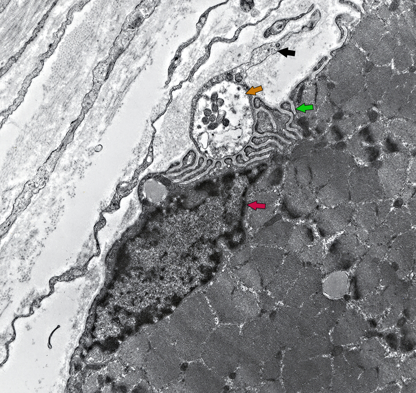
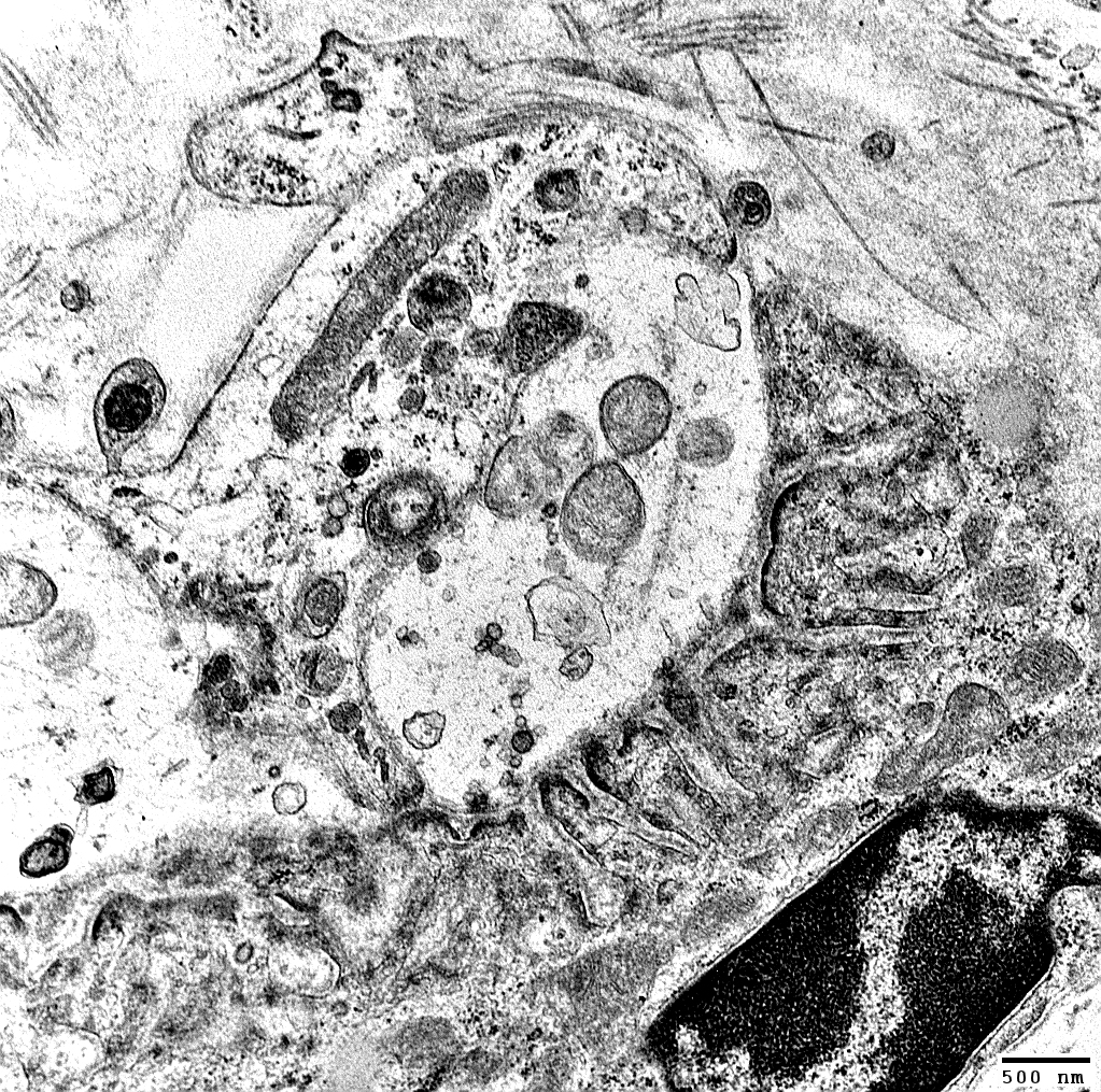
Presynaptic Axon Terminal: Brown Arrow
Vesicles: Clustered near post-synaptic folds
Mitochondria: Present in axon terminal cytoplasm
Terminal Schwann Cell (Telodendroglia): Black Arrow
Surrounded by pale Basal lamina
Post-synaptic Folds: Green Arrow
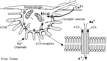
AChRs: Concentrated at top of folds, near nerve terminal
Na+ Channels: Concentrated at bottom of folds
Note Basal lamina layer within folds
Acetylcholinesterse: Located in Basal lamina
NMJ Myonucleus: Red Arrow
Molecular program has specificity for NMJ molecules
Endomysial Fibroblasts (Left): Long, thin cell processes
Muscle Fiber (Bottom Right): Sarcomeres cut in cross-section; Lipid droplets (2)
Also see: Esterase stain
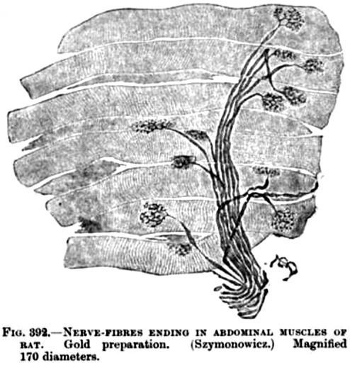
NMJs: Species differences
Human
Size: Small (20 μM)
Postsynaptic folding: Extensive
Morphology: Fragmented ("Cat's paw")
AChR clusters: Irregular
Mouse
Morphology: Pretzel-shaped
Size: Large (30 μM)
Compact
AChR clusters: Few branches
Neuromuscular Junction: Ions & Molecules
 |
|
7/1/2025