BULBOSPINAL MUSCULAR ATROPHY
Patterns of Pathology: Muscle
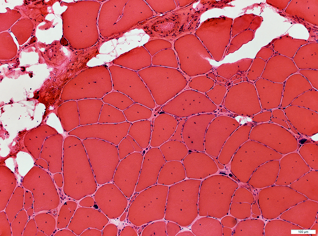
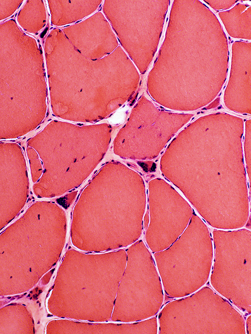
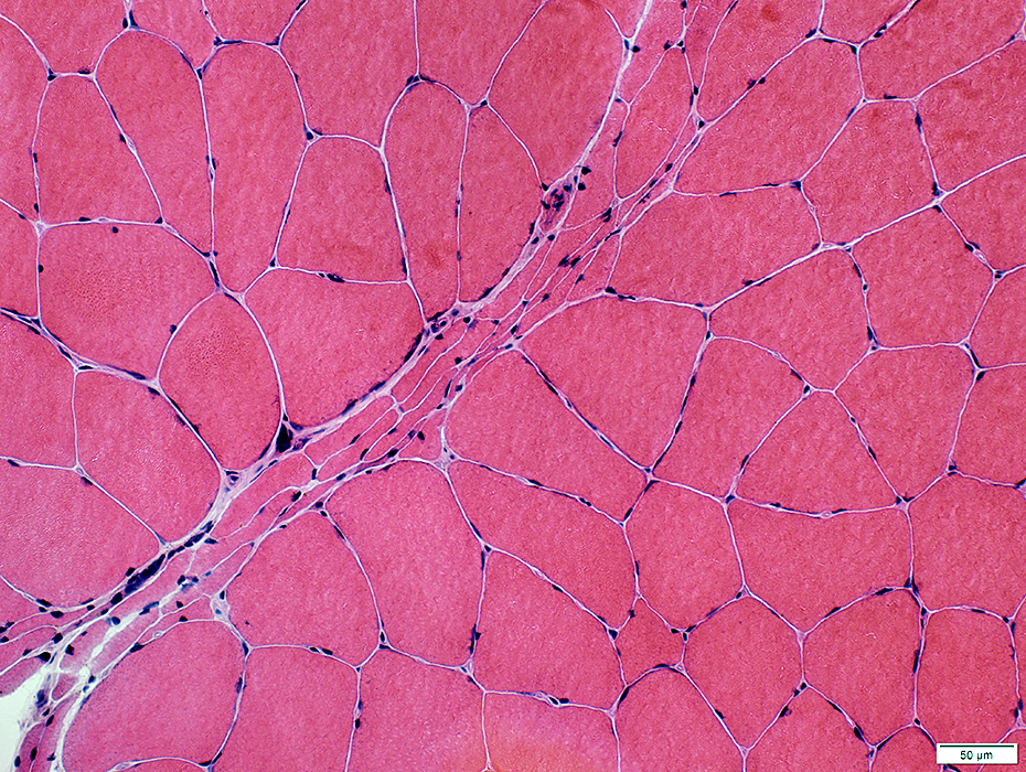
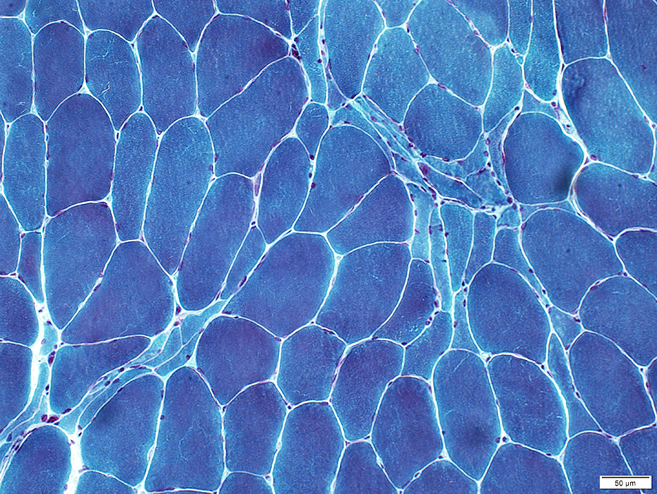
H & E stain |
|
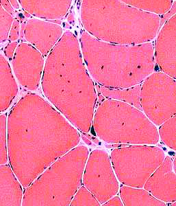
Muscle Fibers Sizes: Varied Hypertrophic Small: Angular or Rounded Grouped or Individual Pyknotic nuclear clumps (Large) Internal architecture Partially fused (Split) Internal nuclei Damage Necrosis: Scattered large fibers Regeneration: Clustered immature fibers |
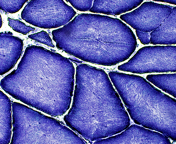
 VvG stain |
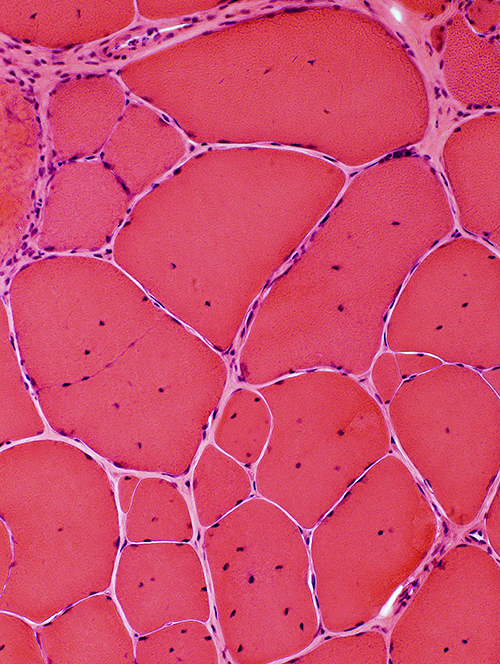
 H & E stain |
 VvG stain |
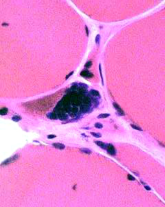
 H & E stain |
 |
 |
 |
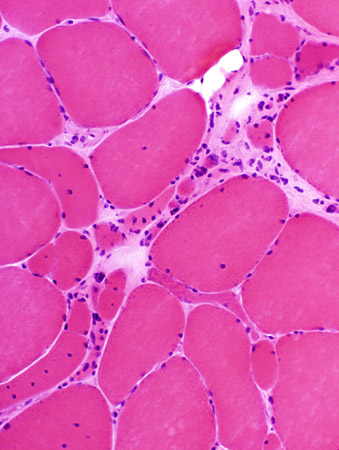
Internal nuclei
Partial, or incomplete, fusion
Hypertrophy
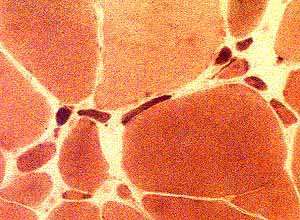
Pyknotic nuclear clumps
|
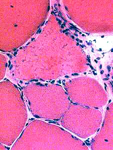
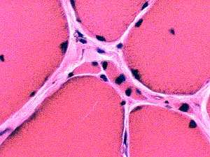
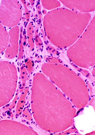
Muscle fibers: Small & Angular, or Pyknotic nuclear clumps |
|
 H & E stain |
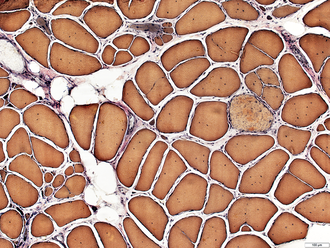
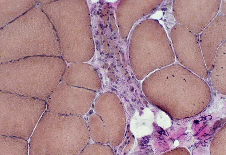
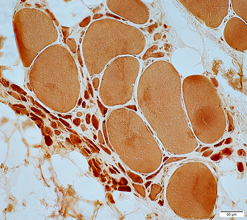
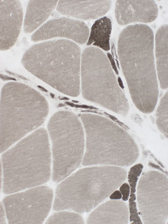
 Esterase stain |
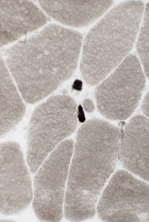
Muscle fibers: Grouped atrophy
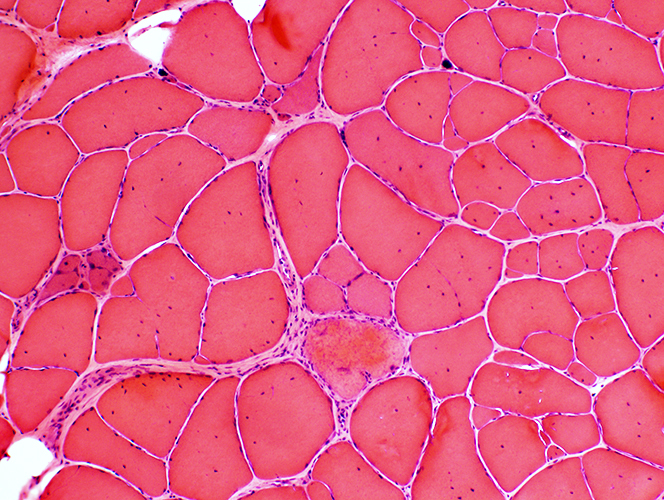
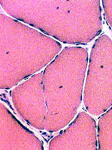
 H&E stain |
 H&E stain |
 H&E stain |
|
|
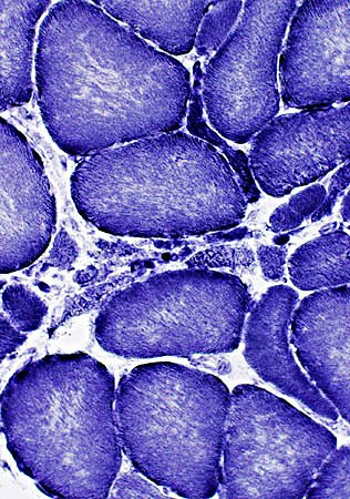
Grouped atrophy Smaller fibers may be rounded or angular Large muscle fibers are hypertrophied | ||
 VvG stain | ||
 Gomori trichrome stain |
 Esterase stain |
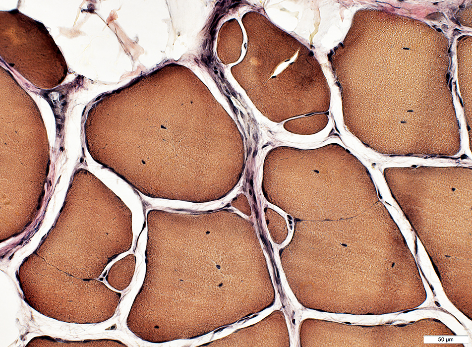
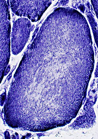
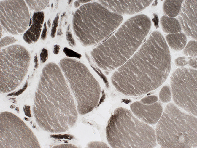
Muscle fibers: Internal architecture
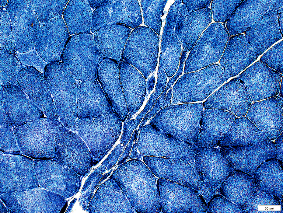
 NADH stain |
 NADH stain |
|
Some small fibers are darkly stained Large muscle fibers Coarse internal architecture; Pale centers "Splitting" (Partial fusion)(Below) | |
 NADH stain | |
 NADH stain |
Muscle: Fiber types
 ATPase, pH 9.4 |
 ATPase, pH 9.4 |
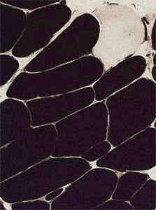 ATPase pH 4.3 stain |
|
Large muscle fibers: Type predominance Types: 1 or Abnormal with pale stain at pH 9.4 & pH 4.3 Small muscle fibers: Either type 1 or 2 Grouped atrophy: Multiple; Small sizes, Mixed fiber types |
 ATPase pH 9.4 stain |
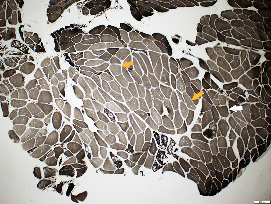 ATPase pH 9.4 stain |
Dark fiber on ATPase pH 9.4 (White arrow) is also dark at ATPase pH 4.3 (Black arrow)
Intermediate fibers at ATPase pH 9.4 are pale at ATPase pH 4.3 (Orange arrows)
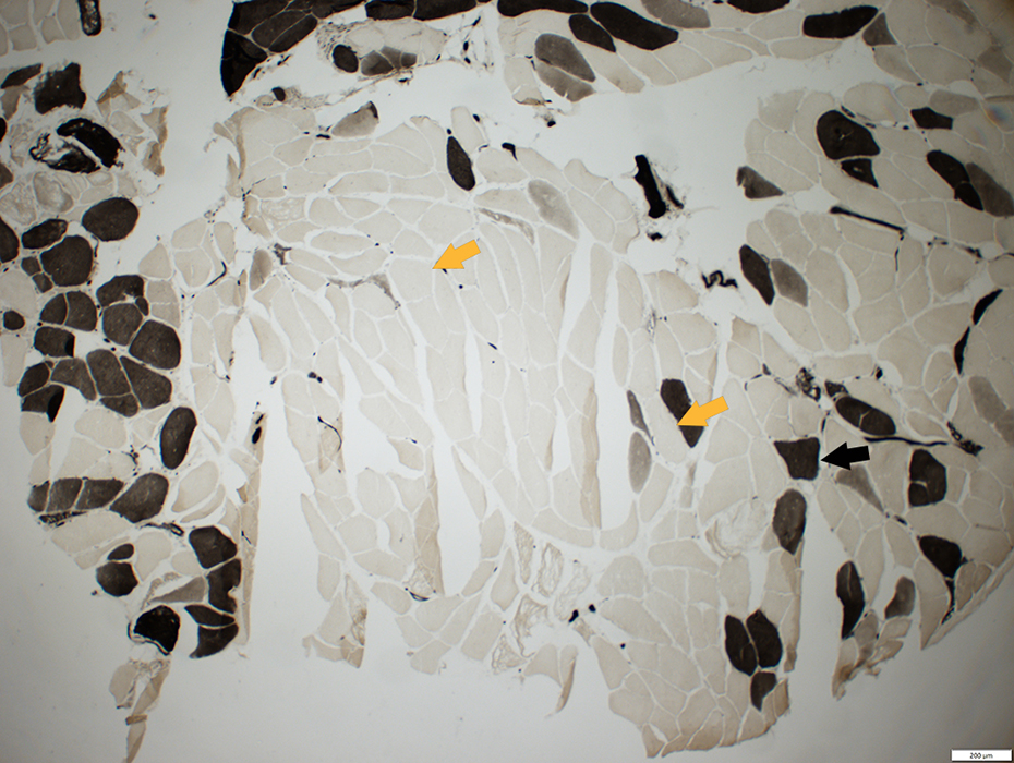 ATPase pH 4.3 stain |
Neuronal Intranuclear Aggregates
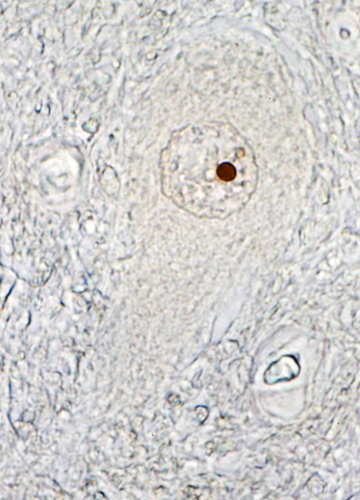 From Mei Li MD Androgen Receptor in Motor Neuron
|
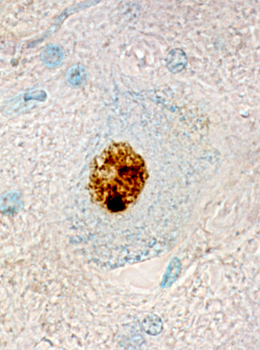 From Mei Li MD Polyglutamine in Motor Neuron
|
Return to Neuromuscular Home Page
Return to Pathology index
Return to BSMA (Kennedy's syndrome)
5/24/2019