Benign Acute Childhood Myositis (BACM)
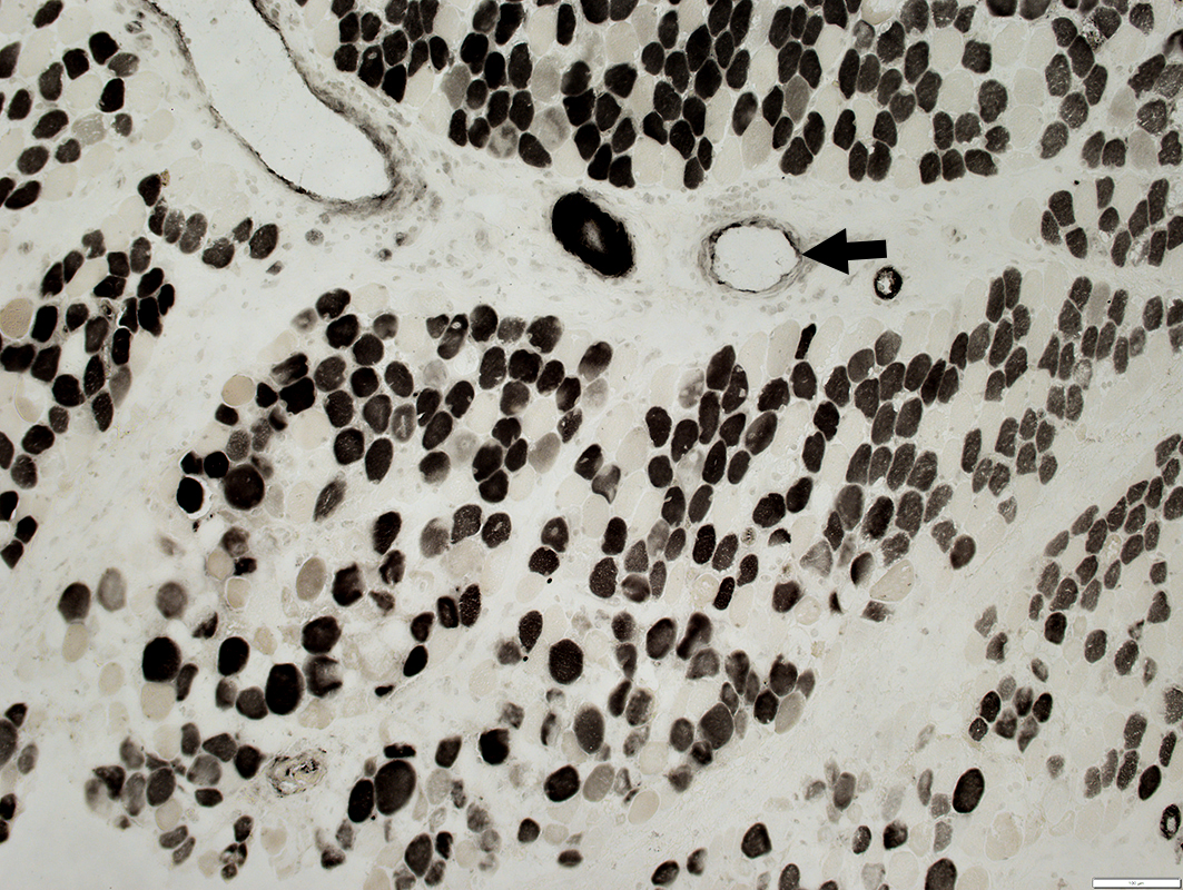
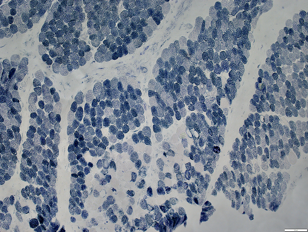
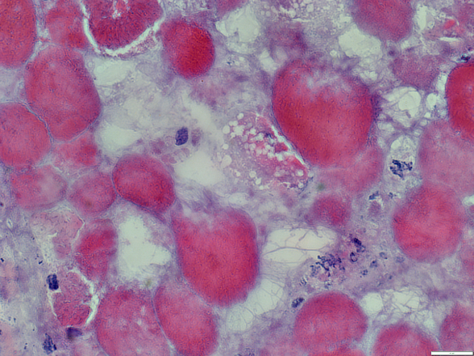
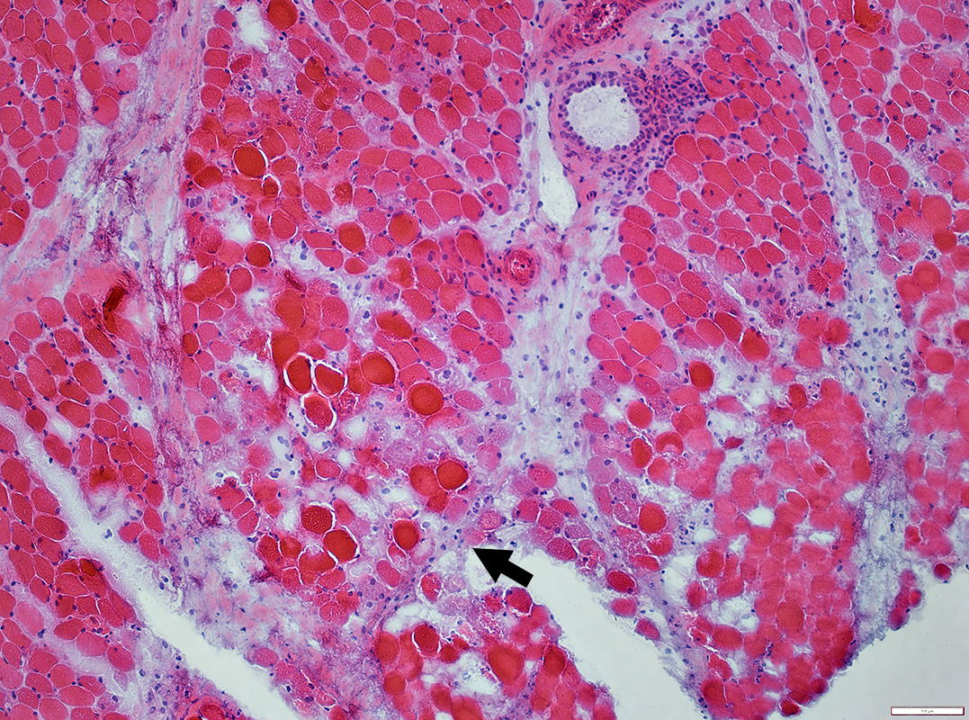 H&E stain |
Large cluster of necrotic fibers (Arrows) with
Cytoplasm: Dark or Very Pale
Internal architecture & Myonuclei: Lost
Perimysium near region of clustered necrosis
Fragmented & Pale connective tissue
Wide
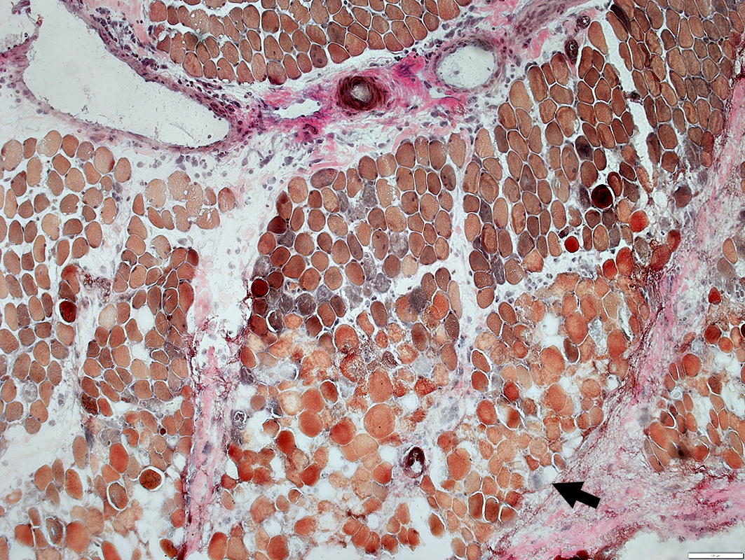 VvG stain |
Necrosis: Regional, Early stage
Large cluster of necrotic fibers (Arrow) with
Cytoplasm: Dark or Very Pale
Internal architecture & Myonuclei: Lost
Perimysium near region of clustered necrosis
Fragmented & Pale connective tissue
Wide
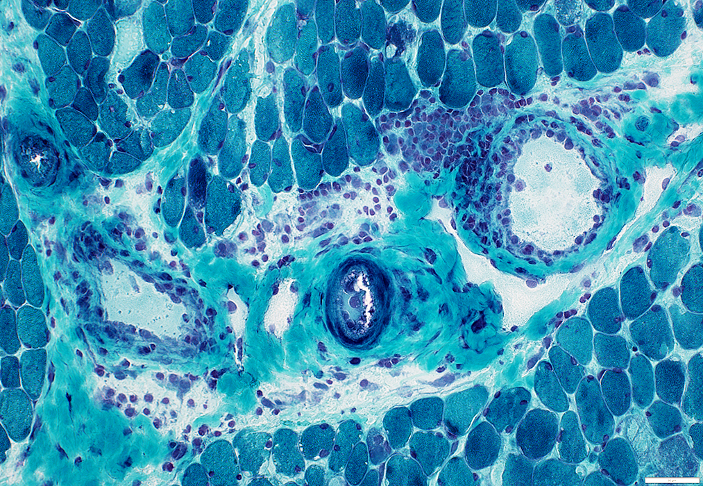
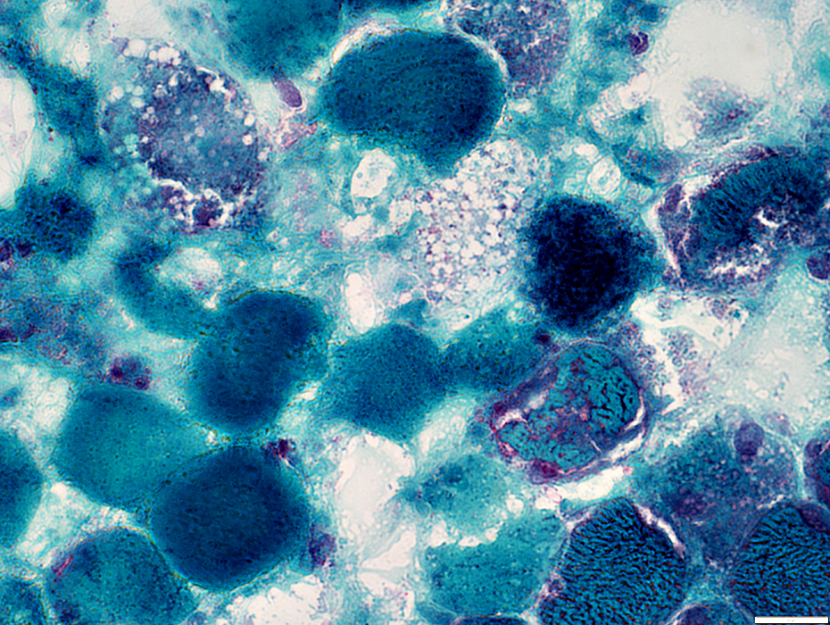
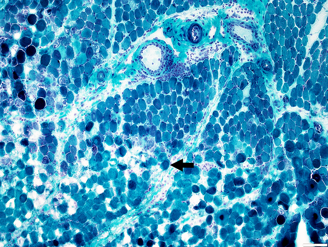 Gomori trichrome stain |
Necrosis: Regional, Early stage
Large region containing very pale necrotic fibers (Arrows)
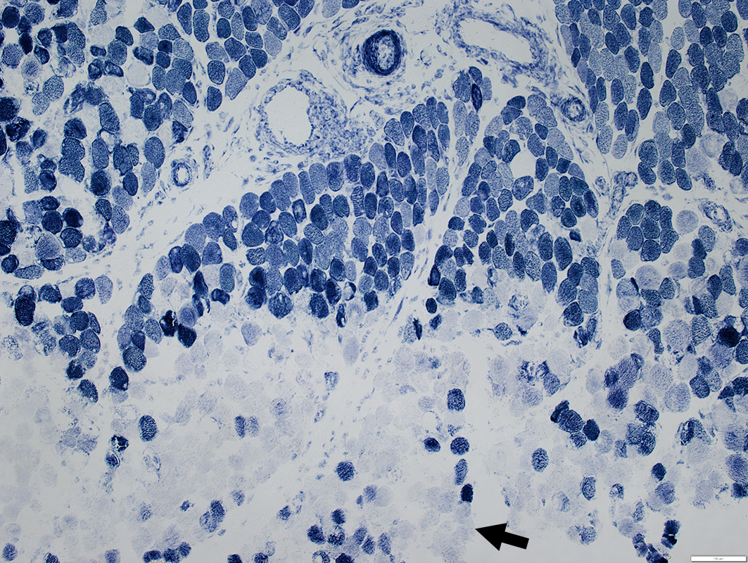 NADH stain |
Necrosis: Regional, Early stage
Scattered fibers in region of necrosis have intermediate staining
Perimysial vein (Arrow): Pale staining wall
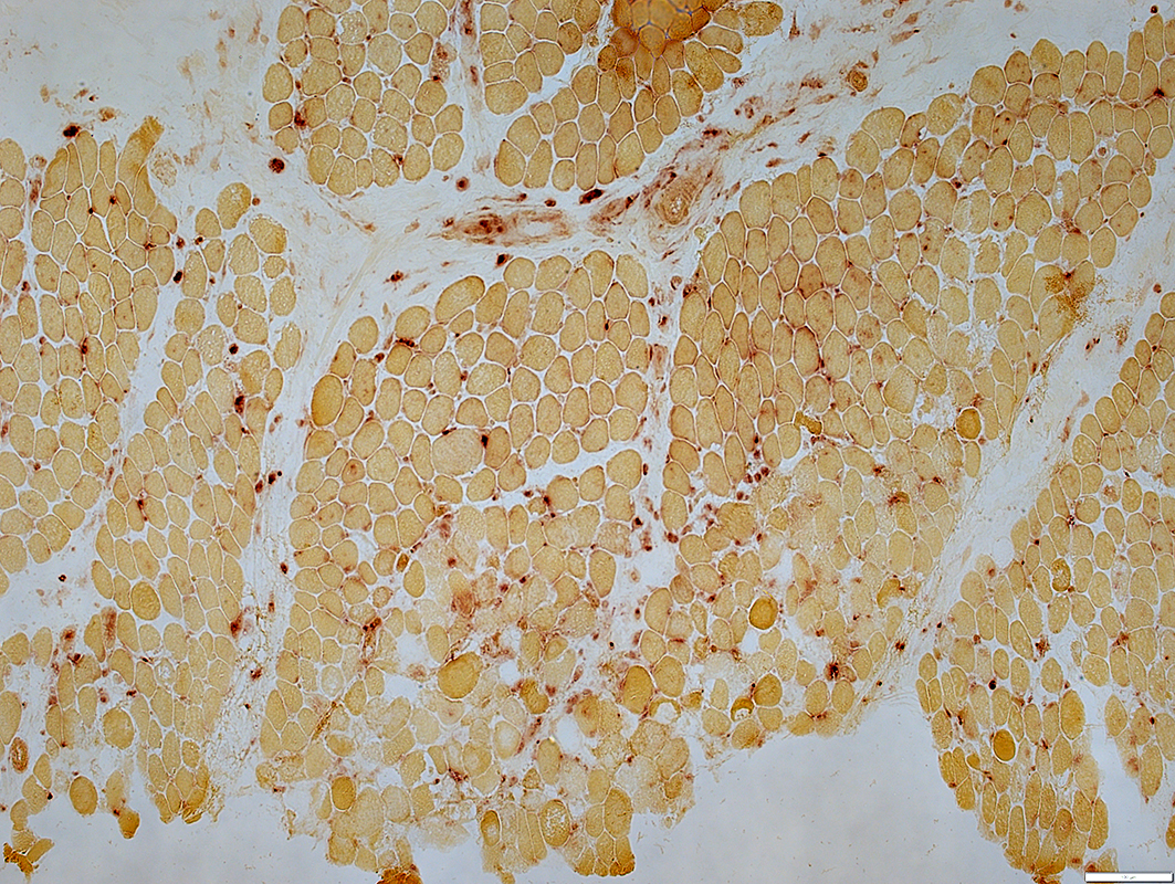
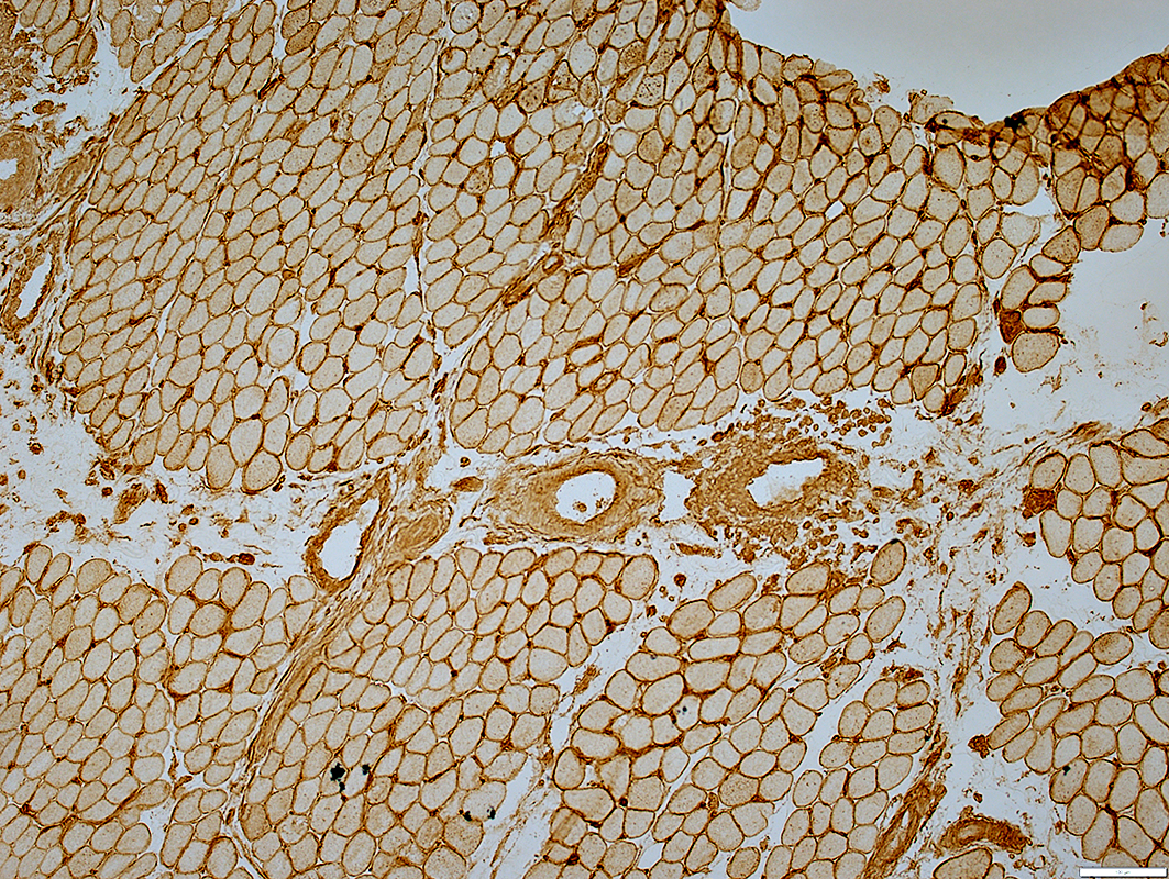
 ATPase pH 4.3 stain |
Necrosis: Regional, Early stage
Histiocytes (Acid phosphatase +): Scattered in perimysium & endomysium
In this early stage, and likely due to an ischemic nature of the process, there is no invasion of muscle fibers by histiocytes
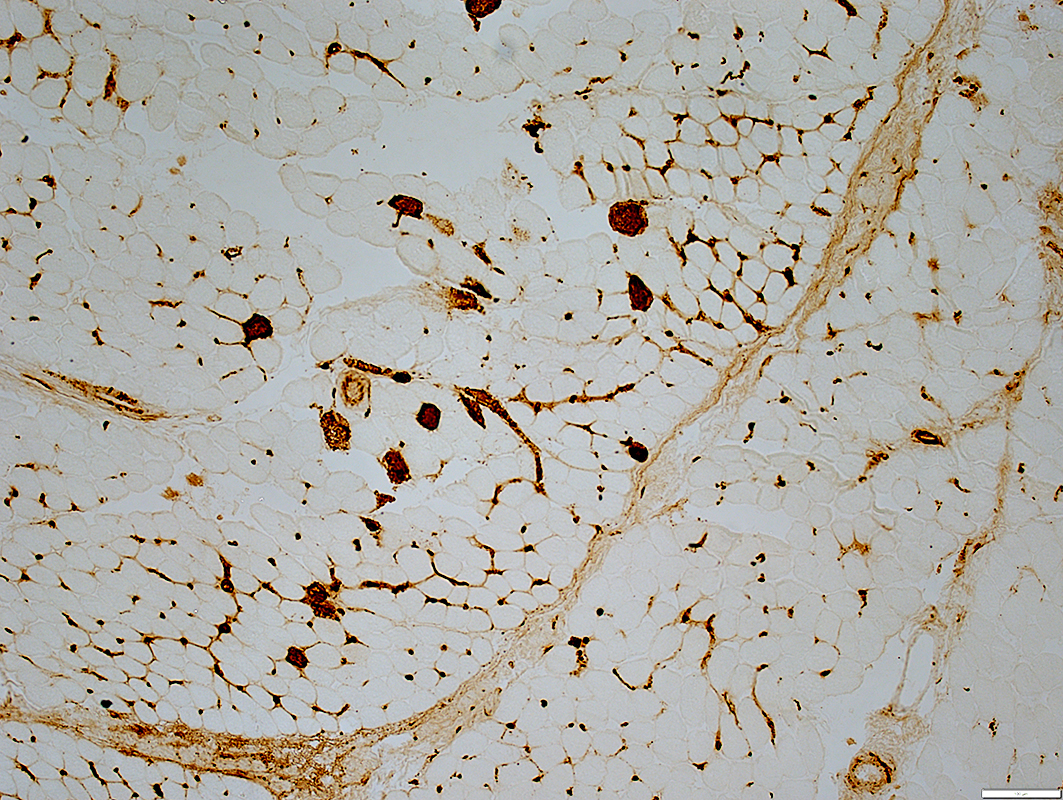
 Acid phosphatase stain |
Necrosis: Regional, Early stage
Perimysium: Only minor staining
Muscle fibers: No staining around fibers in region of necrosis
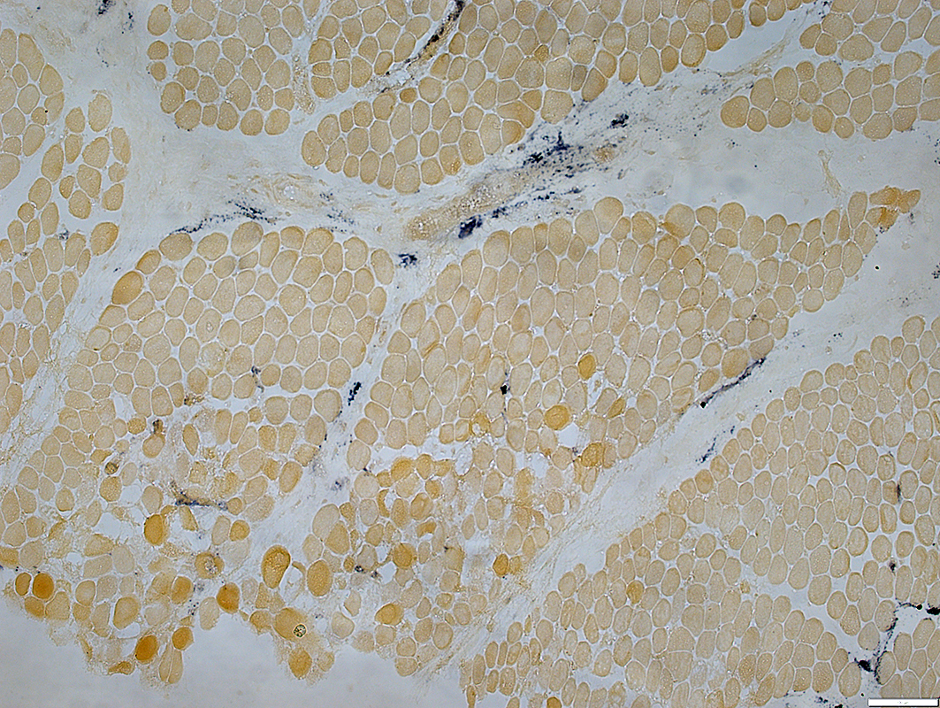 Alkaline phosphatase stain |
Necrosis: Regional, Early stage
 COX stain |
 SDH stain |
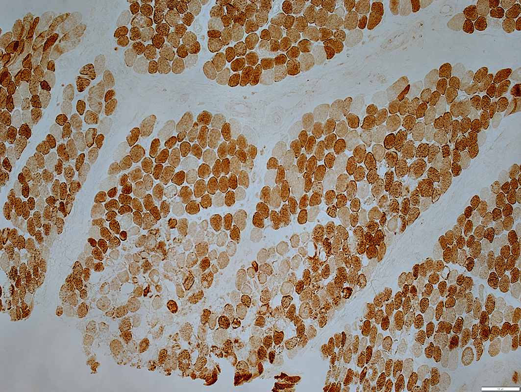
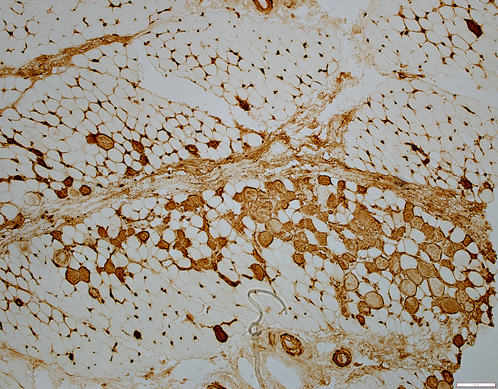
Necrosis: Regional, Early stage
Muscle fibers outside region of necrosis have increased MHC1 on their surfaces
 MHC I stain |
 C5b-9 stain |
C5b-9
Region of clustered necrosis (Above): Stains cytoplasm of many muscle fibers
Necrotic muscle fibers
Are mostly located near avascular perimysium
Endomysial capillaries: Many stained for C5b-9
Perimysial connective tissue: Diffusely stained for C5b-9
Region with only scattered muscle fiber necrosis (Below)
C5b-9 stains many endomysial capillaries
Perimysial connective tissue: Some areas stained for C5b-9
 C5b-9 stain |
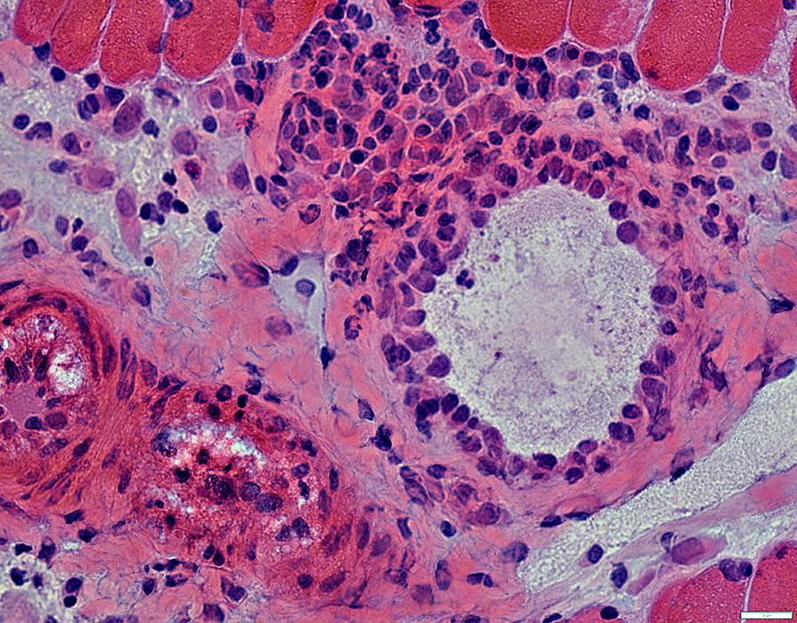
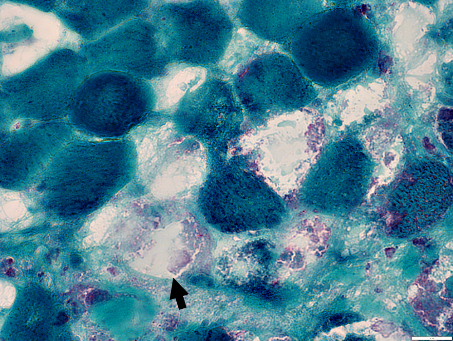
 Gomori trichrome stain |
Surrounded by damaged perimysium populated by increased cellularity (probably histiocytes).
 H&E stain |
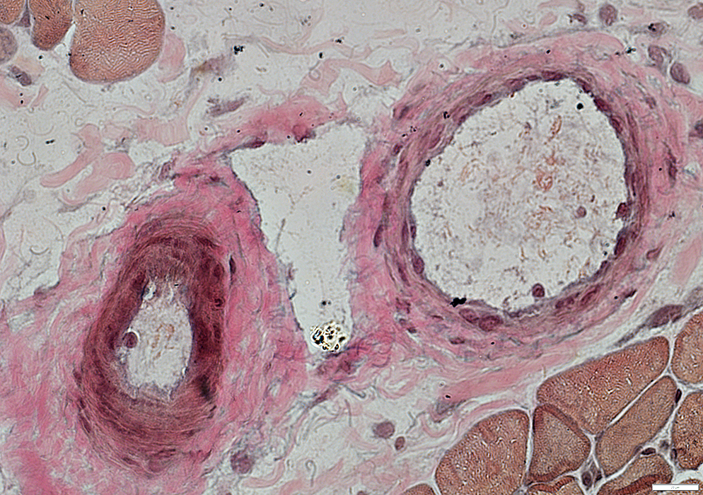 VvG stain |
Vessel walls are abnormal with loss of fibrils.
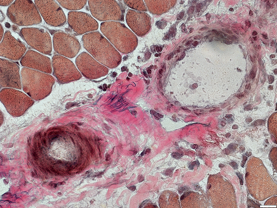 VvG stain |
 H&E stain |
Early necrotic fibers: Have "Dark" necrosis with dark stained featureless cytoplasm and no myonuclei
Some non-necrotic fibers (Bottom Right) have retained sarcoplasmic reticulum staining in cytoplasm
Later necrotic fibers: Pale, fragmented cytoplasm with no associated histiocytes
 Gomori trichrome stain |
Necrosis, Later stage
Many Pale-stained fibers (Arrow) with: Fragmented cytoplasm; No associated histiocytes
Scattered "Dark" Necrotic Fibers are also present
 Gomori trichrome stain |
Return to: BACM
See: RIIM
Return to: Neuromuscular Home Page
11/10/2022