Myopathy with NMO & Aquaporin-4 (AQP4) Antibodies
|
Capillaries Histiocytes Muscle fibers Aggregates & Vacuoles Cytoplasmic bodies C5b-9 deposition MHC Class I Pathology patterns Perimysium |
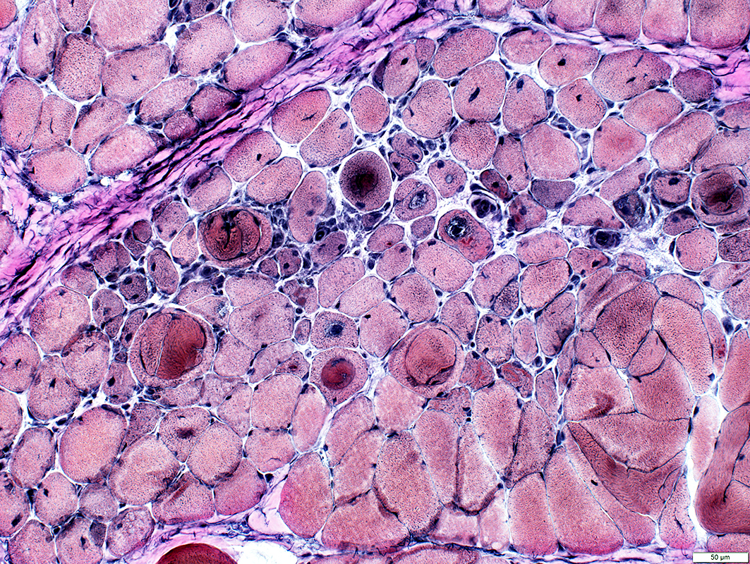
Muscle Fibers
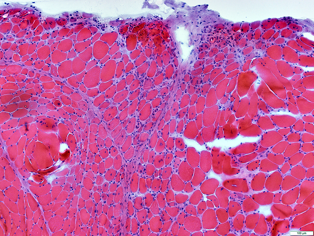 H& E stain |
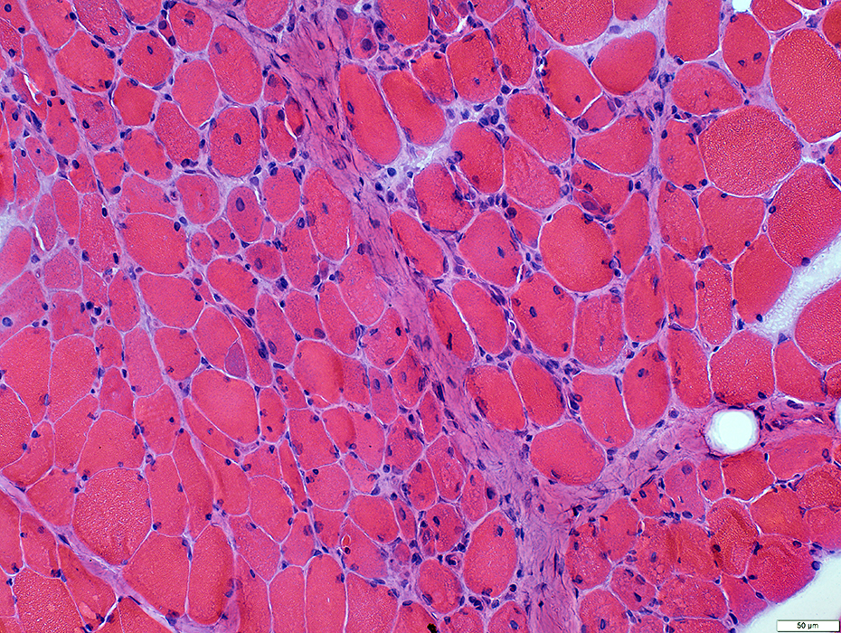
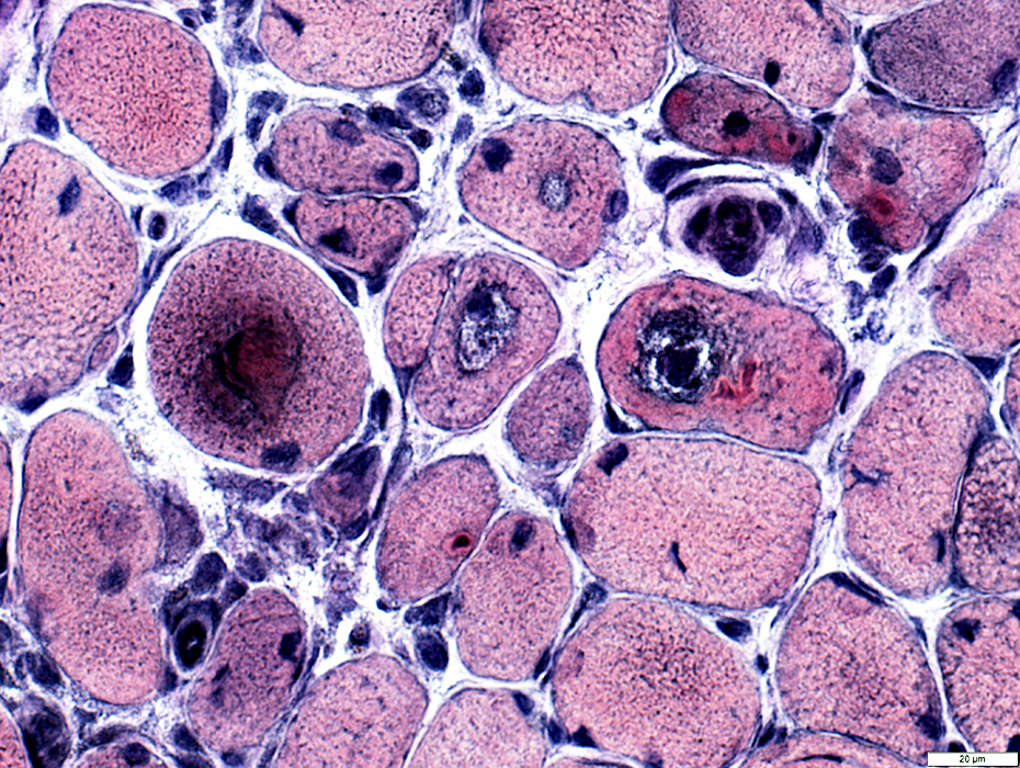
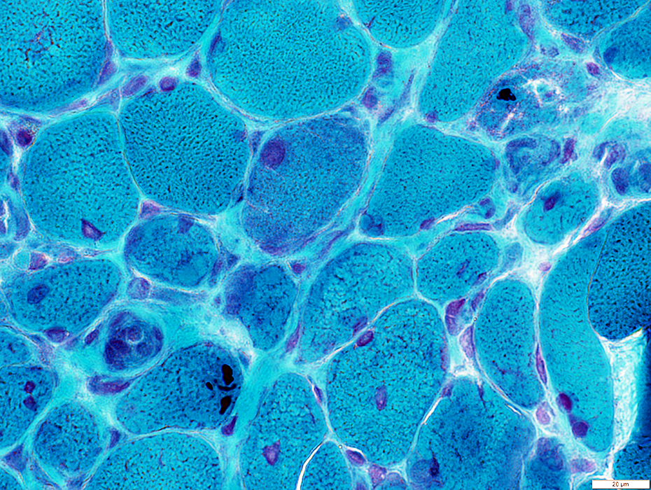
Atrophy & Immaturity
More pathology at edges of fascicles
 H & E stain |
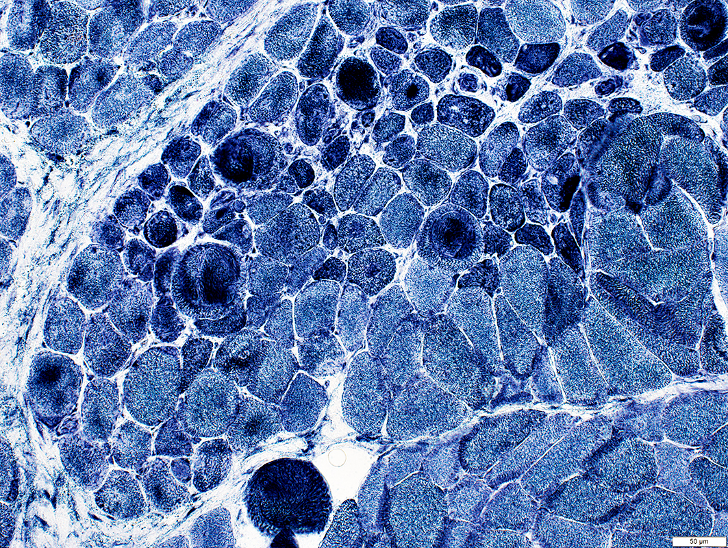
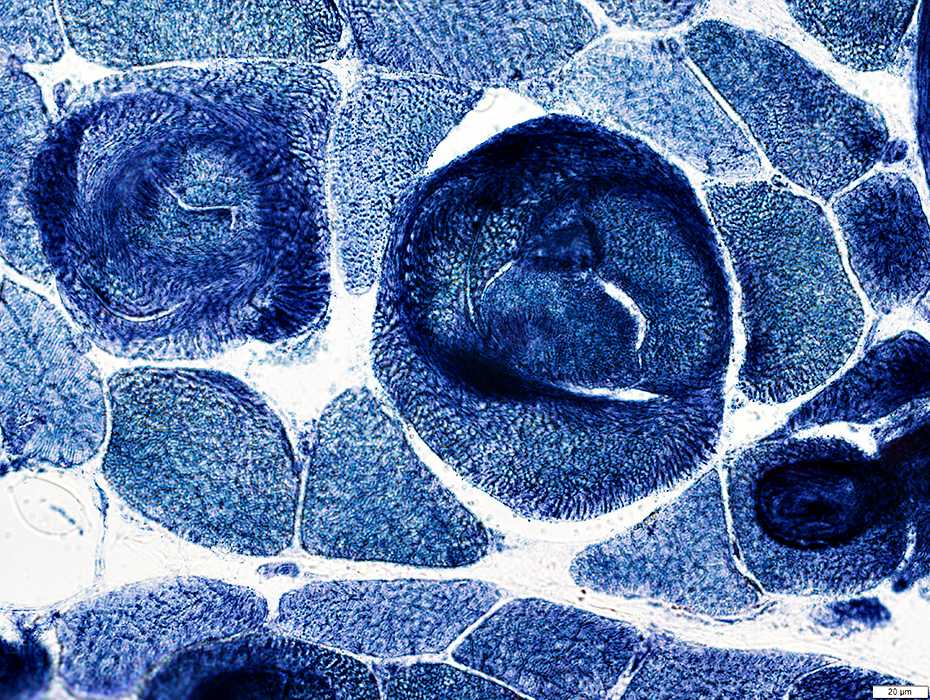
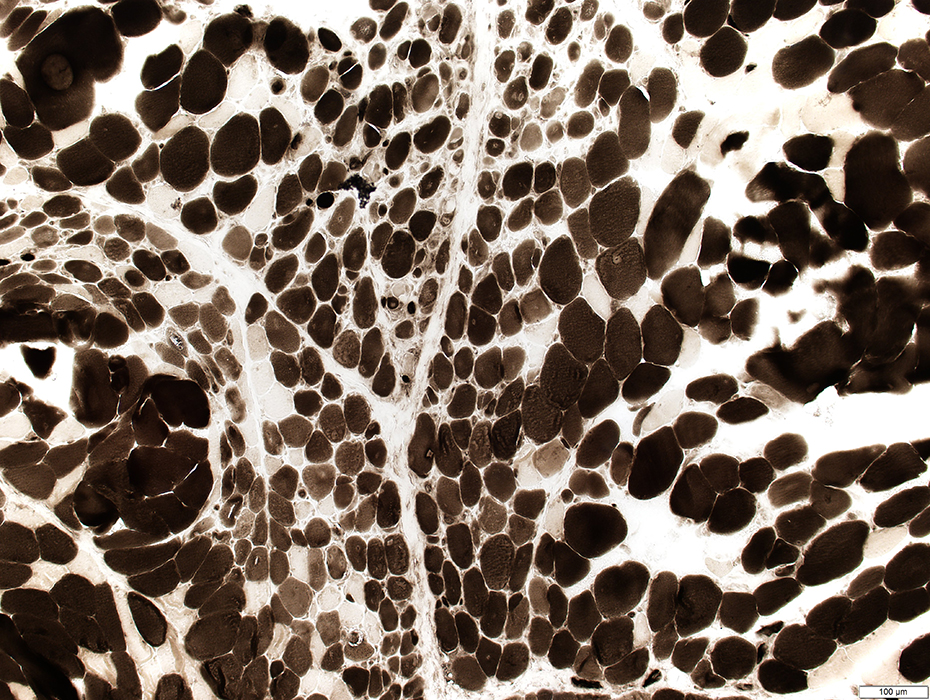 ATPase pH 4.3 stain |
Perimysial Connective Tissue
 Alkaline phosphatase stain |
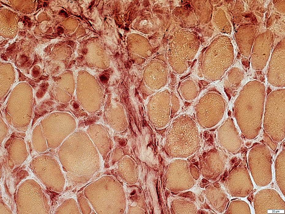
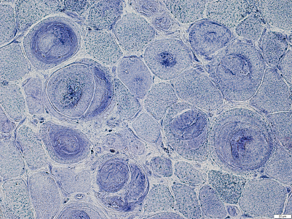
Acid phosphatase: Stains cells in perimysium & endomysium
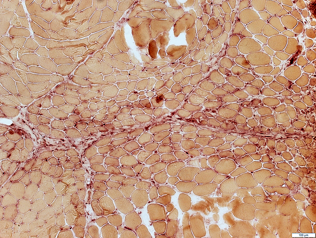 Acid phosphatase stain |
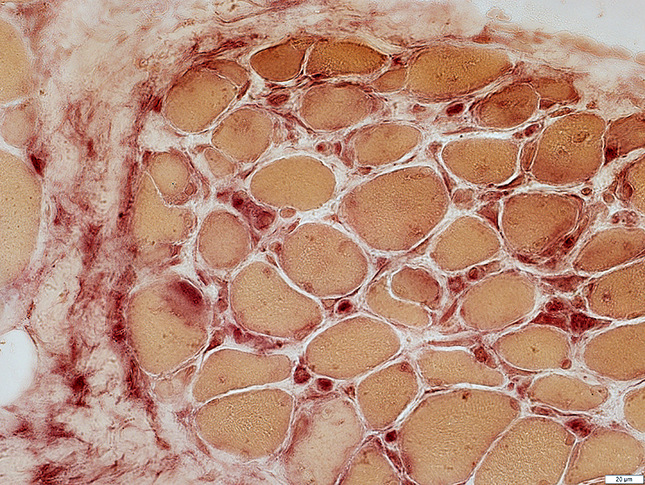 Acid phosphatase stain |
Acid phosphatase cells scattered in endomysium & perimysium
 Acid phosphatase stain |
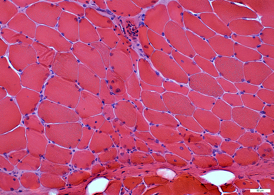
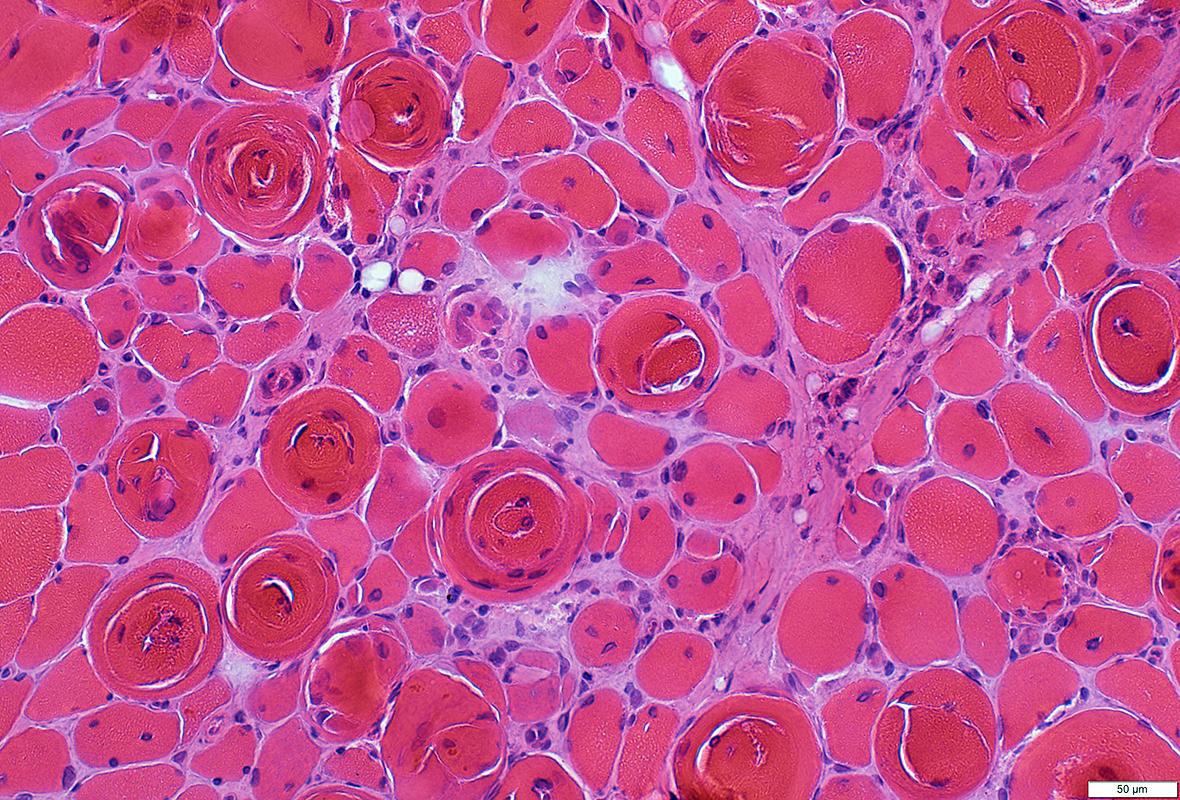
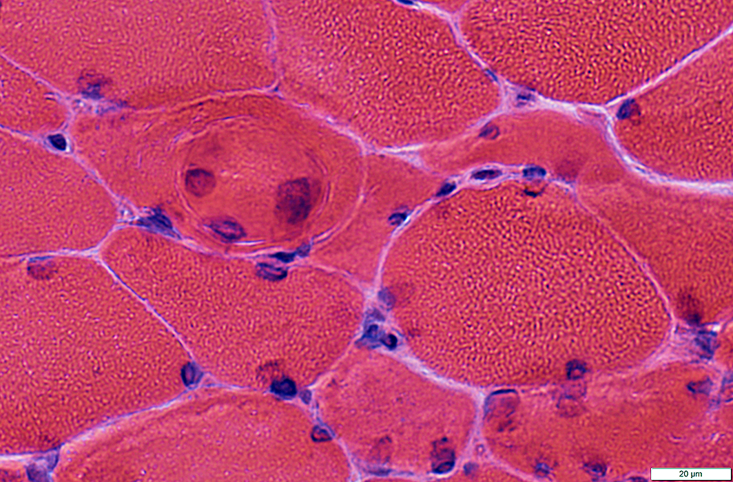
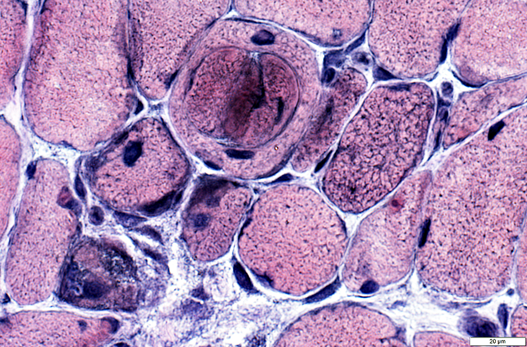
Muscle Fiber Pathology
 H & E stain |
Often immature (Basophilic)
Internal nuclei
Internal architecture
Sworled
Vacuoles & Aggregates
 VvG stain |
 NADH stain |
 H & E stain |
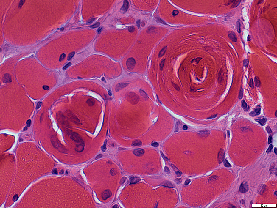 H & E stain |
 H& E stain |
 NADH stain |
 AMPDA blue stain |
 VvG stain |
 VvG stain |
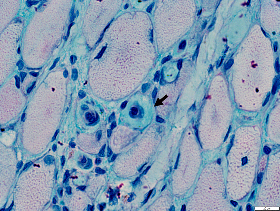
Cytoplasmic bodies
 Gomori trichrome blue stain |
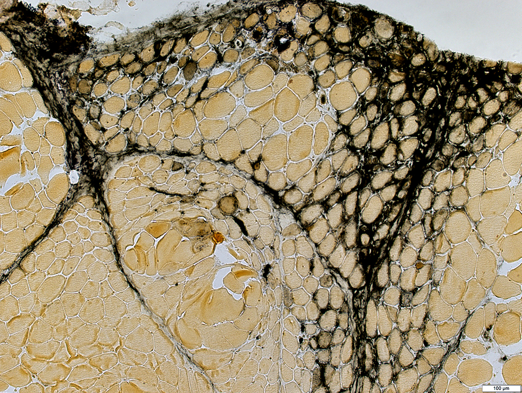
Capillary pathology
Some capillaries are large & have thickened basal lamina (Arrow)
There is a regional loss of endomysial capillaries in some areas
 Alcian blue stain |
 UEA1 stain |
Regional loss of endomysial capillaries, some perifascicular
UEA I also abnormally stains the surfaces of some muscle fibers
 UEA1 stain |
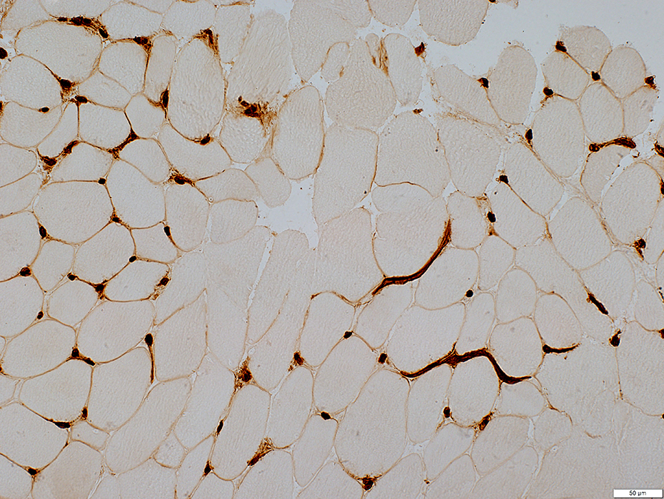
C5b-9 deposition
Connective tissue: Endomysium & Perimysium
Endomysial capillaries: Scattered
Muscle fibers: Patchy on surfaces
 C5b-9 stain |
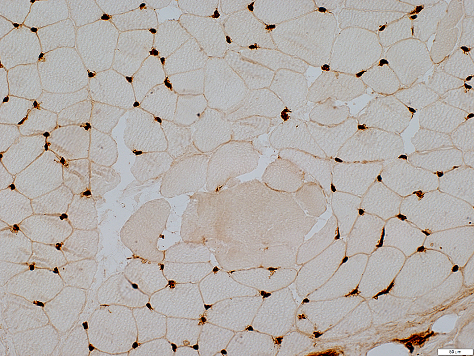
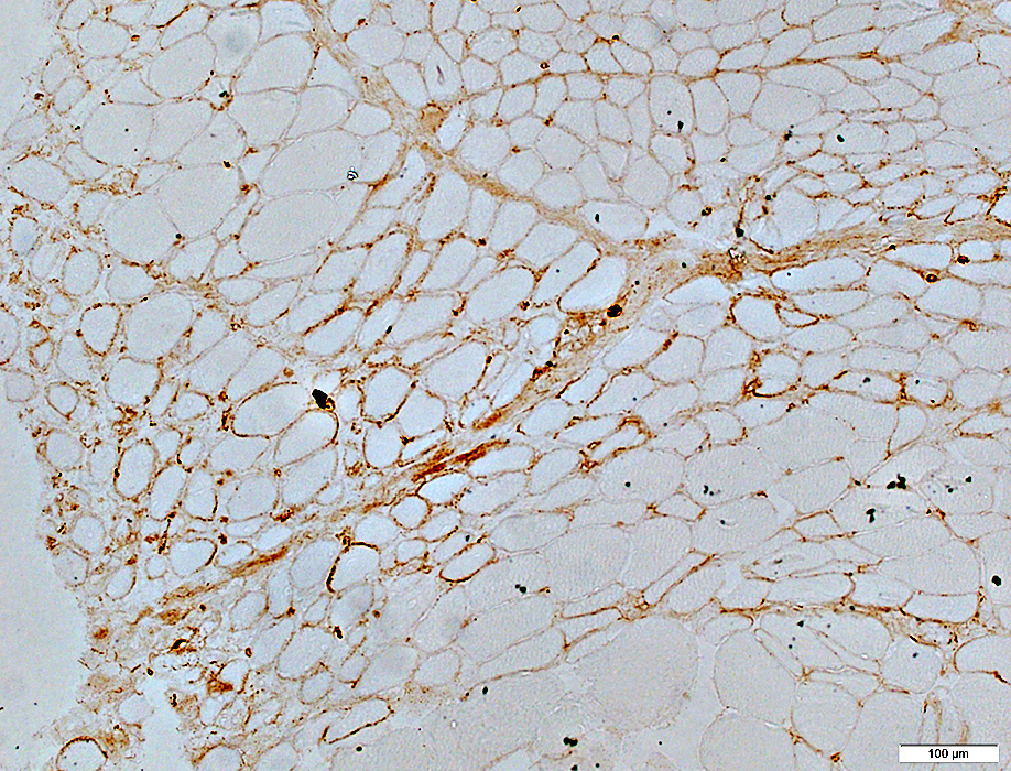
MHC-I upregulation by muscle fibers
In regions with muscle fiber atrophy
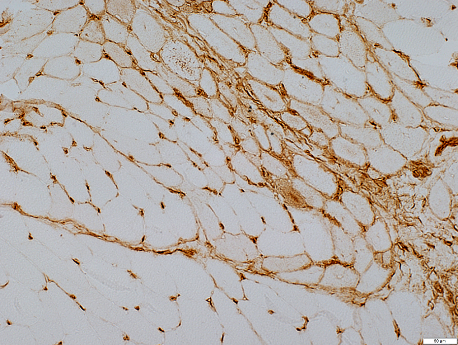 MHC1 stain |
Return to Neuromuscular Home Page
Return to Muscle autoantibodies
2/20/2021