Immune Myopathy with Serum IgG vs HMGCR
|
HMGCR pathology Active myopathy Muscle fibers Immature fibers Lipid Necrosis Nuclear pathology Vacuoles: 1; 2 Connective tissue Perimysium Epimysium Immune pathology Mild pathology Chronic myopathy Younger onset |
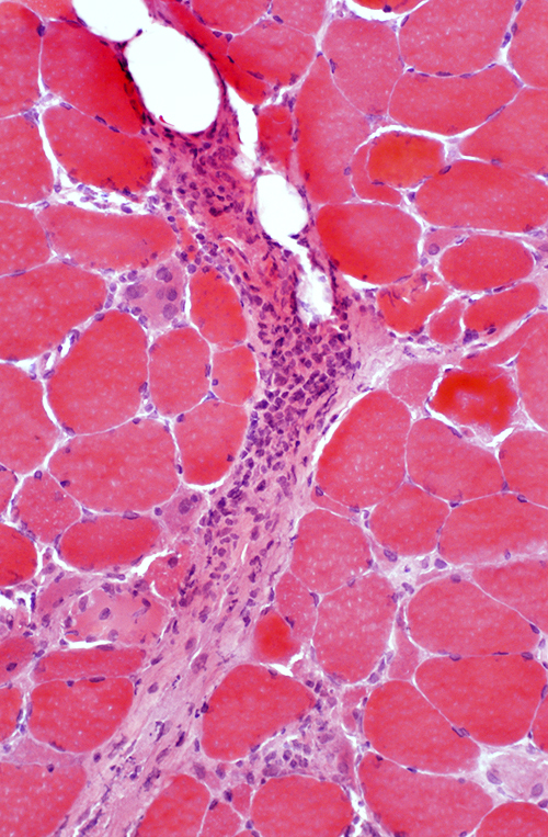
Active Myopathy with Perimysial Pathology (IMPP)
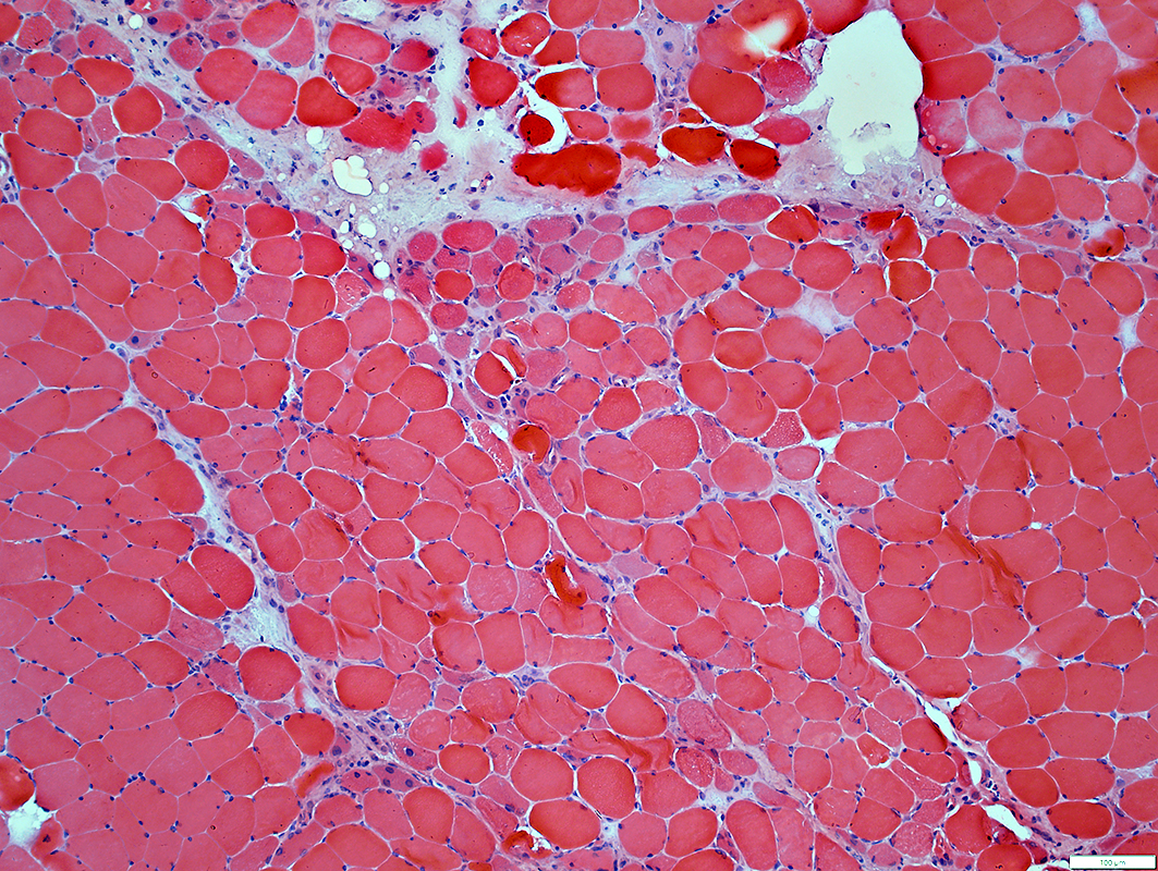 H&E stain |
Muscle fibers
Necrosis & Regeneration: Scattered or at Edge of fascicles
MHC1 upregulation: Diffuse or Perifascicular
Perimysium
Structure: Damaged
Histiocytic cells
Alkaline Phosphatase stain
 VvG stain |
 Alkaline Phosphatase stain |
 C5b-9 stain |
 MHC Class 1 stain |
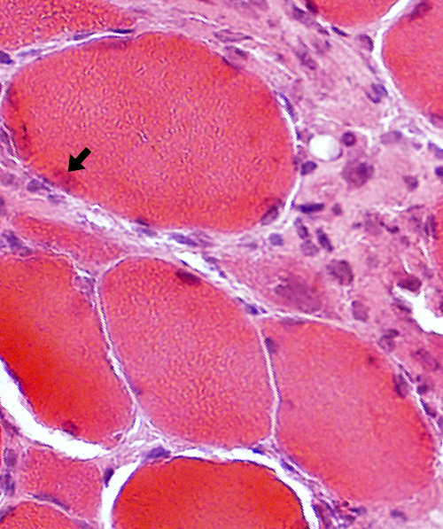
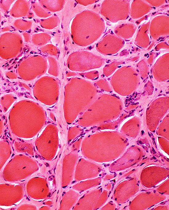
Myopathy, Ongoing
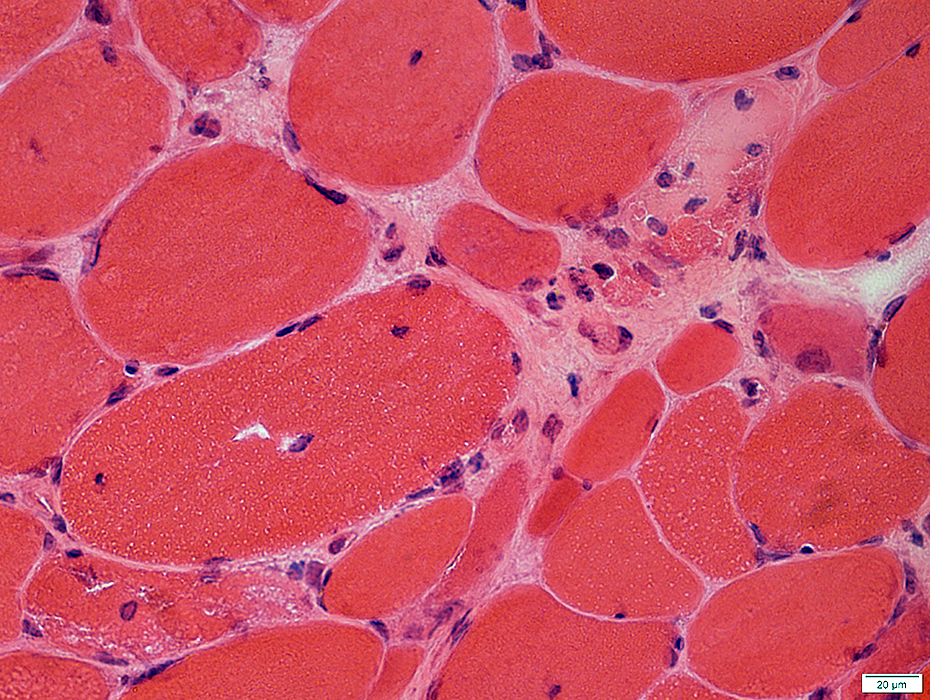
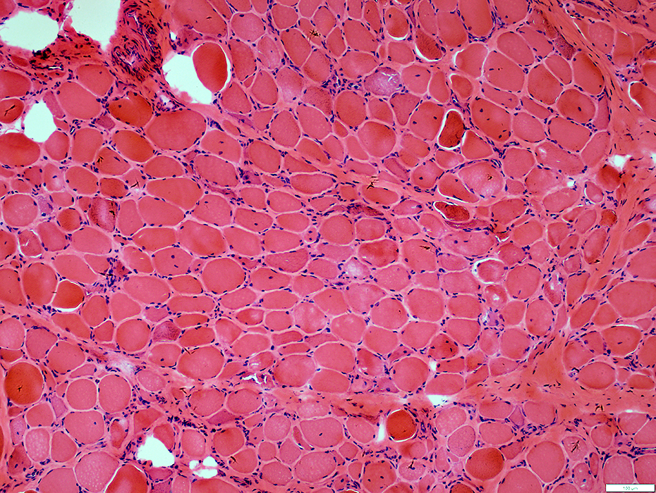
 H&E stain |
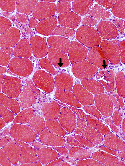
| Necrotic (Arrows) & Regenerating muscle fibers: Scattered | |
 H&E stain |
 H&E stain |
 H&E stain |
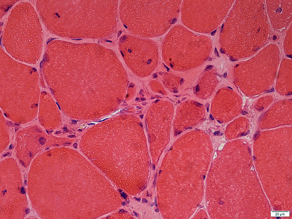
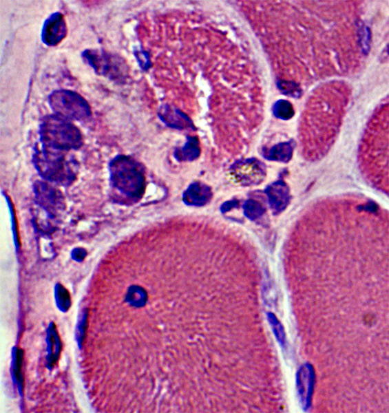
Necrosis & Regeneration
|
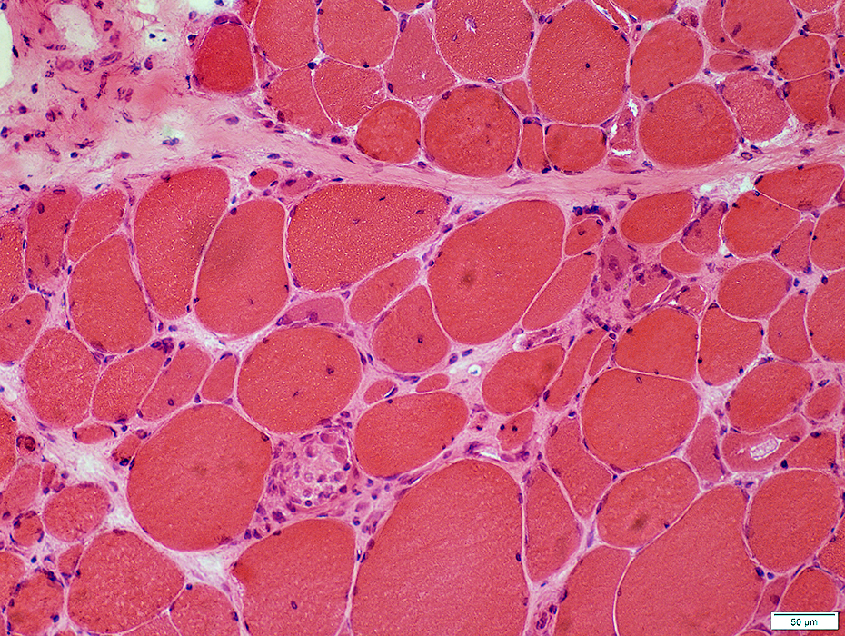
Variable Damage: Region with small muscle fibers & increased endomysial connnective tissue
|
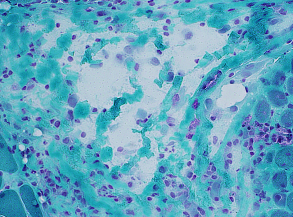
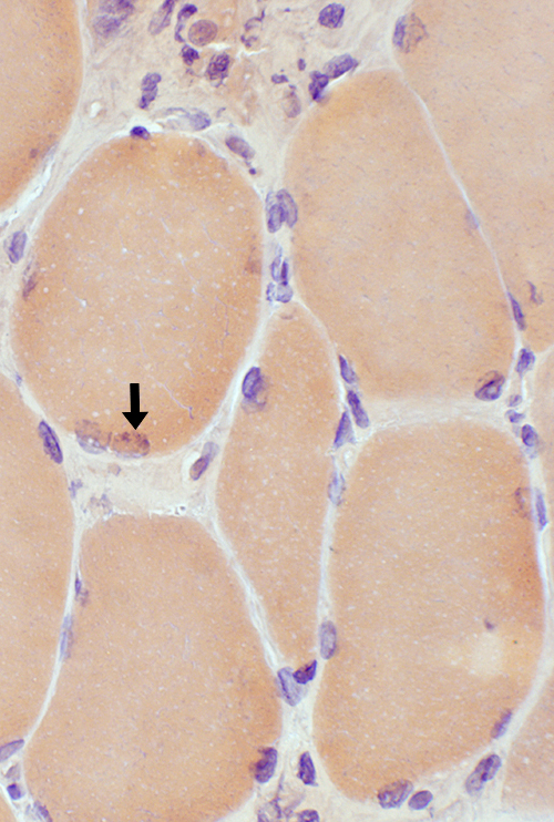
Necrotic muscle fibers are pale and infiltrated by cells (Arrow)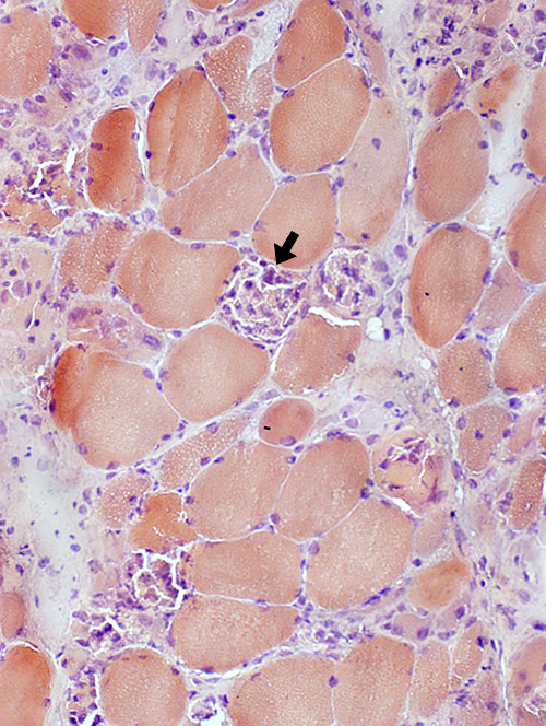 Congo red stain |
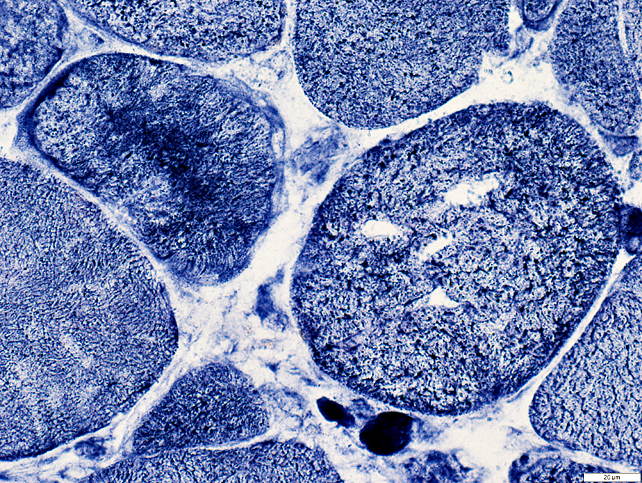
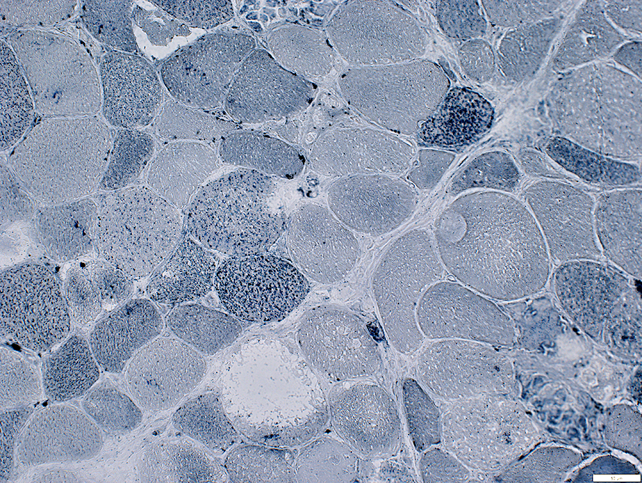
Necrotic muscle fibers: No or Pale staining (Dark arrow). Immature muscle fibers (Small): Dark stained (White arrow) 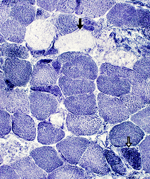 NADH stain |
 Acid phosphatase stain Necrotic muscle fibers Replaced, or infiltrated, by histiocytic (red) cells |
 Acid phosphatase stain |
 Acid phosphatase stain |
Necrotic Muscle Fibers
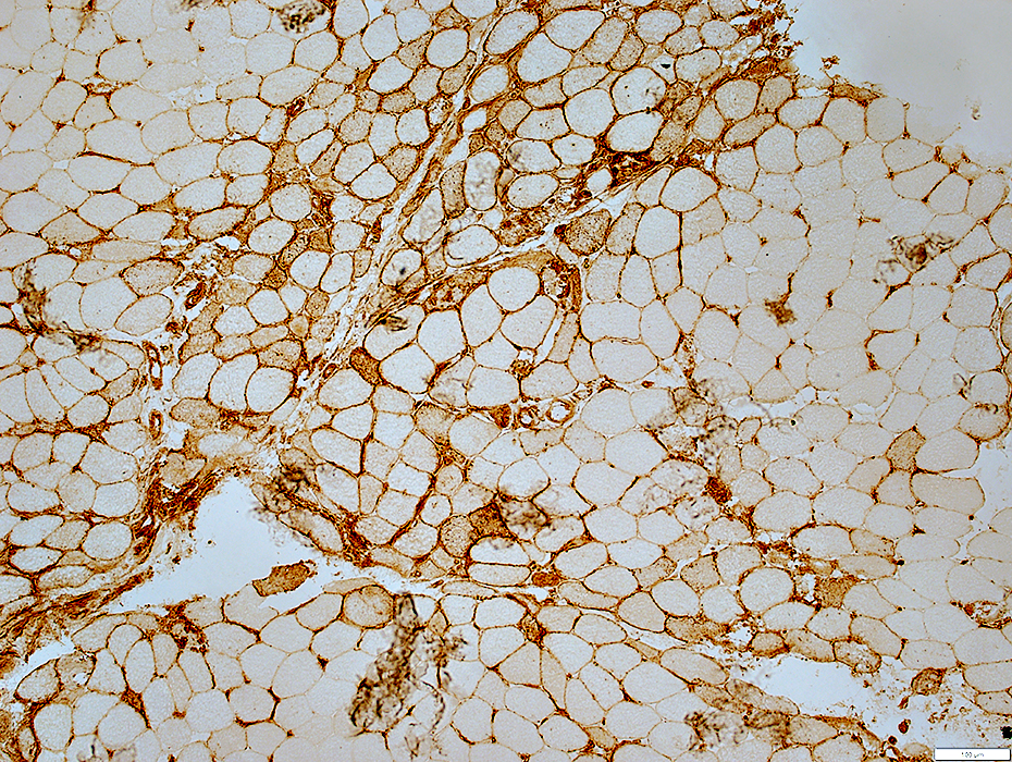
C5b-9 stains: Cytoplasm of necrotic fibers near perimysium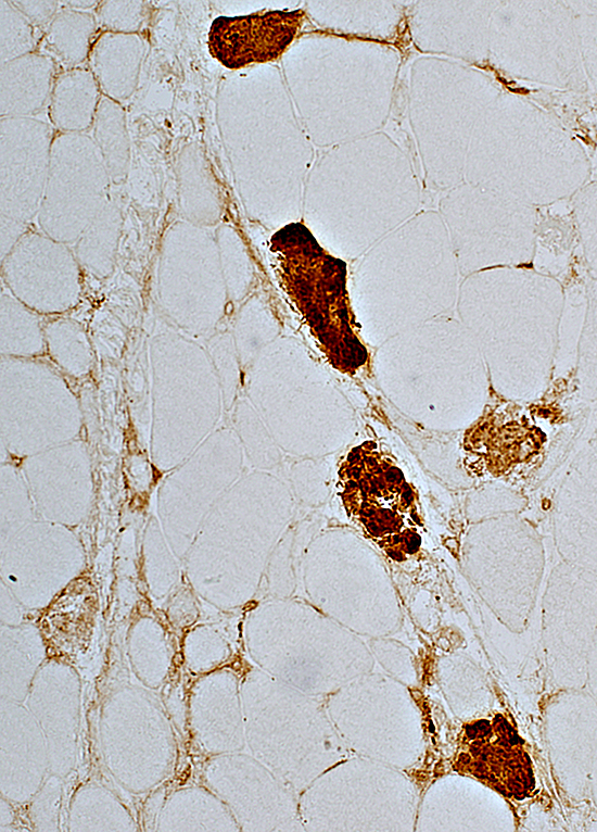 C5b-9 stain |
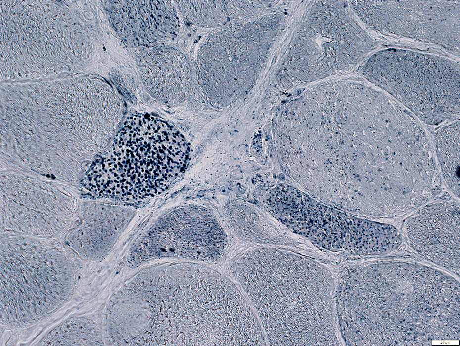
NADH stains: Smaller fibers near perimysium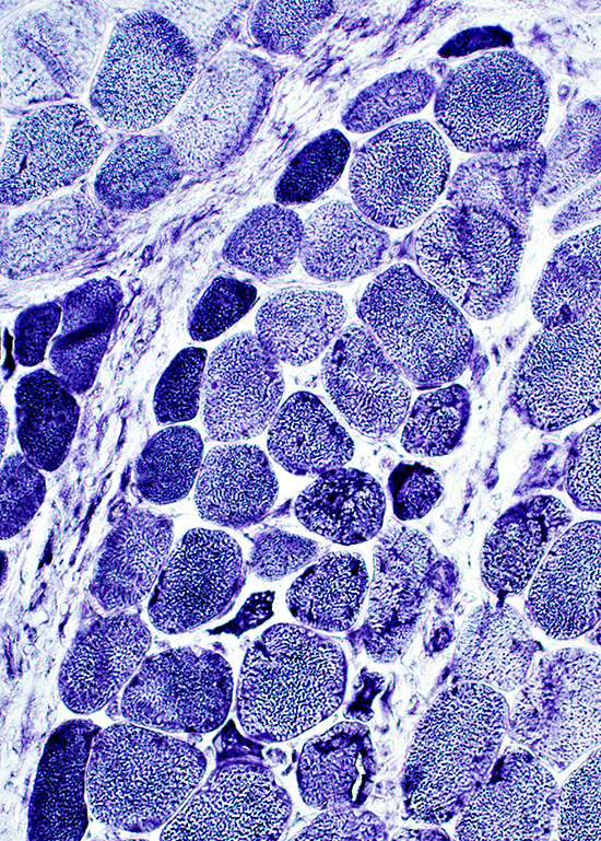 NADH stain |
 C5b-9 stain |
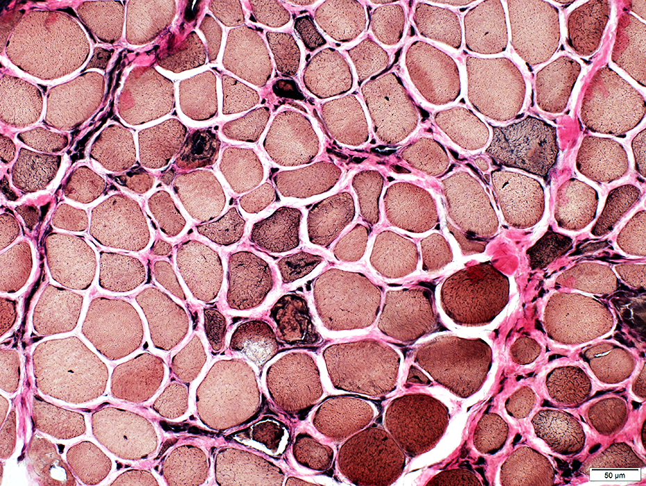
Vacuoles: Myopathy with HMGCR antibodies
 H&E stain |
 Gomori trichrome stain |
 VvG stain |
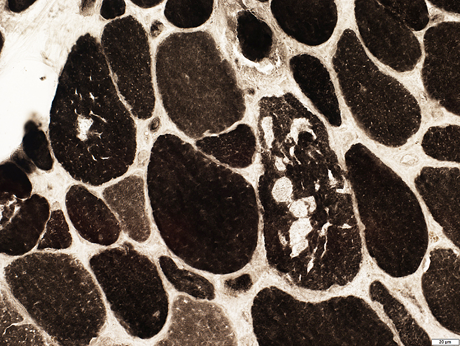
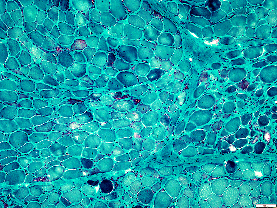
 NADH stain |
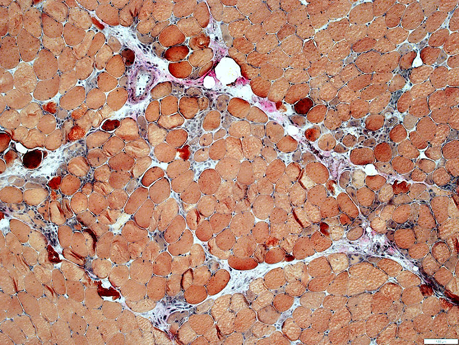
 ATPase pH 9.4 stain |
 ATPase pH 4.3 stain |
 ATPase pH 4.3 stain |
HMGCR MYOPATHY: Perimysium
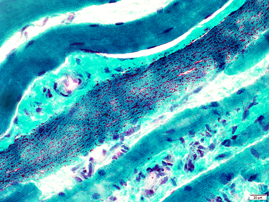
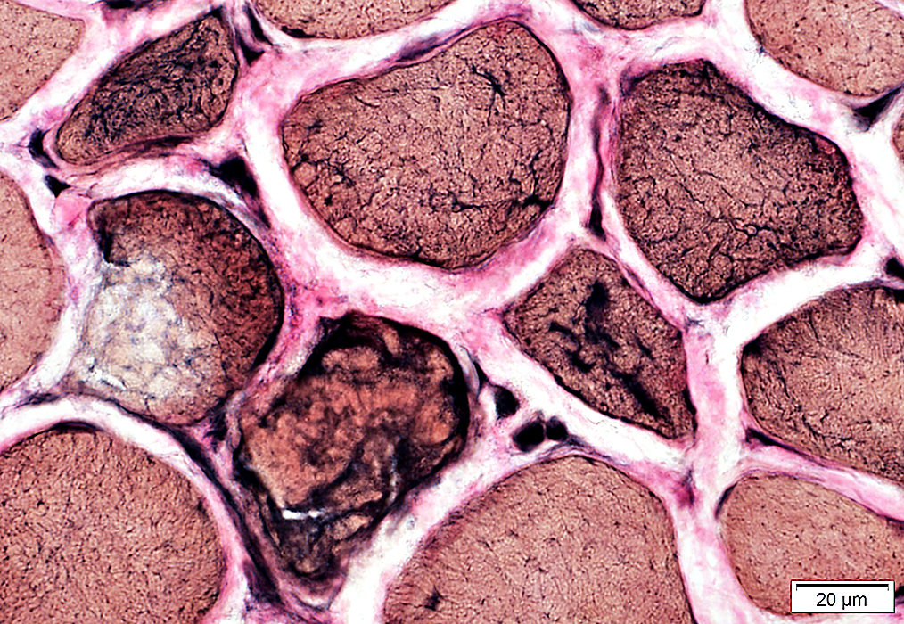
 Gomori trichrome stain |
Perimysial structure
Fragmented & IrregularScattered, Large, Acid phosphatase+ cells: Present in some regions
 Gomori trichrome stain |
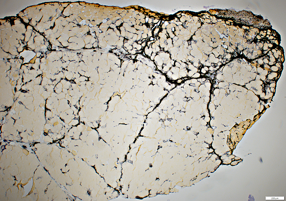
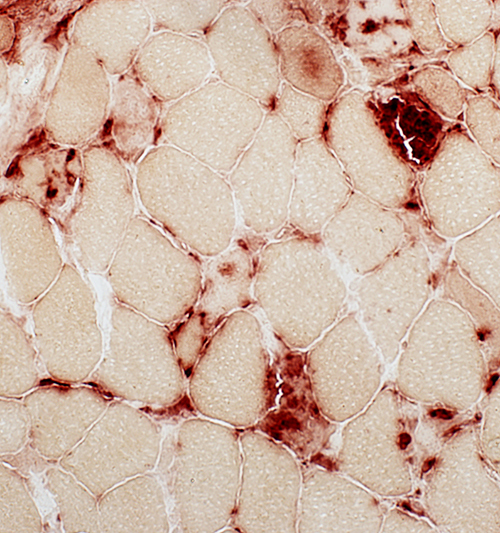
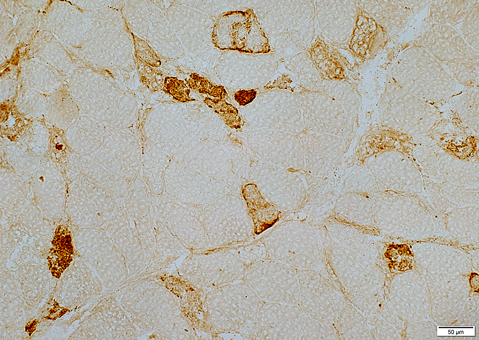
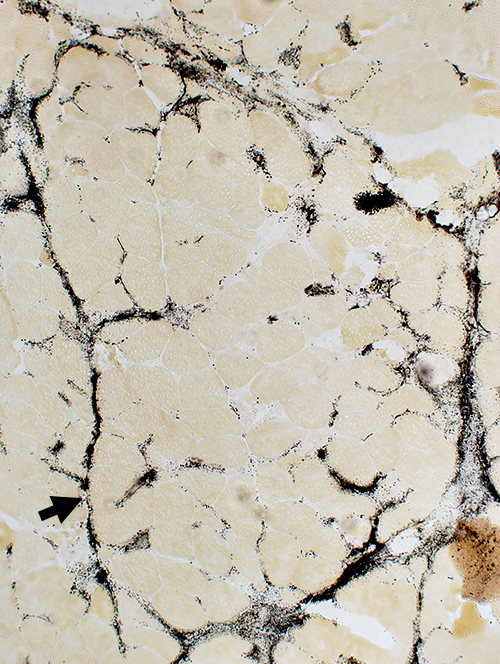
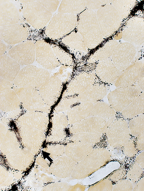
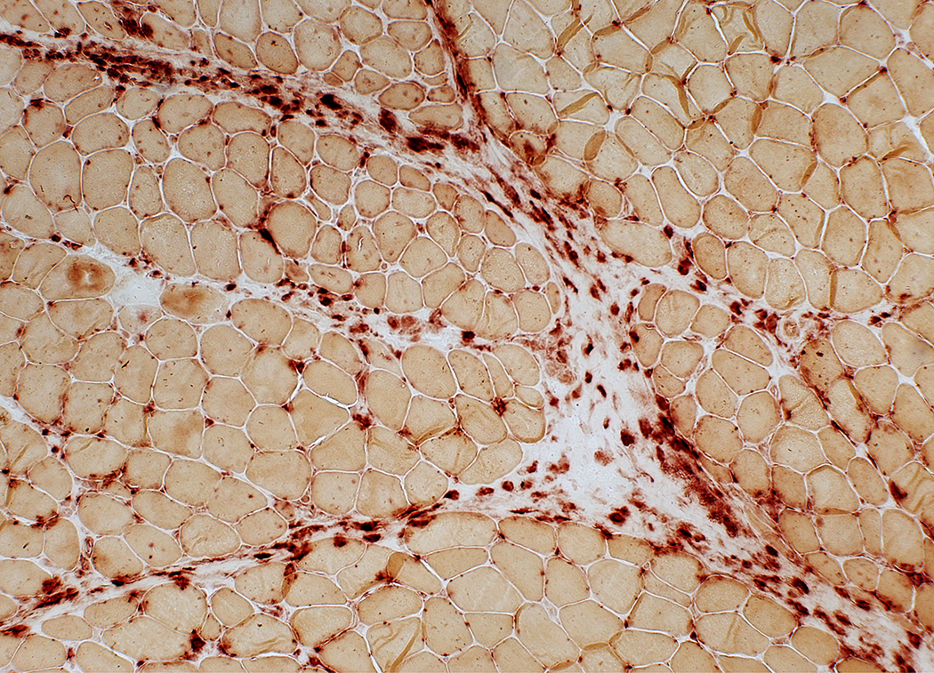
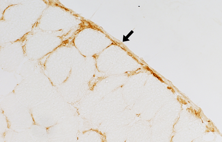
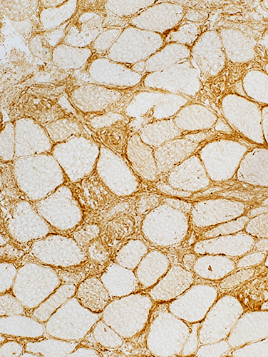
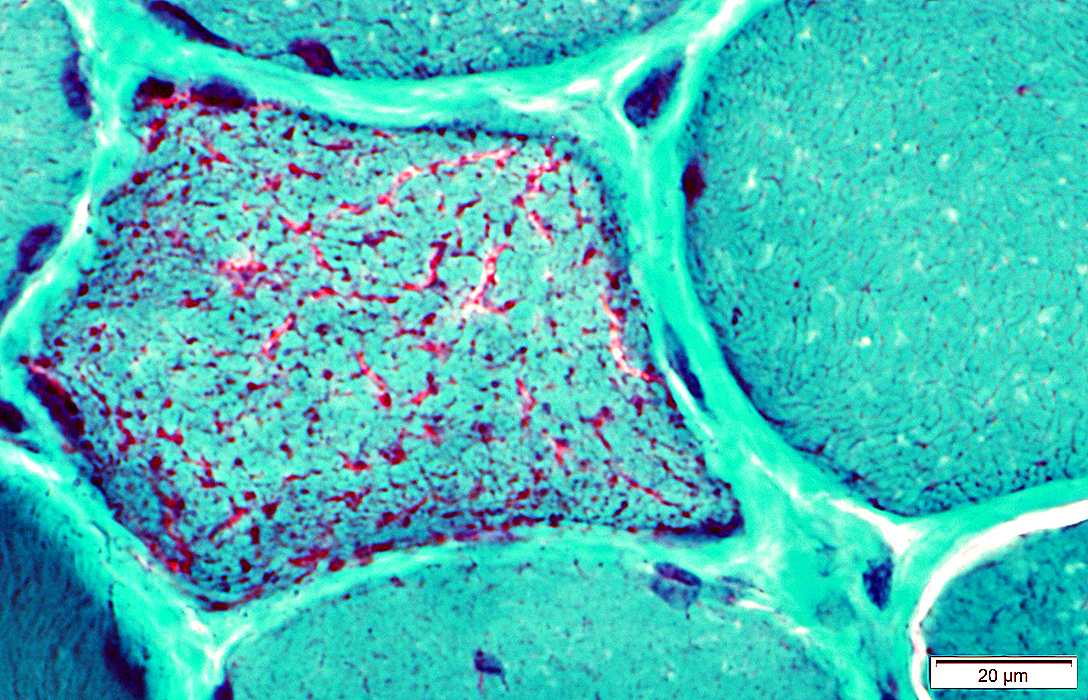
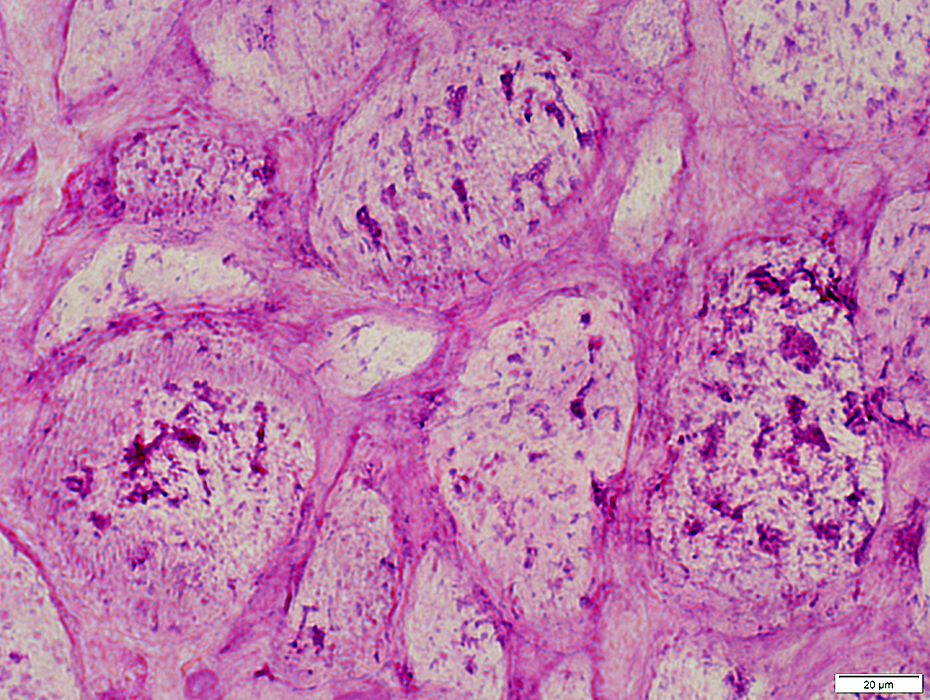
Perimysium: Stains for alkaline phosphatase
Alkaline phosphatase stains:Perimysial connective tissue (Arrows)
Cytoplasm of immature, or regenerating, muscle fibers
 Alkaline phosphatase stain |
 Alkaline phosphatase stain |
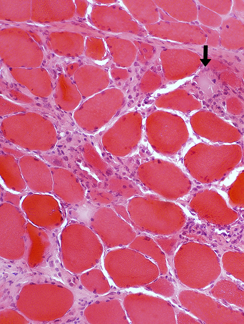
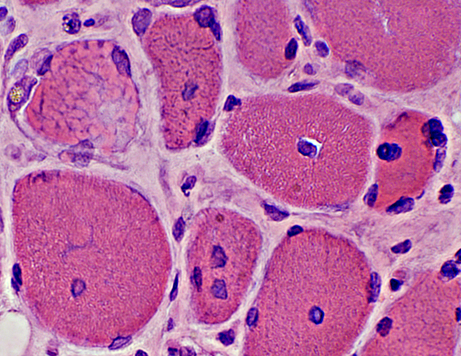
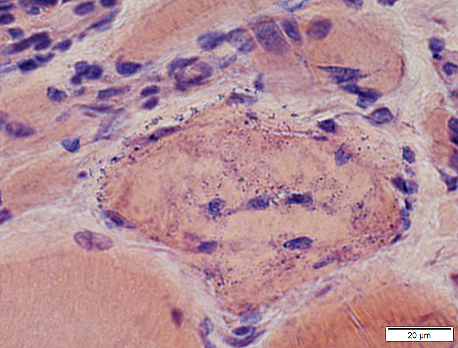
Perimysial cells & Scattered necrotic muscle fibers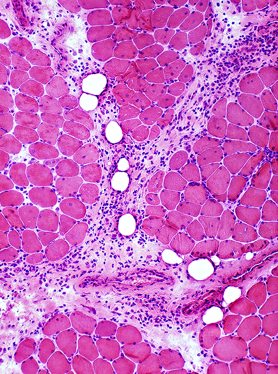 H&E stain |
 H&E stain |
 Acid phosphatase stain |
Perimysial cells:
|
 Acid phosphatase stain |
Perimysial Cells: Large & Irregular shapes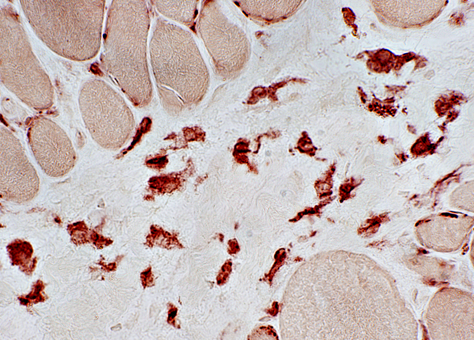 Esterase stain |
CD4 cells
Immune cells: Epimysial (Arrow) & Endomysial
 CD4 stain |
Immune Cells: Perimysial (Arrow), Endomysial & Associated with Necrotic muscle fibers
 CD4 stain |
Lipid
 Sudan Black stain |
Normal in most muscle fibers (Above)
May be increased in scattered Muscle fibers (Below)
 Sudan Black stain |
 Sudan Black stain |
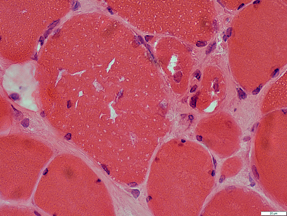
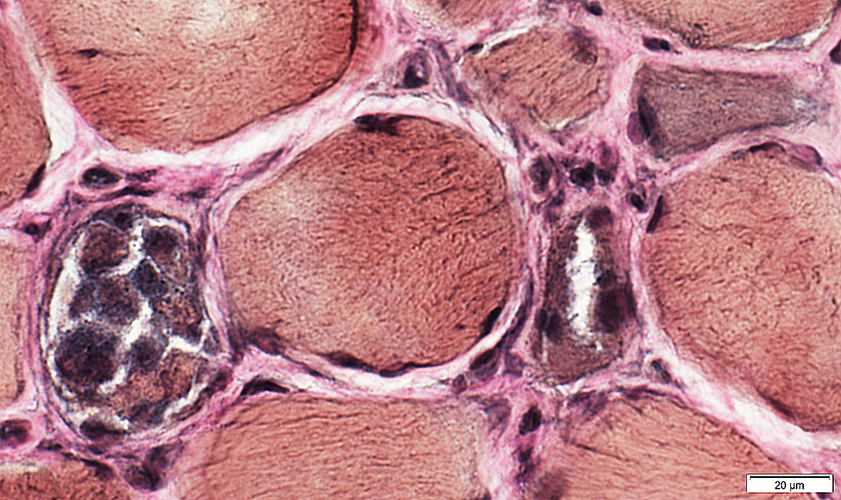
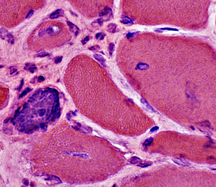 H&E stain MYONUCLEAR PATHOLOGY: Enlargement, Irregular shapes & Clusters |
 Congo red stain Myonuclei (Abnormal) in intact muscle fibers Large Irregular shapes (Arrows) Clustered May contain inclusions |
 H&E stain |
 H&E stain |
 H&E stain |
 Congo red stain |
 Congo red stain |
|
C5b-9 Deposition: In cytoplasm of scattered necrotic muscle fibers (Arrow) 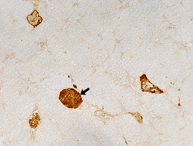 C5b-9 stain |
CD4 Cells: In Necrotic muscle fibers & Near endomysial capillaries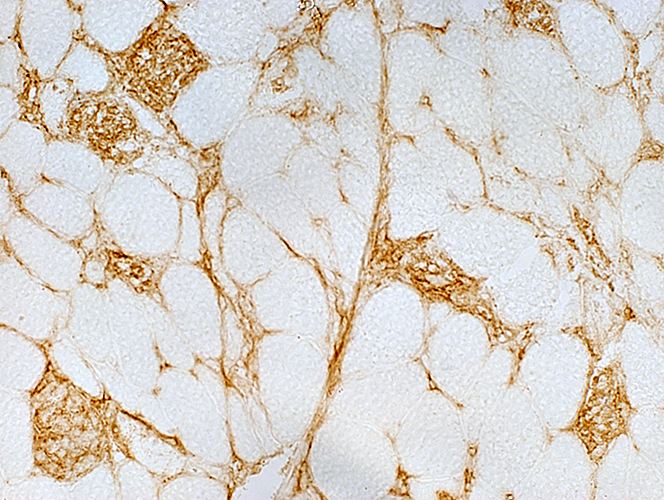 CD4 stain |
Ulex stained capillaries: Normal numbers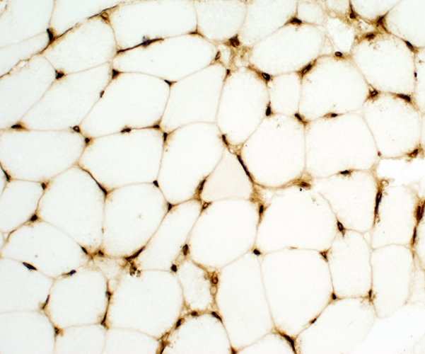 Ulex (UEA1) stain |
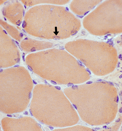
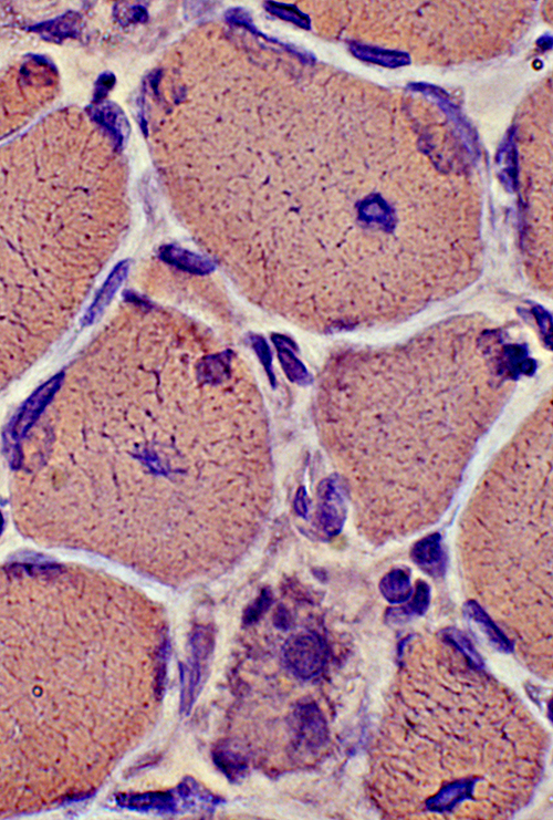
HMGCR MYOPATHY: EARLY or MILD PATHOLOGY
Occasional Necrotic Muscle Fiber (Arrow)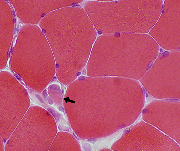 H&E stain |
Large or Irregular Nuclei (Arrow)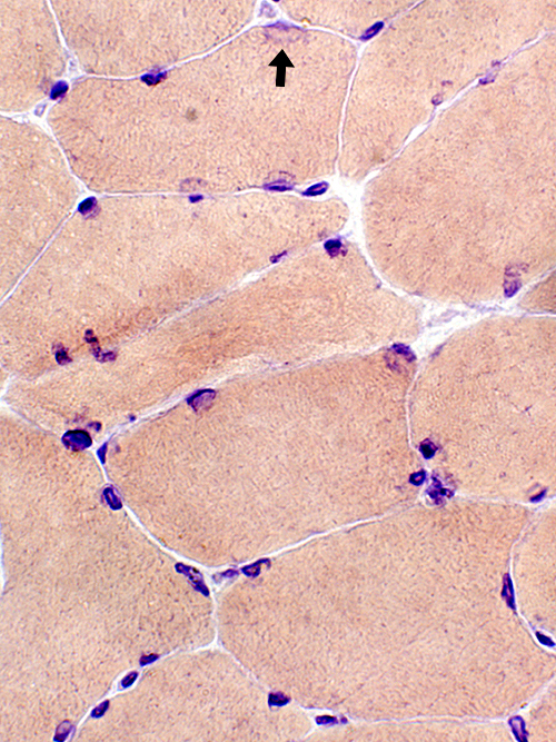 Congo red stain |
Large Nuclei (2nd patient)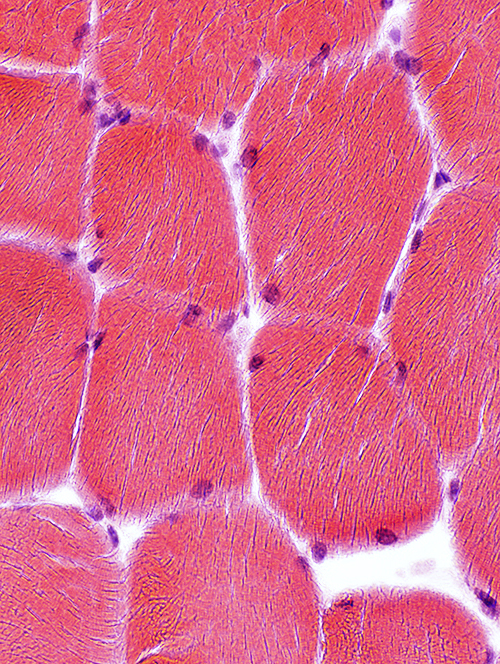 H&E stain |
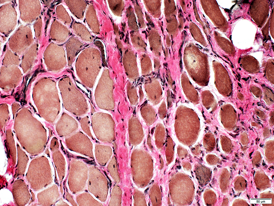
HMGCR Antibody: Chronic Myopathy, Young onset, Poor response to treatment
 H&E stain |
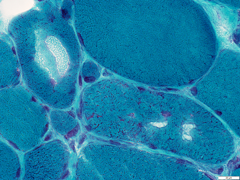
 Gomori Trichrome stain |
 VvG stain |
 VvG stain |
Muscle fibers
Internal architecture: May have aggregates
Size: Varied
Regenerating muscle fibers: Scattered
Endomysial connective tissue: Increased
Severity of pathology: May vary among fascicles
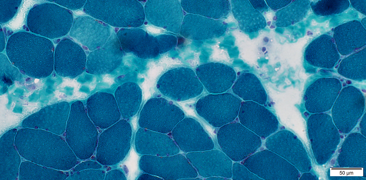
 VvG stain |
|
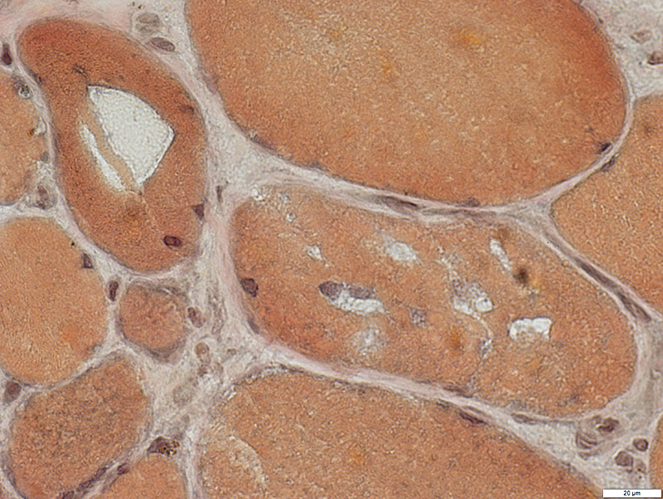
Myopathic features Muscle fibers Size: Varied; Hypertrophy & Atrophy Endomysial connective tissue: Increased 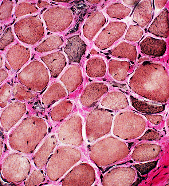 VvG stain |
 H&E stain |
 Gomori trichrome stain |
Inclusions & Vacuoles: Red stained
 Gomori trichrome stain |
 Gomori trichrome stain |
 VvG stain |
Necrosis (Left, Above)
Vacuoles (Right, Above)
Aggregates & Abnormal internal architecture (Below)
 VvG stain |
|
Muscle fiber pathology Vacuoles Aggregates Basophilic granular debris |
 Congo red stain |
 Congo red stain |
 PAS stain |
Aggregated glycogen
Internal architecture: Coarse or Dark
Necrosis: Some fibers have very pale-stained cytoplasm (Below)
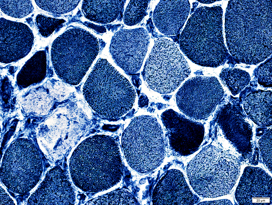 NADH stain |
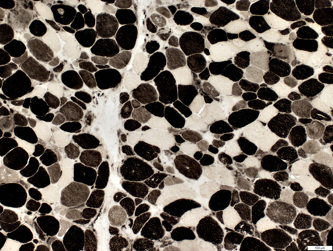
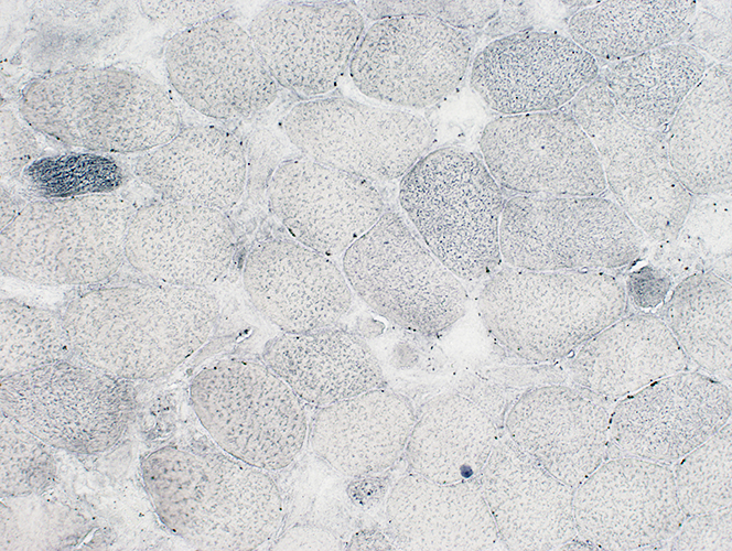
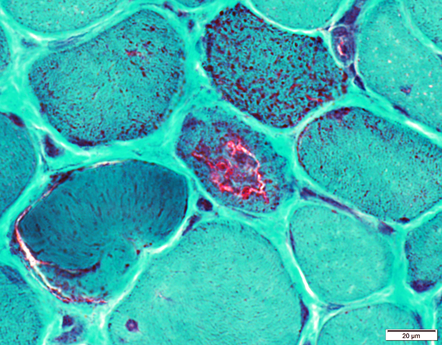
Immature muscle fibers (Intermediate staining)
Abundant
Scattered
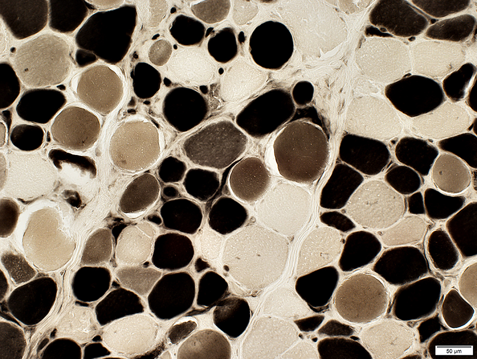 ATPase pH 4.3 stain |
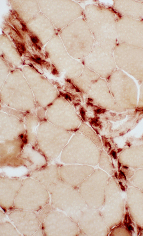
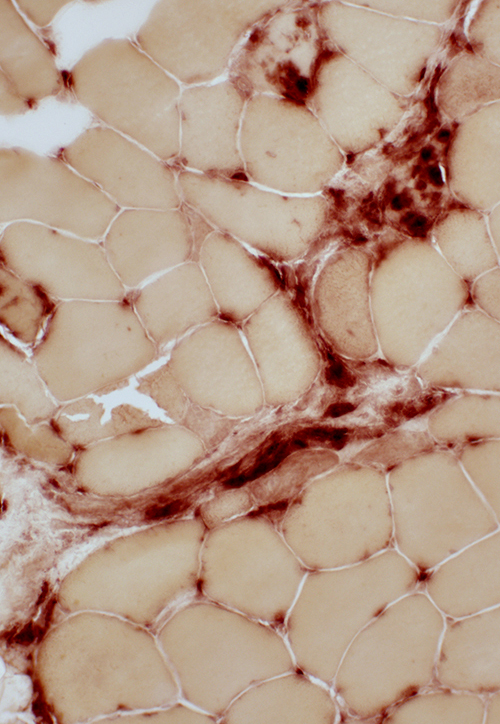
Perimysial Pathology
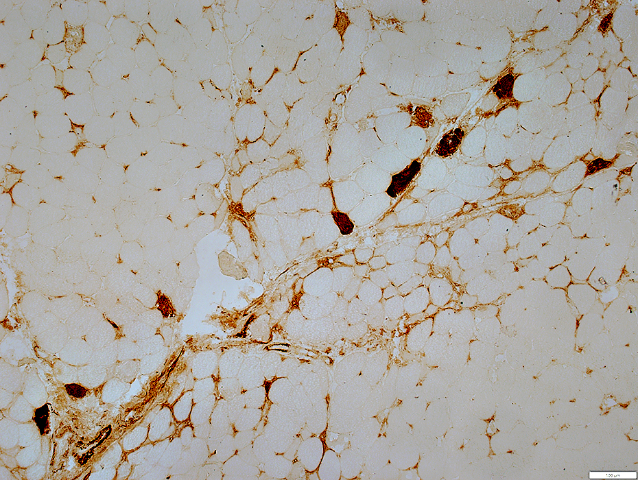
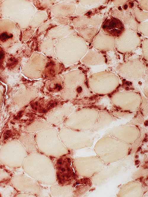
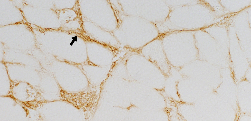
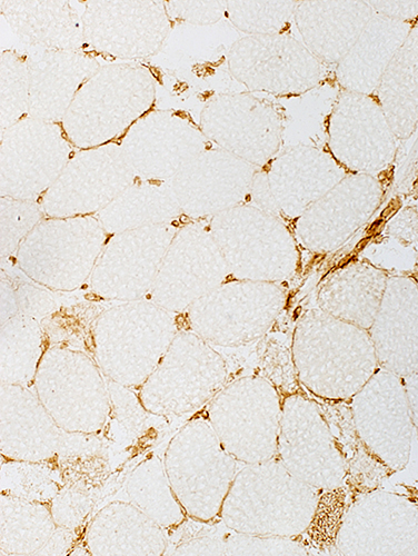
Perimysial staining by Alkaline phosphatase
Immature muscle fibers: Cytoplasm is Alkaline phosphatase +
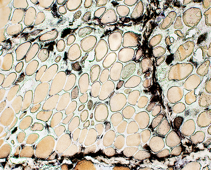 Alkaline phosphatase stain |
|
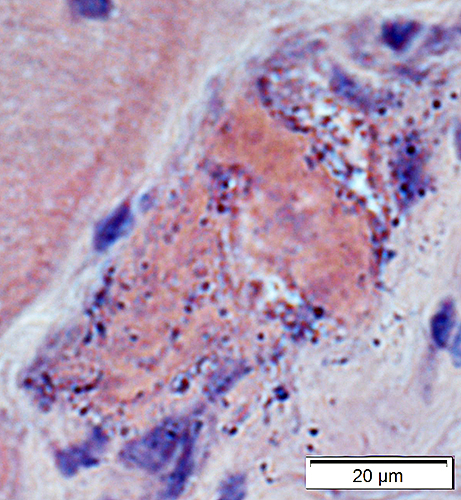
Histiocytic cells (Red) Scattered in perimysium Necrotic muscle fiber: Replaced by histiocytic cells 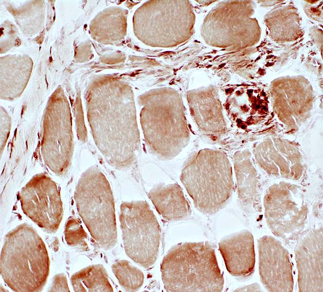 Acid phosphatase stain |
Return to Inflammatory myopathies
Return to HMGCR myopathy
3/7/2024