SCLEROMYXEDEMA
Patterns of pathology in muscle
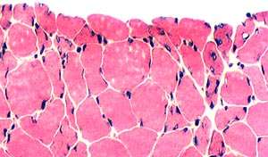 H & E stain |
 H & E stain |
Atrophic muscle fibers
|
|
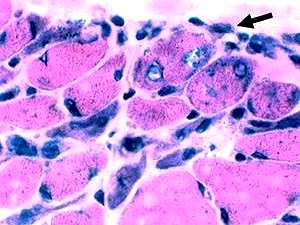
 H & E stain |
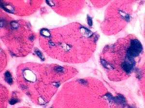
 H & E stain |
|
Vacuoles: Rimmed
|
|
 H & E stain |
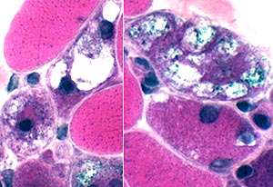
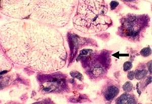 PAS stain |
| Cells in perimysium: Large; PAS positive | |
Return to Neuromuscular Home Page
Return to Pathology index
Return to Scleromyxedema
7/18/2019