Neuromas 1,2
|
Neuromas (Sural nerve) Aberant fascicles Minifascicles Nerve branch Regenerated axon clusters Neuroma (C5 root): Distal to lesion Axon sprouts GAP43 Axons Perineurium Also see Blocked axon regeneration Axon swellings |
Regeneration: Aberrant Fascicles
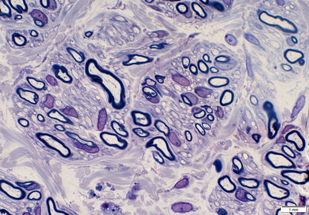
 H&E stain Minifascicles Varied sizes & orientations  H&E stain |
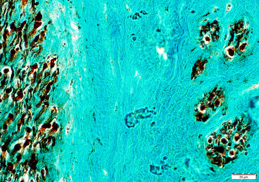
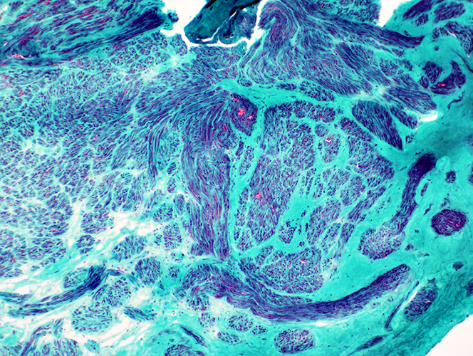 Gomori trichrome stain Post-regeneration: Small fascicles with varied size and orientations 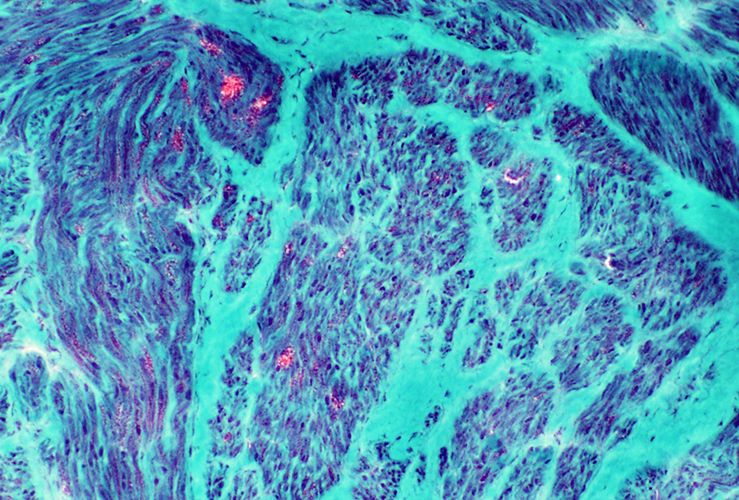 Gomori trichrome stain Neuroma: Multiple small fascicles containing myelinated axons 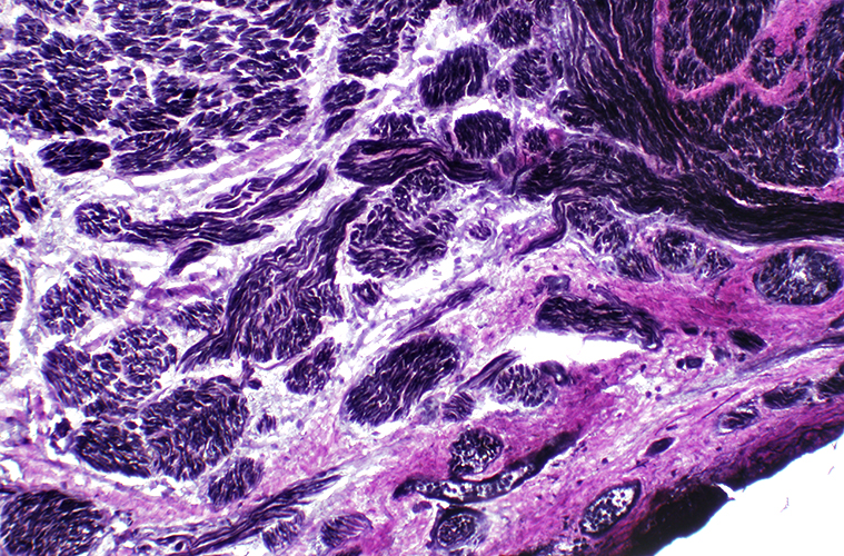 VvG stain |
 Neurofilament stain |
Contain myelinated & unmyelinated axons
 Neurofilament stain |
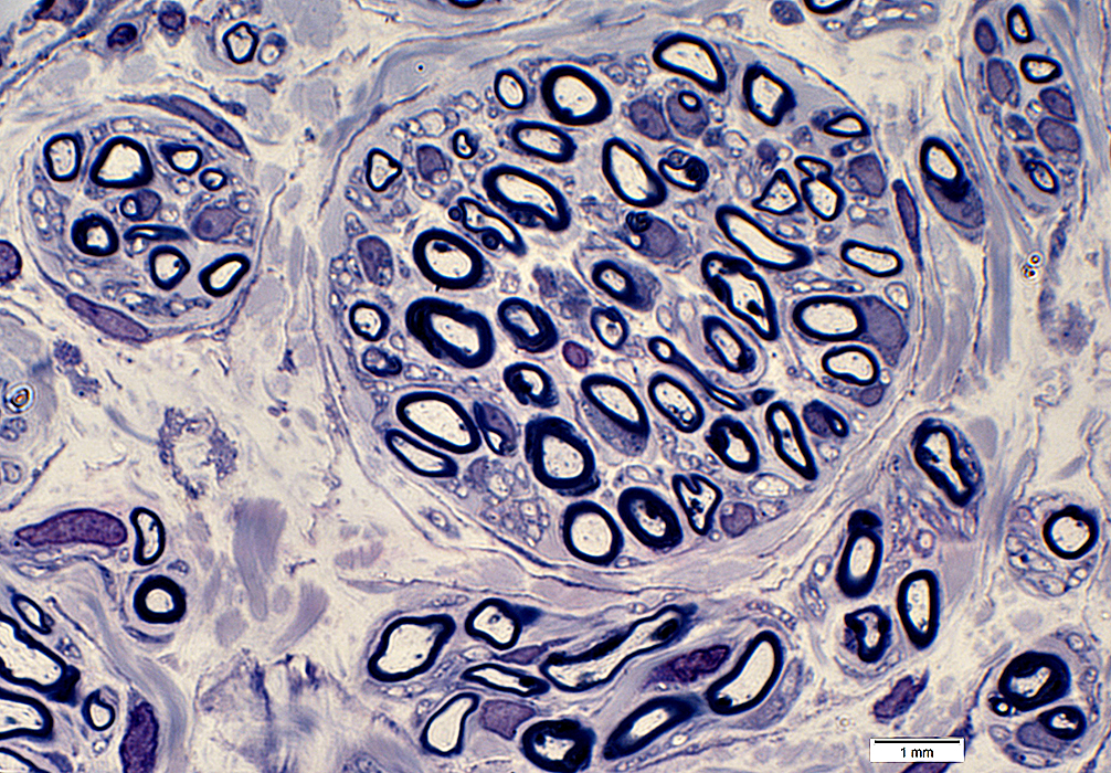
Regenerated Axon Clusters within Neuroma
|
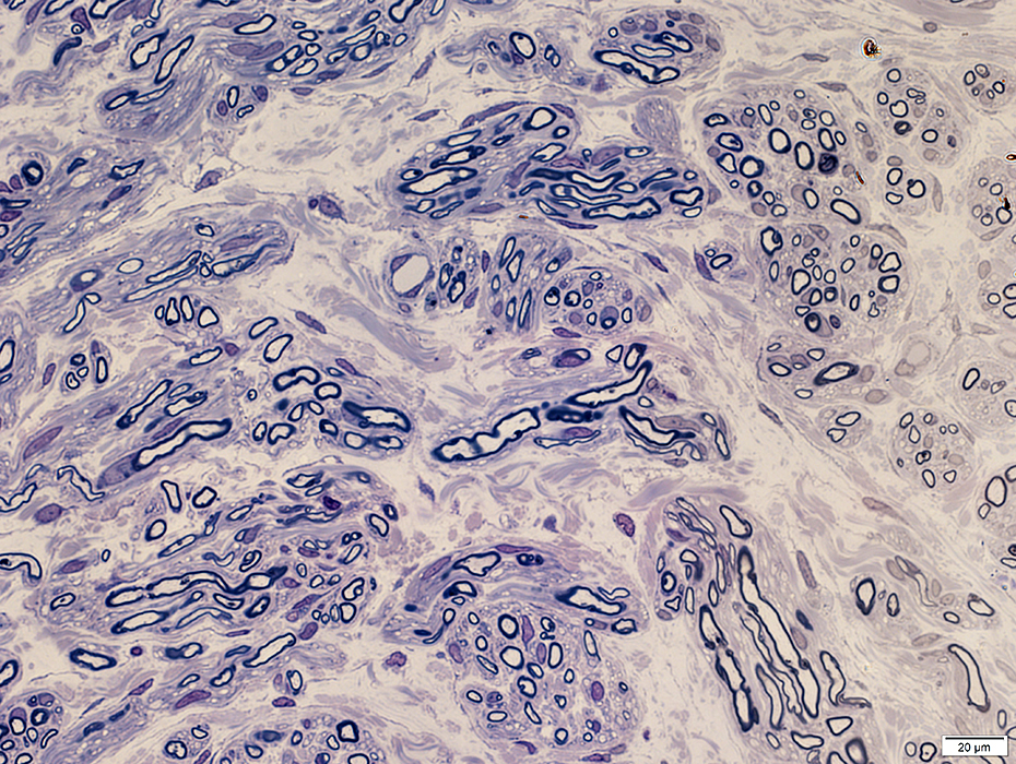
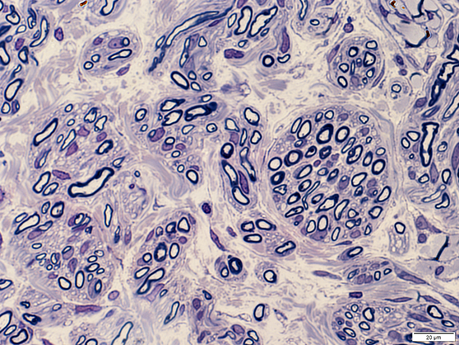
Varied sizes & orientations of axon clusters (fascicles)
|
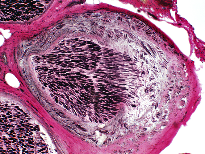
Neuroma
Axons in fascicles tend to be thinly myelinated for their size
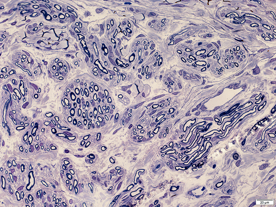
Small minifascicles contain 1 to 30 myelinated axons
|
Neuroma
|
Varied numbers of myelinated & non-myelinated axons in fascicles
Varied numbers of axons in fascicles
|
Neuroma
Several small fascicles separated by connective tissue
Each fascicle is surrounded by perineurium, 1 or several layers
|
 From: R Schmidt |
Contents
Thinly myelinated axons
Clusters of Schwann cell processes (Büngner bands)
Collagen
Surrounded by
Perimysial cell/Fibroblast process
 From: R Schmidt |
 From: R Schmidt |
Contents
Thinly myelinated axons
Clusters of Schwann cell processes (Büngner bands)
Collagen
Surrounded by
Perimysial cell/Fibroblast process
 From: R Schmidt |
 From: R Schmidt |
|
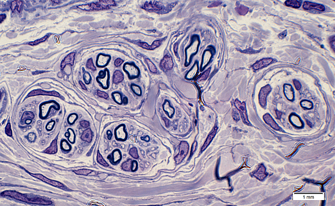
Neuroma: Minifascicles Small minifascicles: Contain 1 or 2 thinly myelinated axons (Top) Fascicle with normal organization (Bottom left) 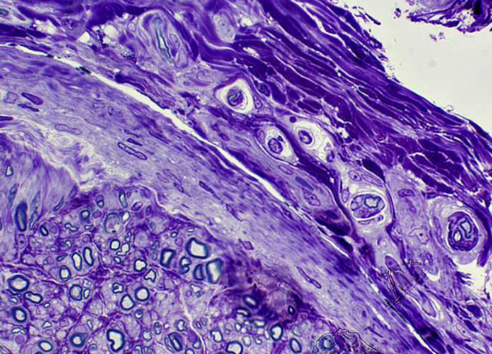 Toluidine blue stain Small minifascicles: Contain thinly myelinated axons 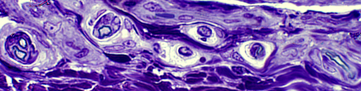
|
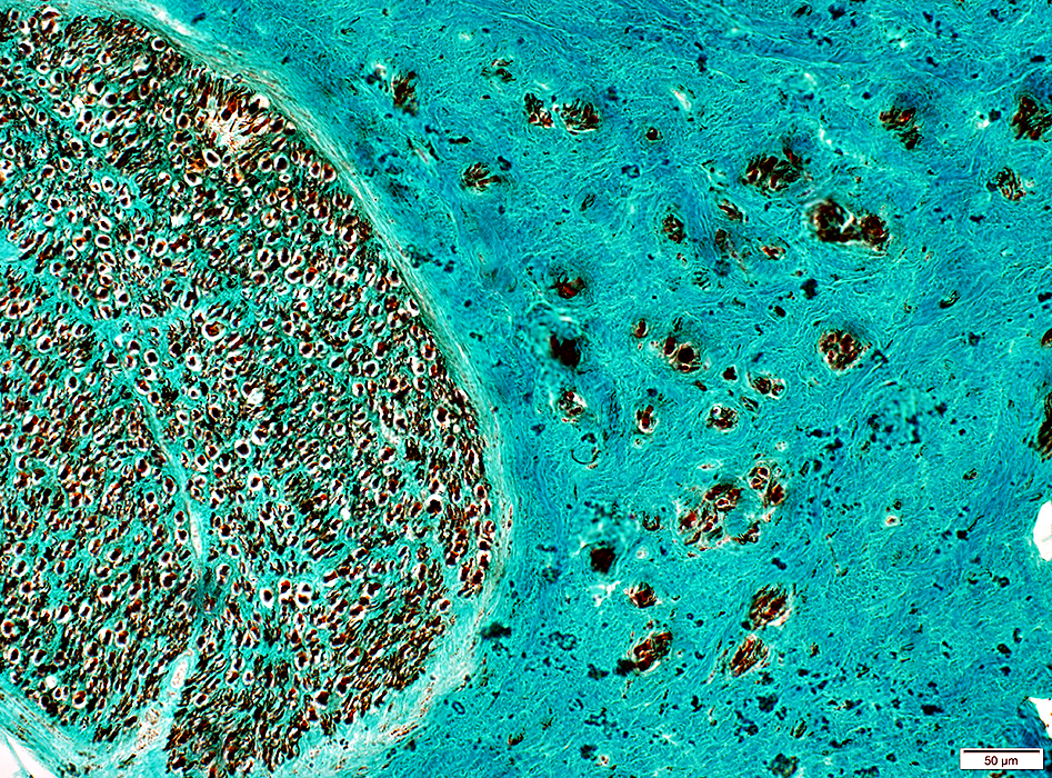
Neuroma: Origin
Neuroma: Nerve branch originating from normal fascicleNormal nerve fascicle (Left); Fascicles with varied size and orientations (Right)
 VvG stain 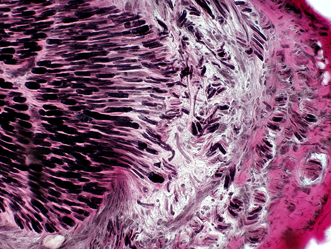 VvG stain 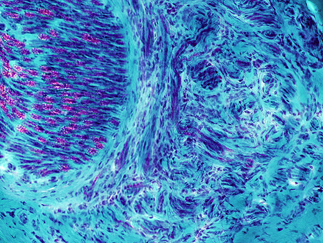 Gomori trichrome stain |
Neuroma (Region along nerve at, or distal to, lesion): C5 Root
|
Neuroma (C5 root) Morphology Axon sprouts GAP43 Perineurium |
 H&E stain |
Persisting nerve structures (Arrows)
Cellular: Heterogeneous region; Cells present in clusters
Contains clusters of myelinated axons
Surrounding connective tissue: Contains aberrantly regenerated axons
Connective Tissue Structure: Moderately dense to pale
Aberrant myelinated axons: Single & Clustered (Below)
 VvG stain |
 Neurofilament stain |
Axon sprouts: Scattered single axons grow into connective tissue (Left)
 NCAM stain |
 H&E stain |
Axon sprouts: Scattered single axons grow into connective tissue (Left)
 H&E stain |
 Gomori Trichrome stain |
Axon sprouts: Scattered single axons grow into connective tissue (Left)
 H&E stain |
Axon sprouts: Scattered single axons grow into connective tissue (Bottom)
 H&E stain |
Axon sprouts: Scattered single axons grow into connective tissue (Below)
 H&E stain |
Neuroma: Axons & Schwann cells
NCAM(r)10X_01apsmb.jpg)
Present in Schwann cells around most axons in Neuroma (Left) & surrounding axon sprouts (Right)
P0(r)sm.jpg) Neurofilament (Green); P0 (Red) |
Present in Schwann cells around some, but not all, axons in & around Neuroma
P0(r)_07apsm.jpg) Neurofilament (Green); P0 (Red) |
 Neurofilament (Green); MBP (Red) |
Small & Intermediate-sized axons
Many have associated MBP+ Schwann cells (Yellow)
 Neurofilament (Green); MBP (Red) |
GAP43(r).jpg) Neurofilament (Green); GAP43 (Red) |
Axons in Neuroma: Contain GAP43 (Yellow) more in peripheral than central areas
GAP43(r)b.jpg) Neurofilament (Green); GAP43 (Red) |
Axon Sprouts
 From: R Schmidt |
Clusters of processes
2 types of contents
Tubulovesicular profiles
Densely distributed in axon processes
Compare to: Axons, large & proximal to nerve lesion
Organelles, including many mitochondria
No surrounding Schwann cell cytoplasm
 From: R Schmidt |
 From: R Schmidt |
Cluster of moderate-sized processes
All contain mainly Tubulovesicular profiles
Densely distributed
No surrounding Schwann cell cytoplasm
 From: R Schmidt |
 From: R Schmidt |
Cluster of processes
Tubulovesicular profiles surrounded by Organelles, including many mitochondria
No surrounding Schwann cell cytoplasm
 From: R Schmidt |
 From: R Schmidt |
Mixed contents: Tubulovesicular profiles & Organelles
No surrounding Schwann cell cytoplasm
 From: R Schmidt |
Neuroma: New Perineurial sheaths formed around small clusters of axons
P0(r).jpg) EMA (Green): P0 (Red) |
EMA (Green) stains scattered perineurial cells & rings of perineurium around small, aberrant regenerated axon (myelin) clusters (Red)
Compare to: Nerve proximal to damage region
P0(r)b.jpg) EMA (Green): P0 (Red) |
Return to Neuromuscular Home Page
Return to Pathology index
References
1. J Am Acad Orthop Surg 2025;33:178-186
2. PLoS One 2018;13:e0200548
6/13/2025