Mitochondrial disease: Cytochrome oxidase deficiency, Children
|
1 month old child 6 month old child Ultrastructure |
COX Deficiency: 6 month old child
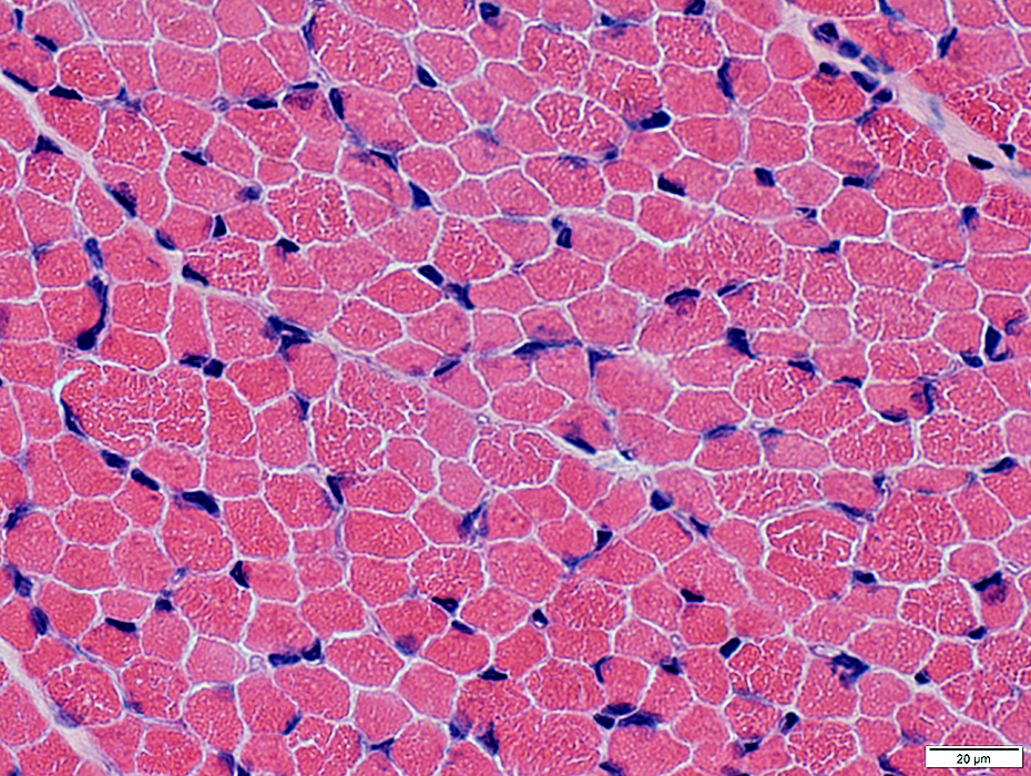 H&E stain |
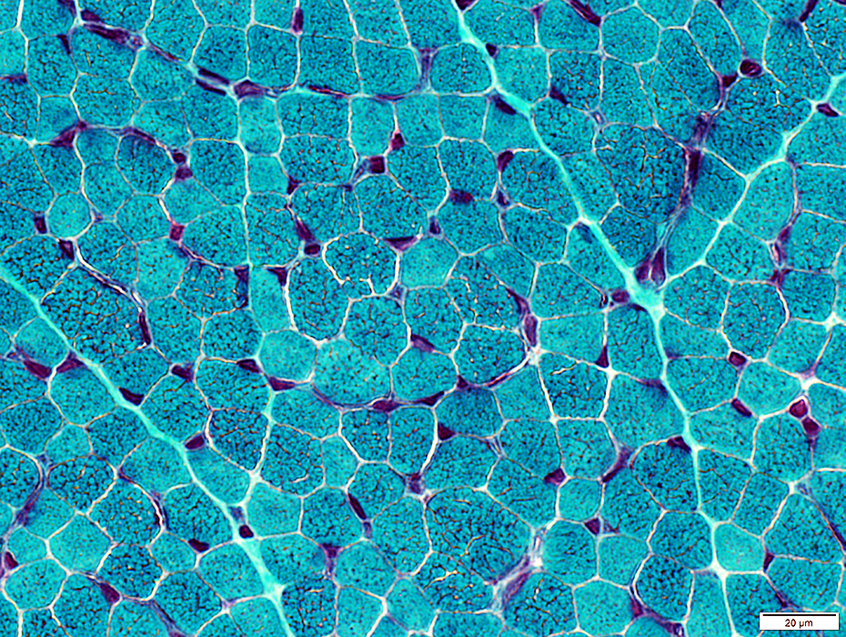 Gomori trichrime stain |
"Checkerboard" pattern of fiber types
"Cracks" & Small round holes are more prominent in type I (pale) fibers
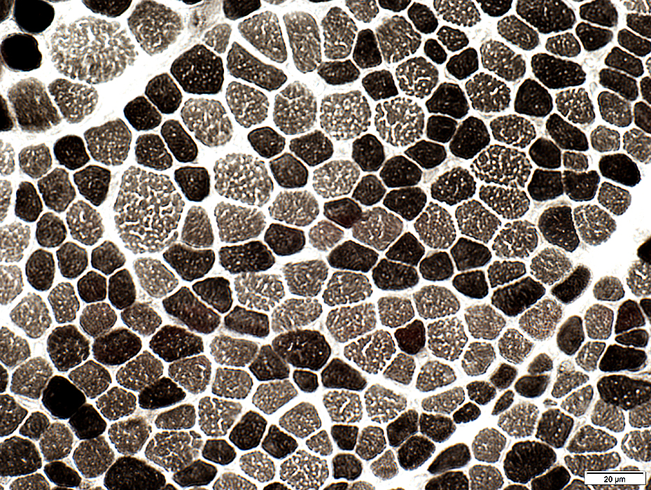 ATPase, pH 9.4 |
Type 2C (Immature; Intermediate-staining), muscle fibers: Increased numbers (> 5%)
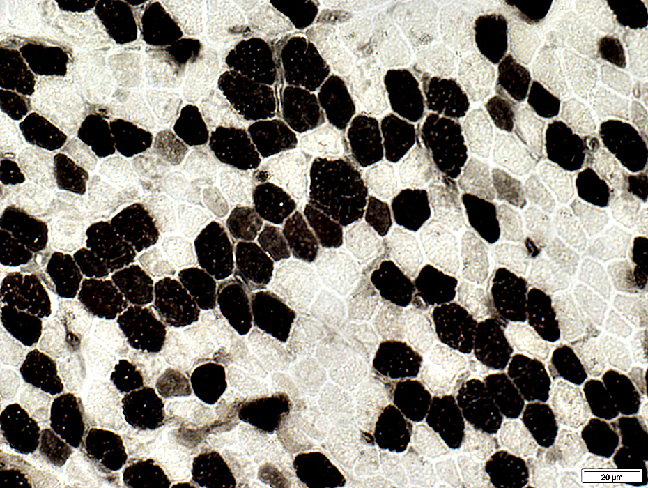 ATPase, pH 4.3 |
 Sudan black |
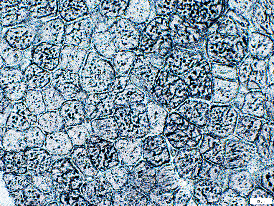
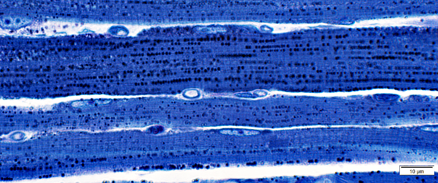 Toluidine blue |
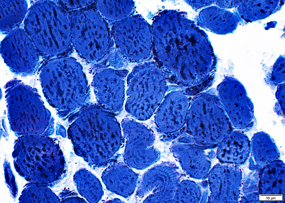 Toluidine blue |
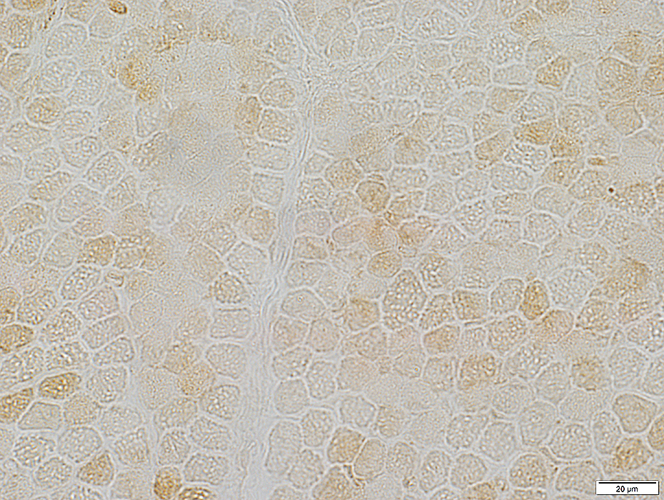
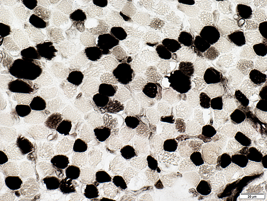
Cytochrome oxidase
Reduced staining in all fibers
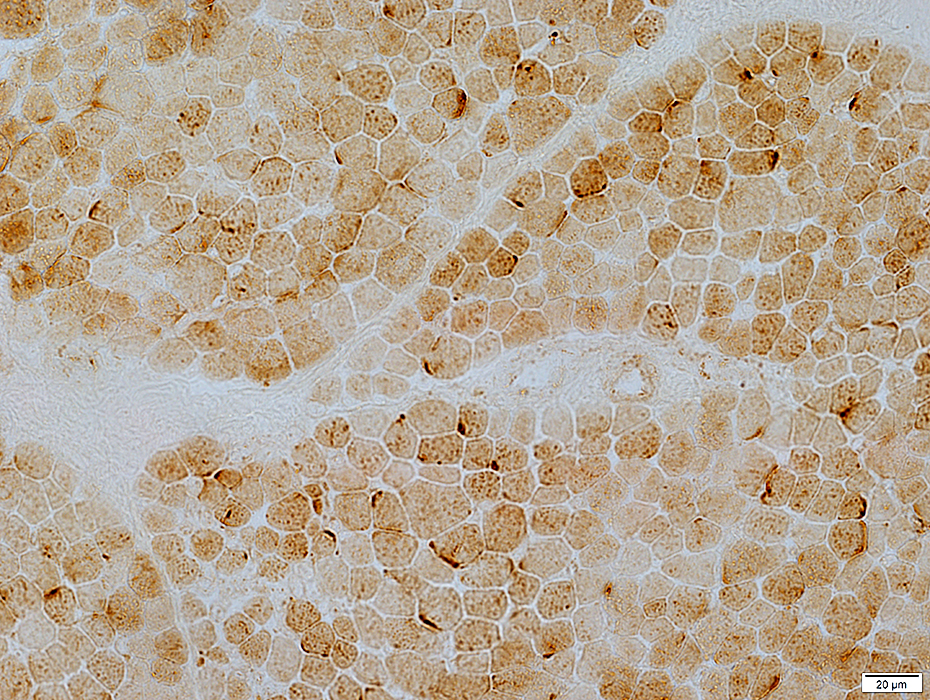 COX stain |
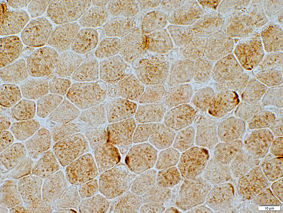 COX stain |
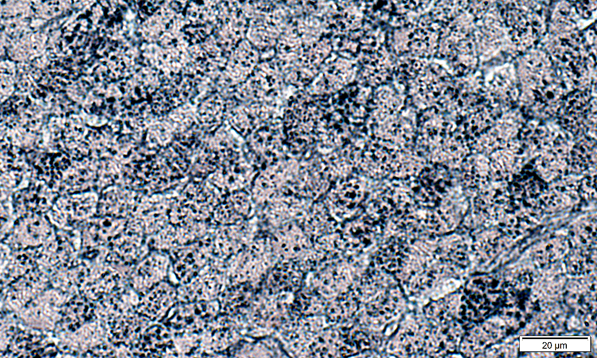
Succinate dehydrogenase (SDH)
Increased staining in many fibers
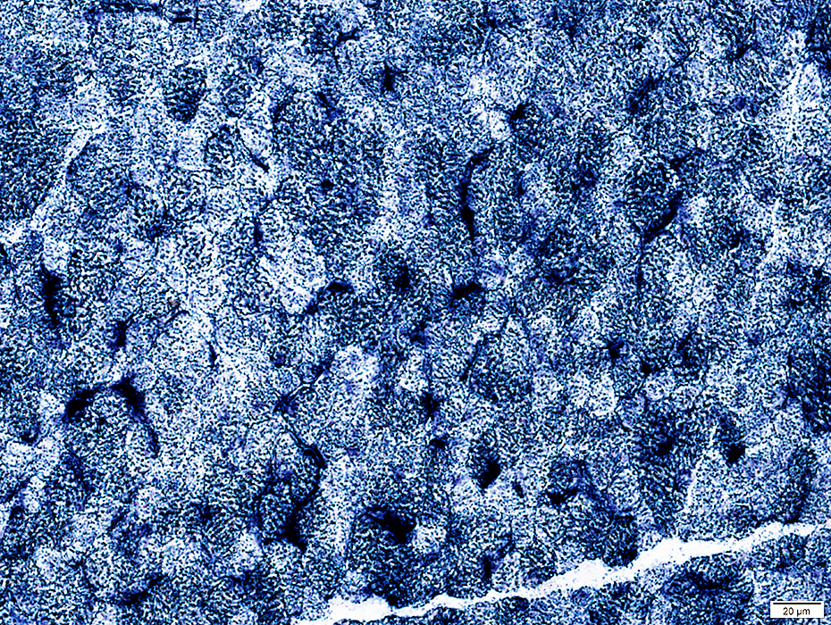 SDH stain |
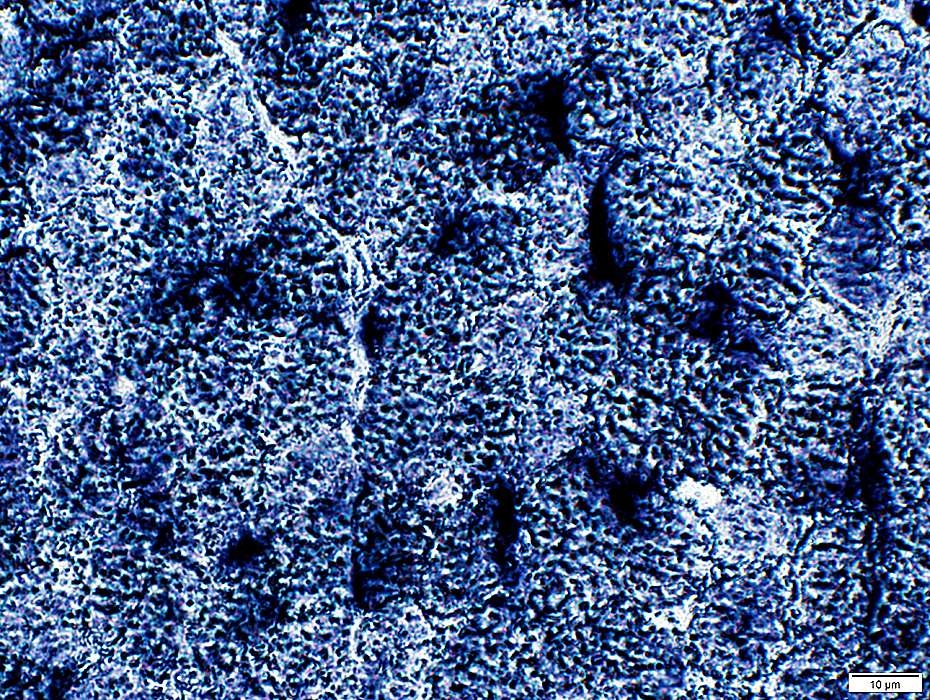 SDH stain |
Mitochondrial Infantile Encephalopathy, Ultrastructure
 From: R Schmidt |
Proliferation
Shapes & structure: Irregular
Sizes: Often large
Lipid droplets
Scattered in muscle fibers
 From: R Schmidt |
 From: R Schmidt |
Proliferation
Shapes & structure: Irregular
Sizes: Often large
Lipid droplets
Scattered in muscle fibers
 From: R Schmidt |
 From: R Schmidt |
Shapes & Structure: Irregular
Sizes: Often large
 From: R Schmidt |
COX Deficiency: 1 month old child
 COX stain |
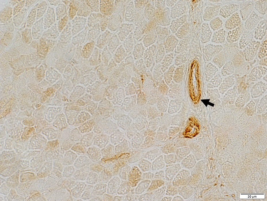 COX stain |
Perimysial vessels (Arrow)
Intrafusil spindle fibers
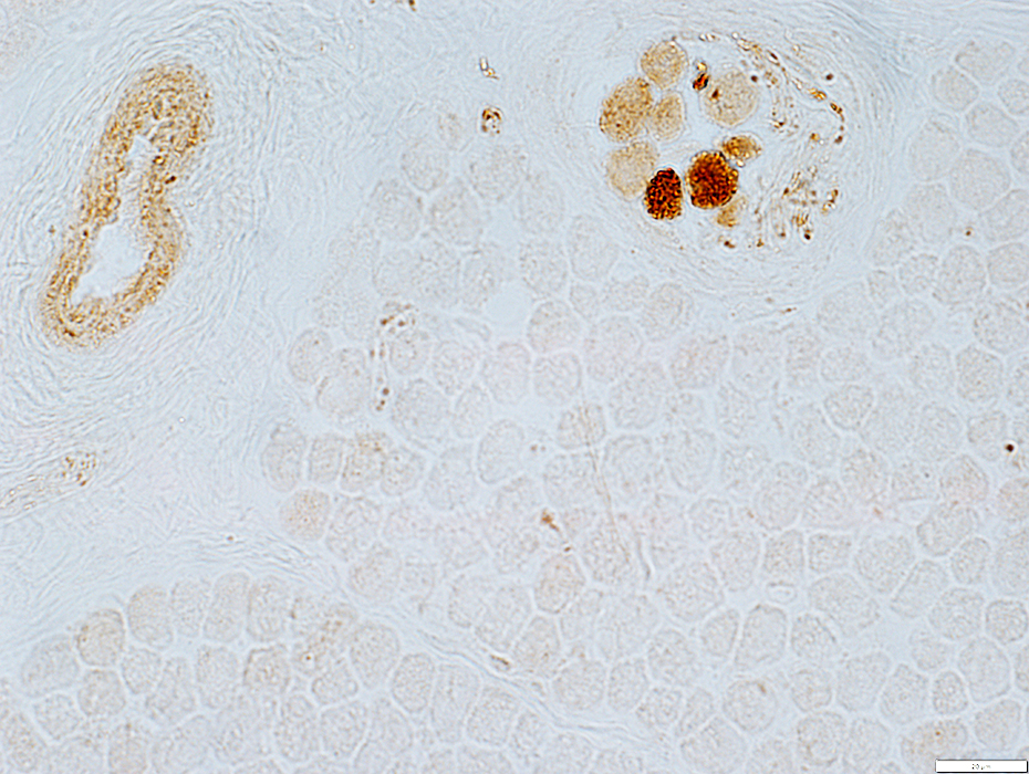 COX stain |
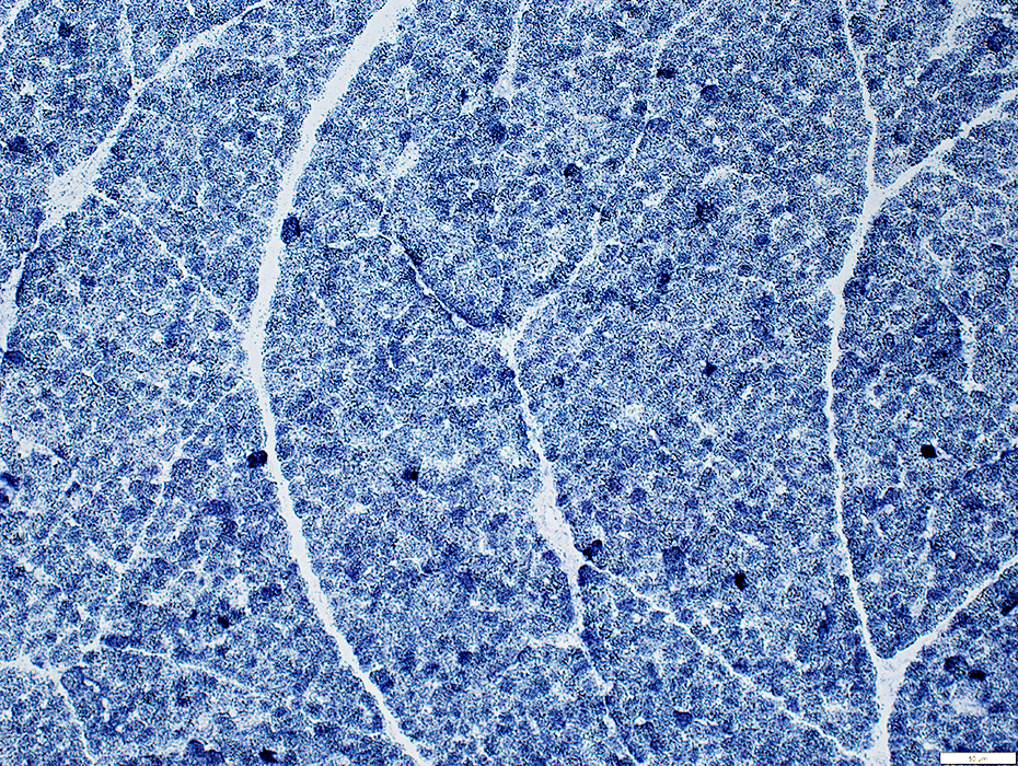 SDH stain |
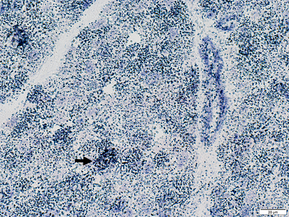 SDH stain |
Type 2C muscle fibers (Intermediate staining): Increased numbers
 ATPase pH 4.3 stain |
Lipid Droplets: Increased size
 Sudan black stain |
Glycogen (PAS staining) is increased in muscle fibers
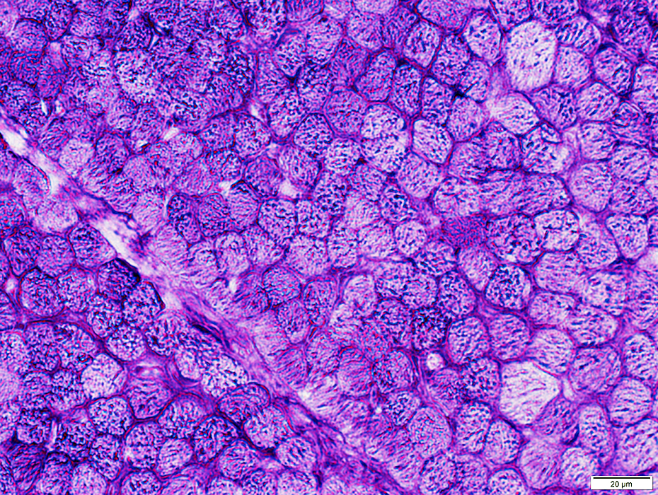 PAS stain |
Return to Mitochondrial pathology
Return to Mitochondrial syndromes
Return to Muscle biopsies
7/8/2025