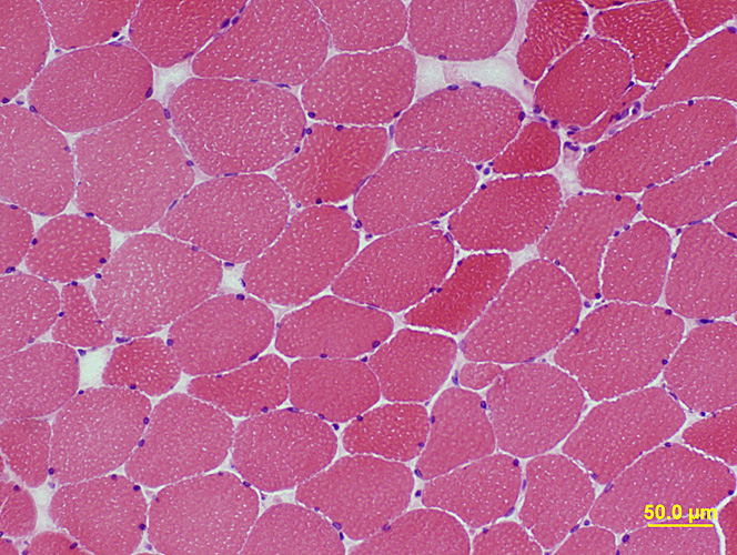
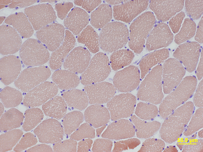
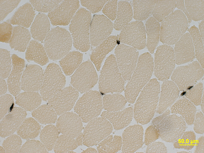
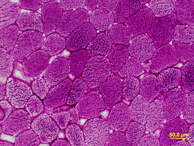
 H&E stain |
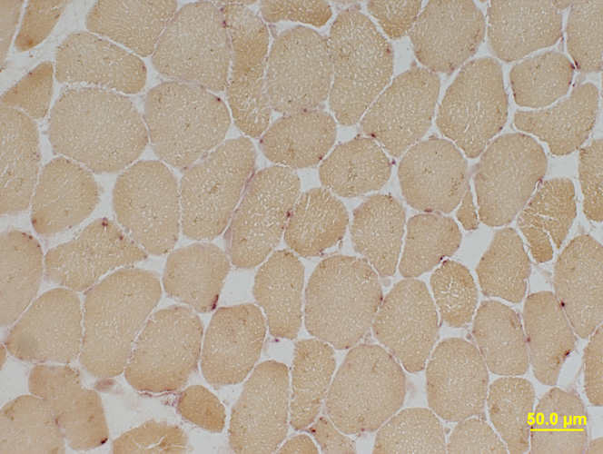
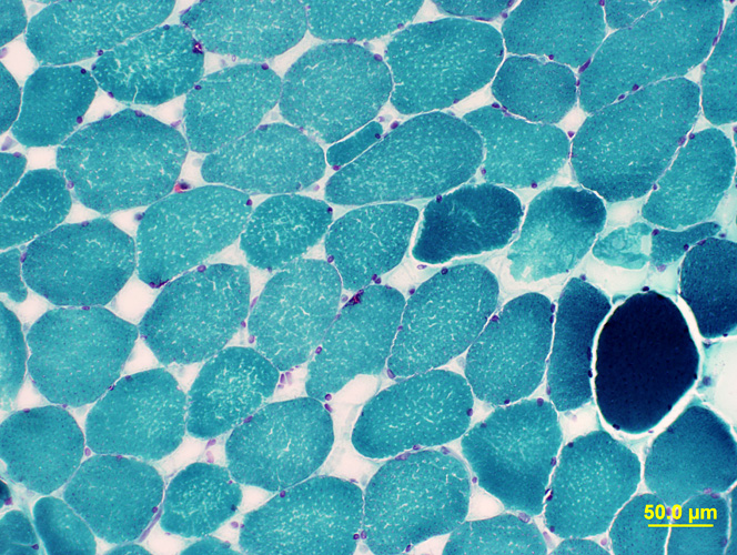 Gomori Trichrome stain |
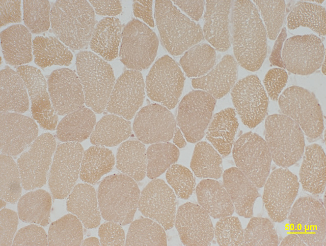
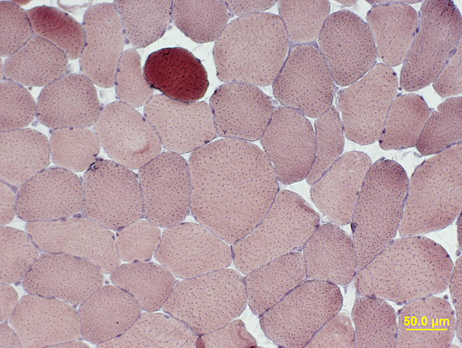 VvG stain |
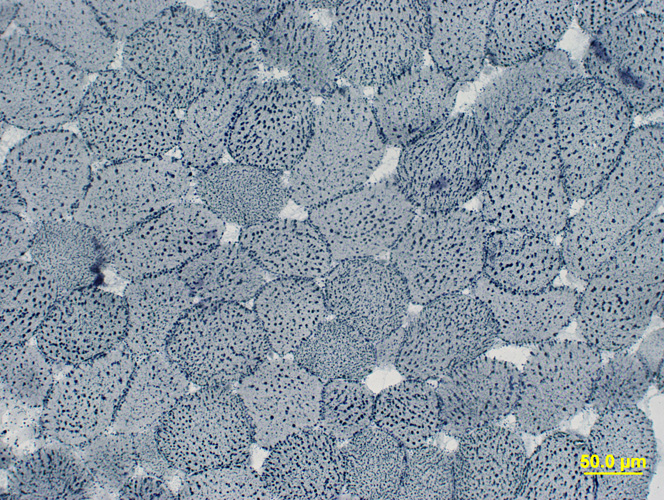
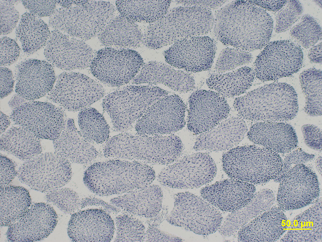
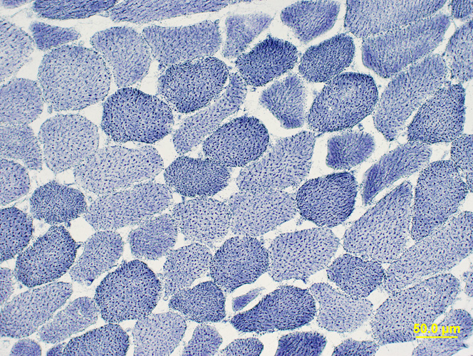 NADH stain |
 Congo Red stain |
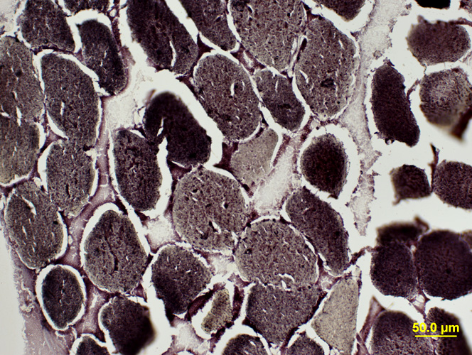
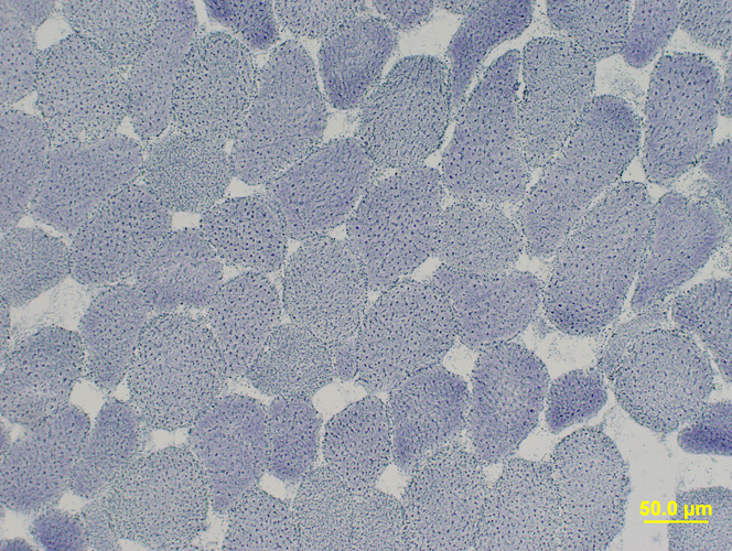
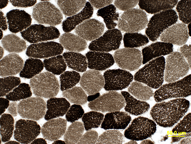 ATPase pH 9.4 stain |
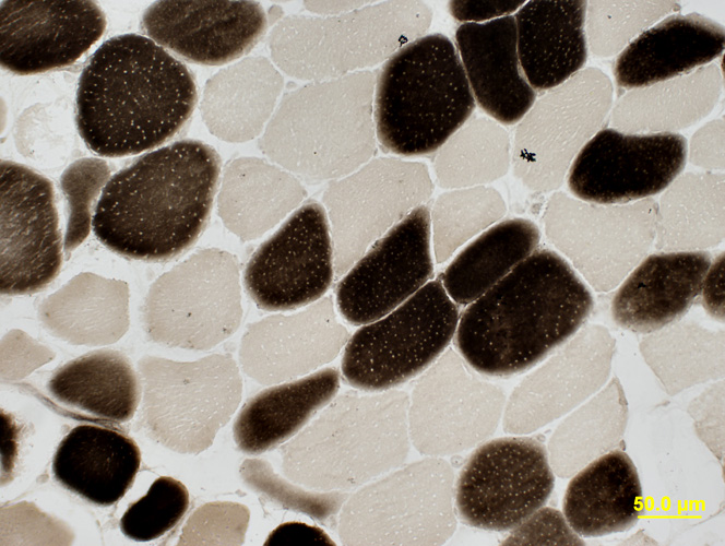 ATPase pH 4.3 stain |
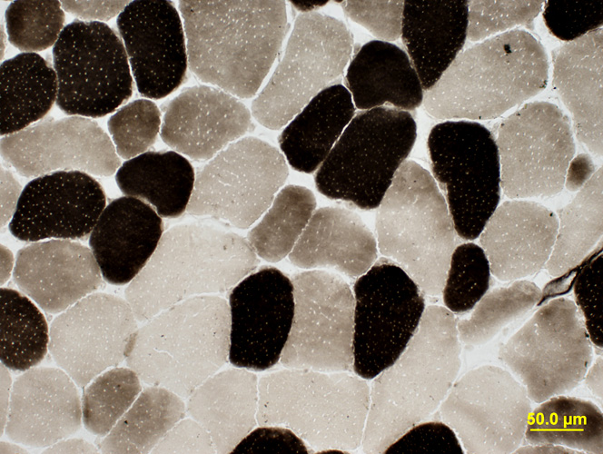 ATPase pH 4.6 stain |
 Alkaline Phosphatase stain |
 Acid Phosphatase stain |
 Esterase stain |
 Sudan Black stain |
 PAS stain |
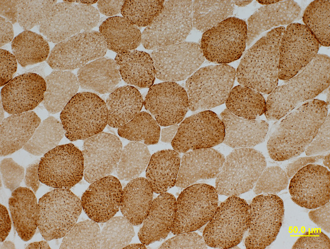
 Succinate Dehydrogenase (SDH) stain |
 Cytochrome Oxidase (COX) stain |
 Phosphorylase stain |
 AMPDA stain |
|
BIOPSY REPORT Clinical history: 29 year-old female with fatigue. Description of Muscle (Microscopic; Sections of snap frozen muscle): H&E stain shows minor variation in muscle fiber size. Endomysial connective tissue and vessels are normal. There is no mononuclear cell inflammation. Muscle fiber internal architecture is abnormal. Most fibers have multiple small internal clear regions. Gomori trichrome and VvGshow similar abnormalities. There is no amyloid or inflammation on Congo red. NADH shows abnormally punctate internal architecture. Alkaline phosphatase, acid phosphatase and esterase are all normal. COX and SDH are show no histologic evidence of mitochondrial pathology. PAS staining of muscle fibers may be mildly increased. Sudan shows abnormally large lipid droplets in most muscle fibers. AMPDA and phosphorylase are present. ATPase (pH 9.4, 4.6 and 4.3) shows no muscle fiber type grouping. There is a rare type IIC muscle fiber. Type I muscle fibers vary from 40 to 80 microns in diameter. Type II muscle fibers vary from 15 to 100 microns in diameter. (Adult normals for both types are 35 to 80 microns.) Type I muscle fibers are 50% of the population. (Normal is 40% to 60%.) Three muscle fiber types are present at ATPase pH 4.6. Interpretation: Abnormal muscle biopsy with lipid storage. This may be related to mitochondrial disease or carnitine disorders. Neutral lipid storage disorders cause lipid accumulation in both fiber types. Mitochondrial oxidative enzyme activities and Coenzyme Q10 levels will be measured. Other workup might include measurement of organic acids and acylcarnitine profile. Mildly increased PAS staining of muscle fibers may occur in mitochondrial disorders. There is selective atrophy of type II muscle fibers. This may be due to disuse, weight loss or systemic disease. |