Ullrich Congenital Muscular Dystrophy
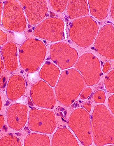
Chronic Myopathic features
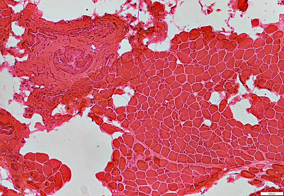 H&E stain |
Muscle is replaced by fat in many regions
Endomysial connective tissue: Generally increased
Muscle fibers: Varied sizes
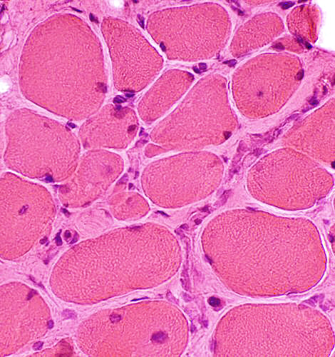
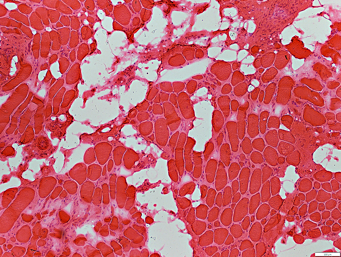 H&E stain |
.
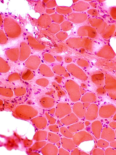 H&E stain |
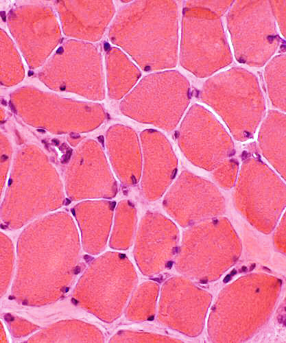 H&E stain |
Chronic myopathy
|
|
H&E stain |
 H&E stain |
Endomysial connective tissue: Increased between fibers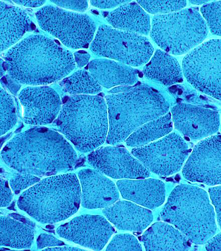 Gomori trichrome stain |
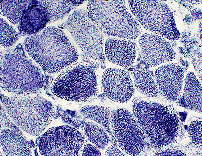
Immature type 2C muscle fiber: Few; Scattered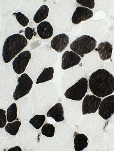 ATPase pH 4.3 stain |
Internal architecture: Coarse
Immature muscle fiber (top left)
 NADH stain |
Endomysial histiocytic cells: Scattered (Arrow)
Small necrotic muscle fiber: Replaced by histiocytic cells (Top)
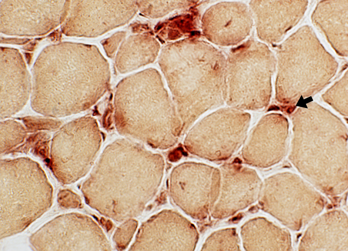 Acid phosphatase stain |
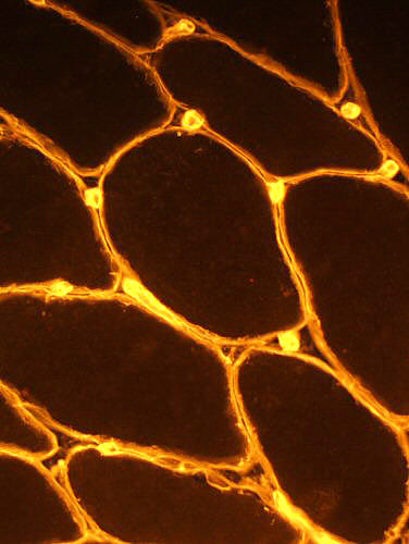
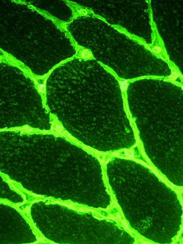
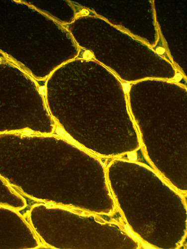
Connective tissue molecules: Collagen VI & IV
 Collagen IV |
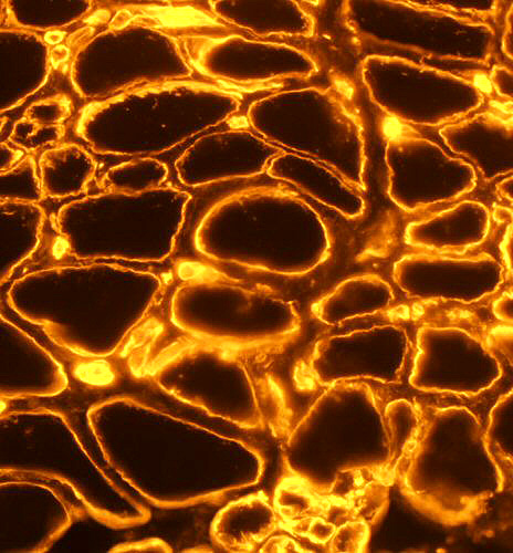
 Collagen VI |
 Collagen VI + Collagen IV |
Collagen Locations: Normal
|
| Collagen Locations: Ullrich | |
 Collagen IV |
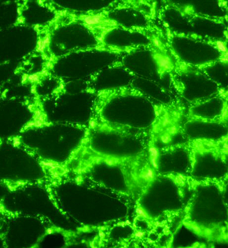 Collagen VI |
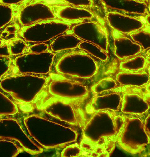 Collagen VI + Collagen IV |
|
Return to Ullrich CMD
Return to Biopsy index
Return to Neuromuscular home page
10/28/2015