TNF Receptor-Associated Periodic Syndrome (TRAPS)
 H&E stain |
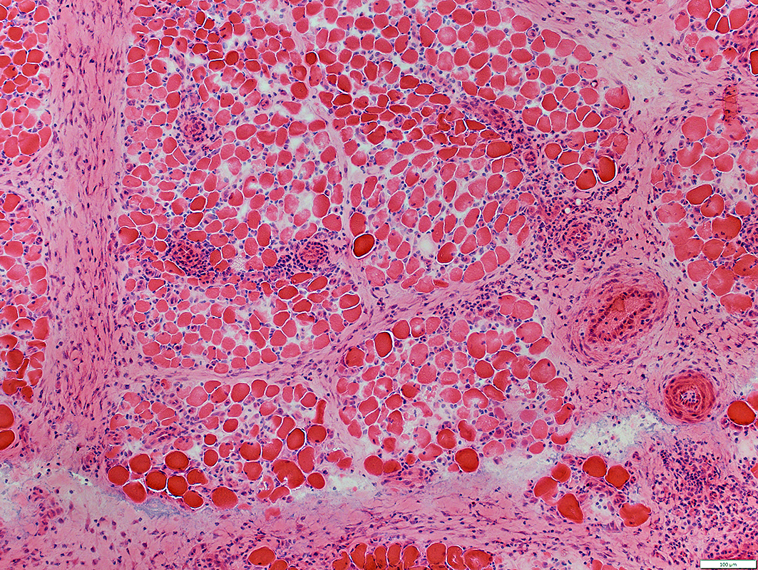
PERIMYSIAL PATHOLOGY
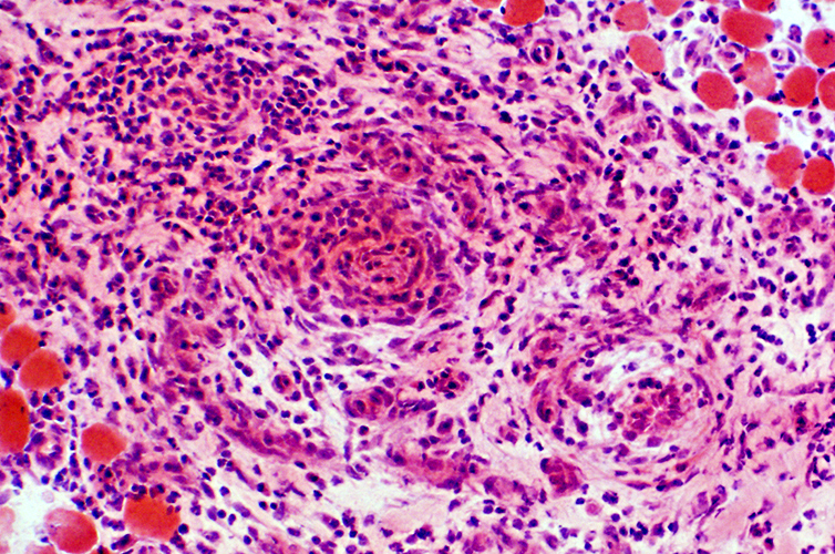
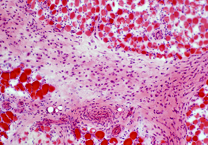 H&E stain Avascular perimysium: Widened; Contains many histiocytes |
 H&E stain Vascular perimysium: Widened; Increased mononuclear cellularity |
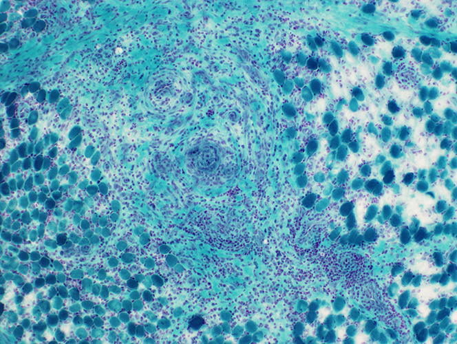 Gomori trichrome stain Vascular perimysium: Widened; Increased mononuclear cellularity |
 H&E stain |
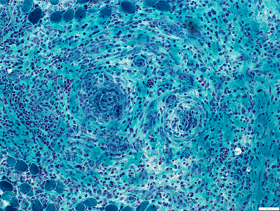 Gomori trichrome stain |
Neovascularization
Many small capillaries in perimysial connective tissue: Endothelium is stained by UEAI lectin
 UEA1 stain |
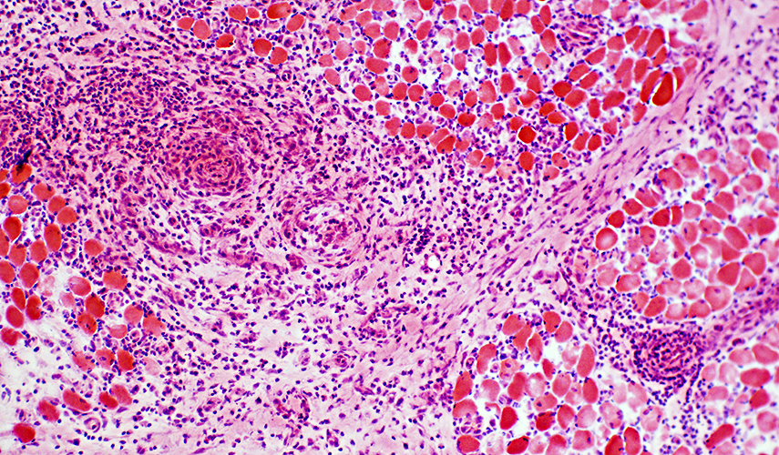
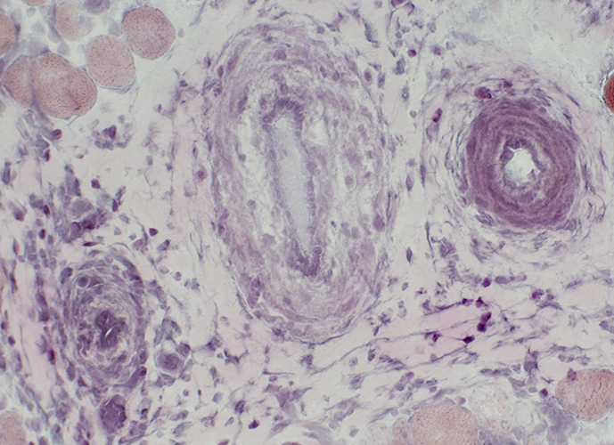
 H&E stain Mononuclear cell inflammation: Surrounds an Intermediae sized perimysial vessel |
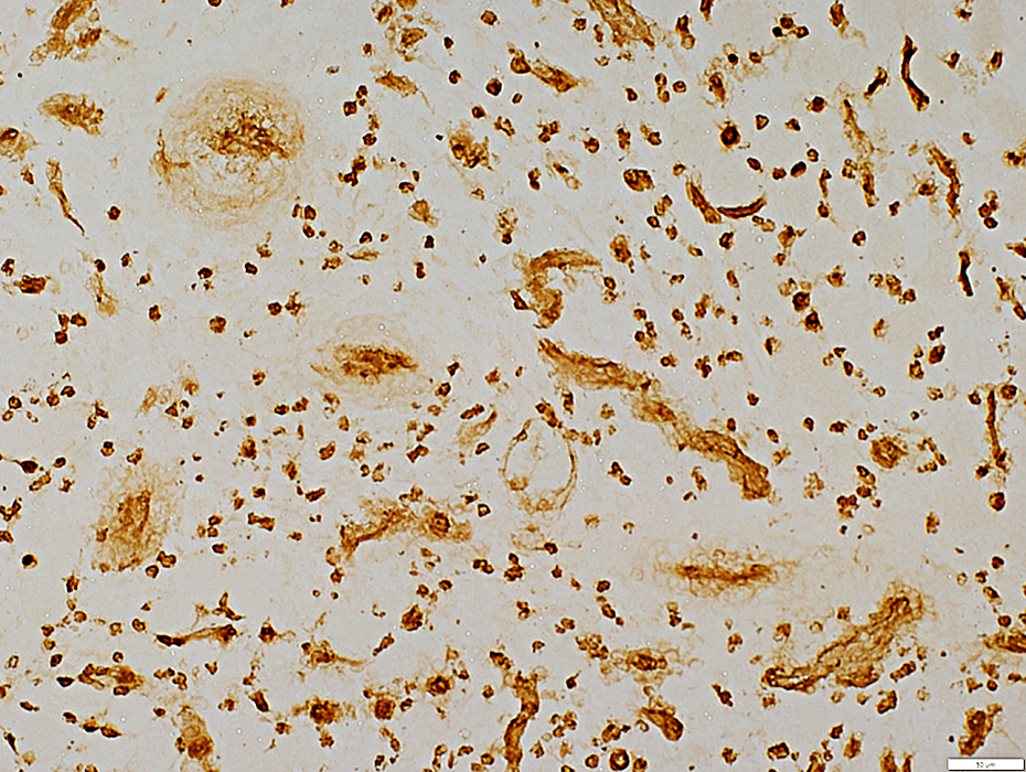
 Alkaline phosphatase stain Alkaline phosphatase stains Perimysial connective tissue & Endomysial capillaries |
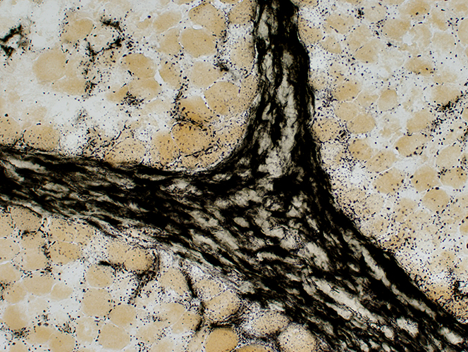 Alkaline phosphatase stain Alkaline phosphatase: Dark staibning of perimysial connective tissue |
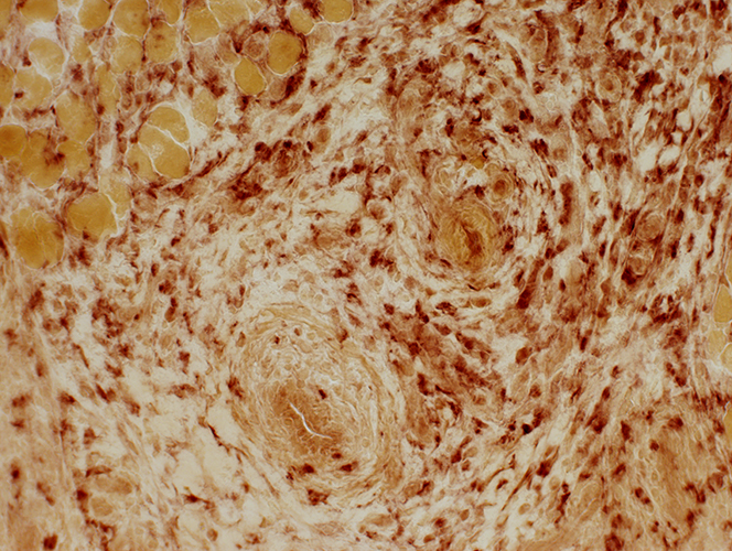 Acid phosphatase stain Acid phosphatase: Cellularity through the perimysium, and extending into endomysial regions, but not concentrated near vessels |
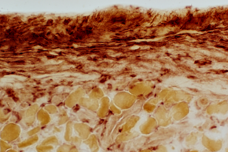 Acid phosphatase stain Acid phosphatase: Histiocytes in thickened epimysial connective tissue surrounding muscle |
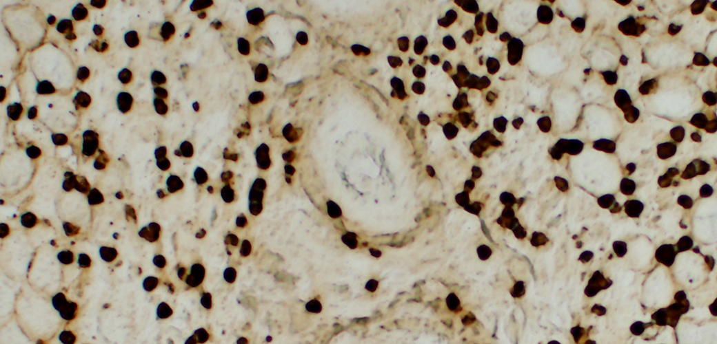 Peroxidase positive perimysial cells |
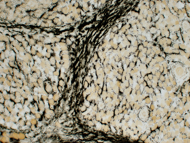
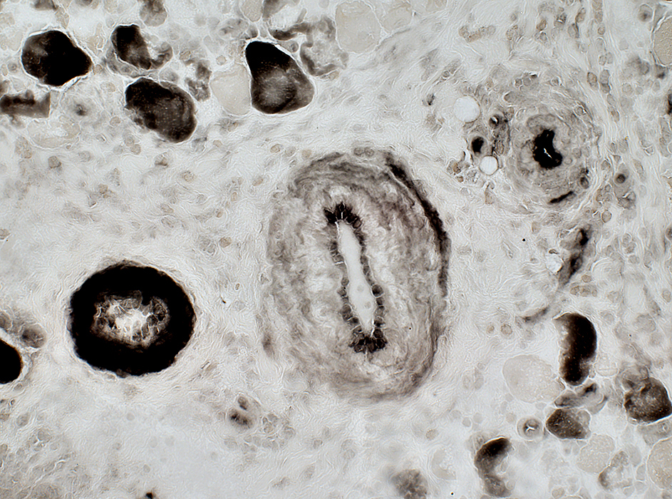
 VvG stain |
Fibrosis: Perivascular
Elastin fibrils: Reduced
Smooth muscle layer: Reduced or Absent
 ATPase pH 4.3 stain |
Return to Inflammatory myopathies
Return to TRAPS
1/31/2024