Sporadic, Late Onset Nemaline Myopathy (SLONM)
Muscle fiber features- Sizes: Varied
- Myonuclei
- Large
- May be clumped
- Internal architecture
- Irregular
- Aggregates
- Nemaline Rods: Smaller, & in fewer fibers, than inherited rods.
- Cytoplasmic bodies
- Sarcoplasmic pads
- Moth-eaten changes
- Fiber types
- Type I: Predominance
- Type 2: Small; Many 2C
- Metabolic/Molecular
- Mitochondrial pathology
- MHC I upregulation: > 50%
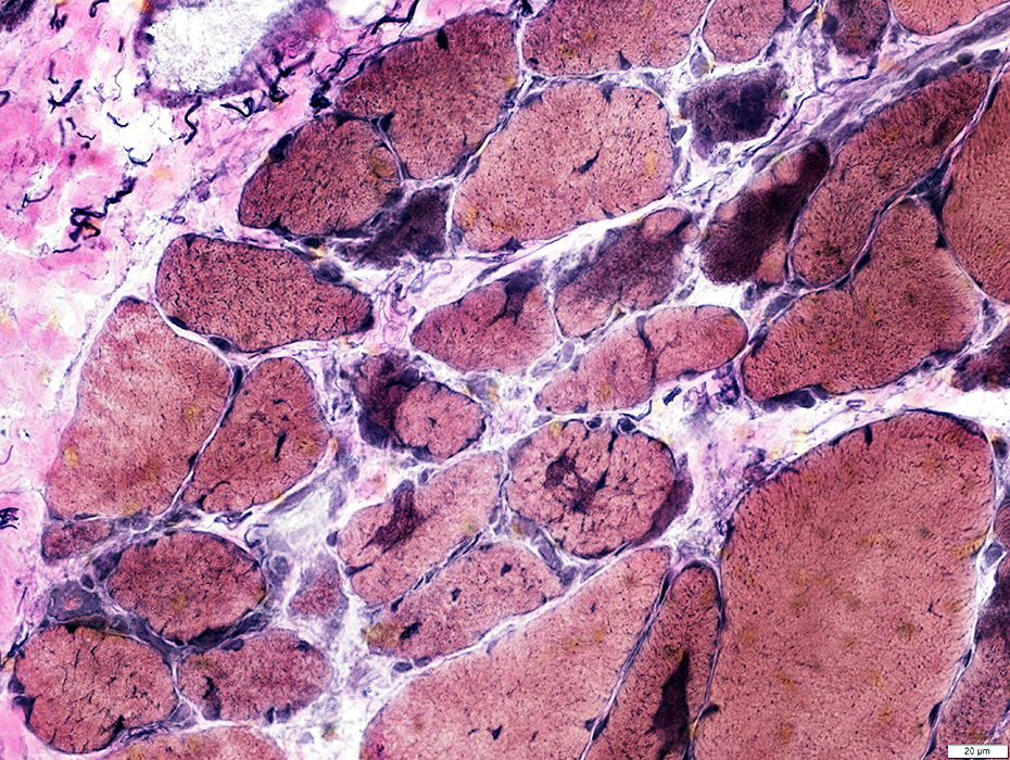
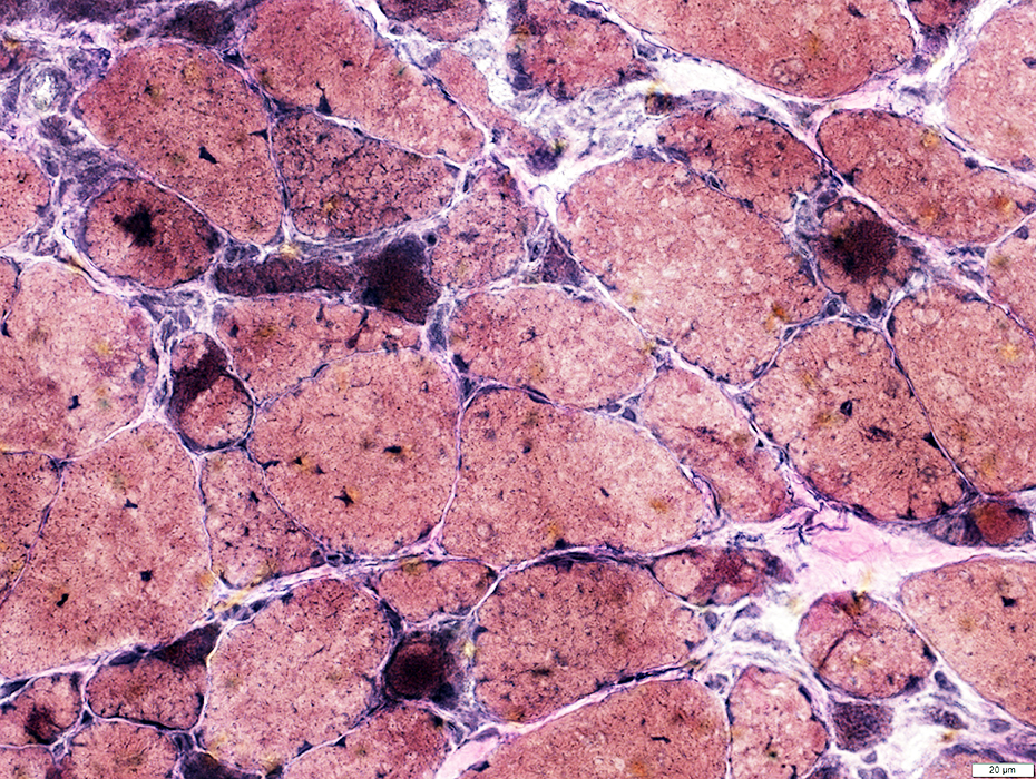
SLONM: Myopathy
Fiber size: Varied
Endomysial connective tissue: Increased
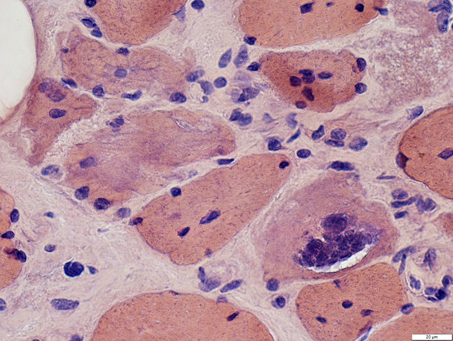
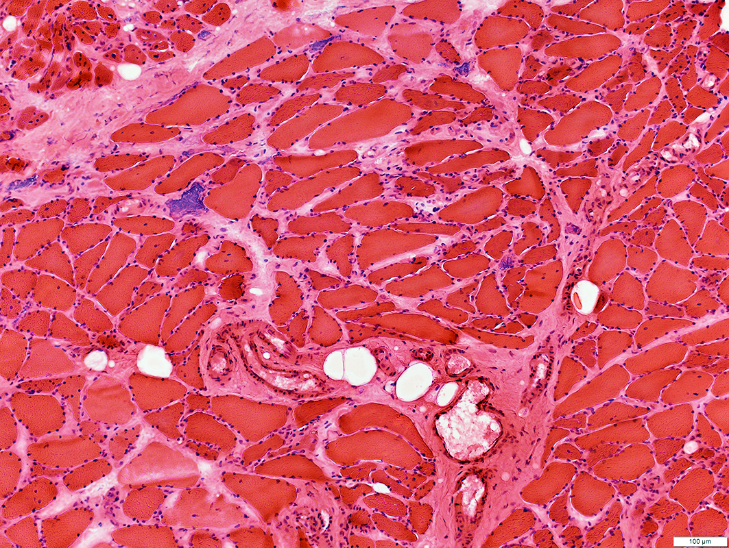 H&E stain |
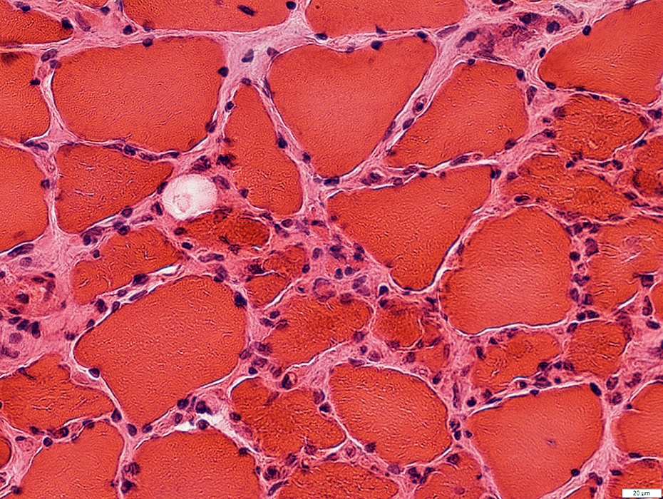 H&E stain |
Large
May be clumped
 H&E stain |
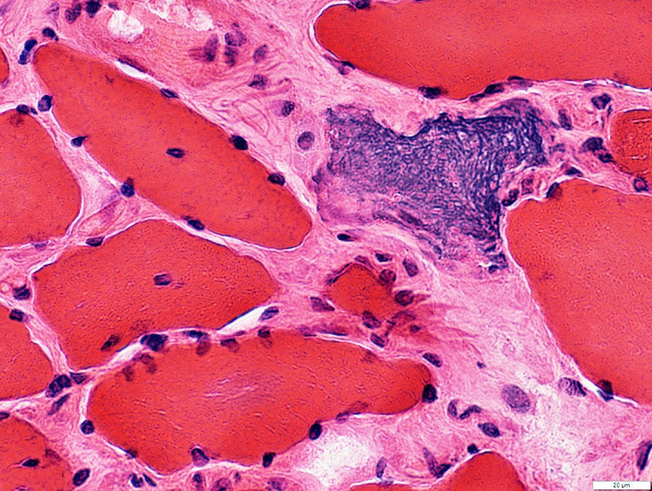
 Congo red stain |
Large
Clumped in some fibers
 Congo red stain |
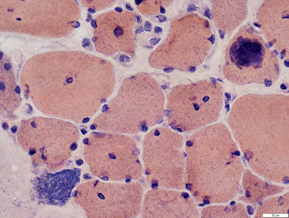
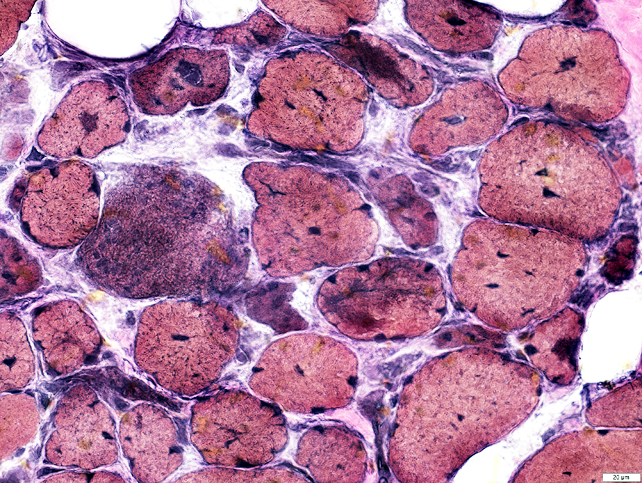
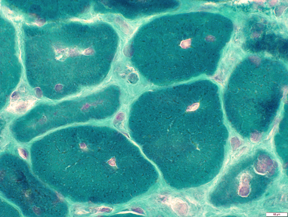 Gomori trichrome stain |
Large
Some fibers: Clumped or Occupy entire myofiber cytoplasm
 Gomori trichrome stain |
Clustered myonuclei in small muscle fiber
Small misoriented sarcomeres are also present
 From R Schmidt |
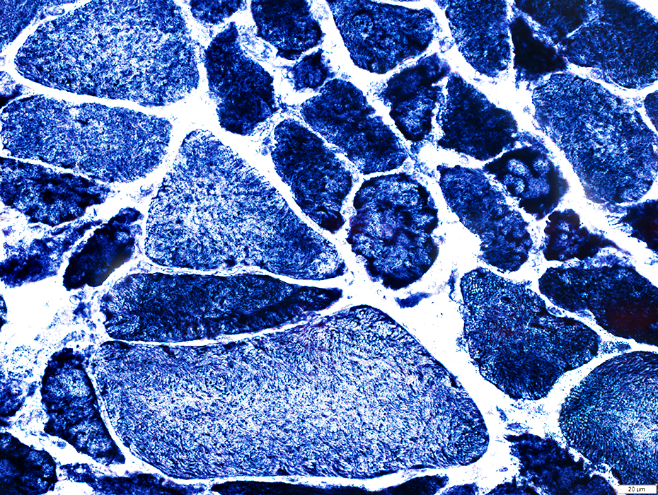
SLONM: Muscle Fiber Internal Architecture
 VvG stain |
Irregular, Coarse Internal architecture
Internal nuclei
 VvG stain |
 VvG stain |
Irregular, Coarse Internal architecture
Internal nuclei
 NADH stain |
Subsarcolemmal pads (Arrow): Contain
Disorganized sarcomeres
Myeloid debris
Small rod-like structures
Myonuclei may be present
 From R Schmidt |
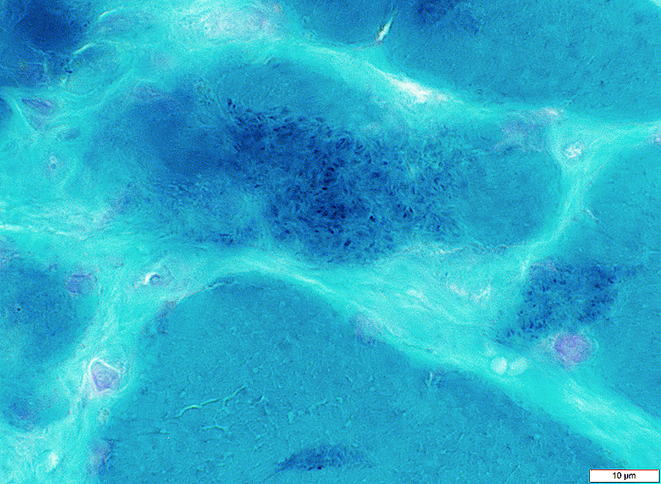
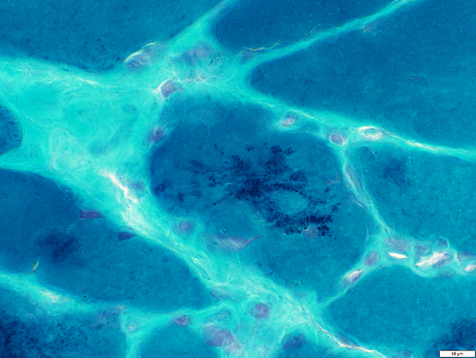
SLONM: Nemaline Rods
Ultrastructure
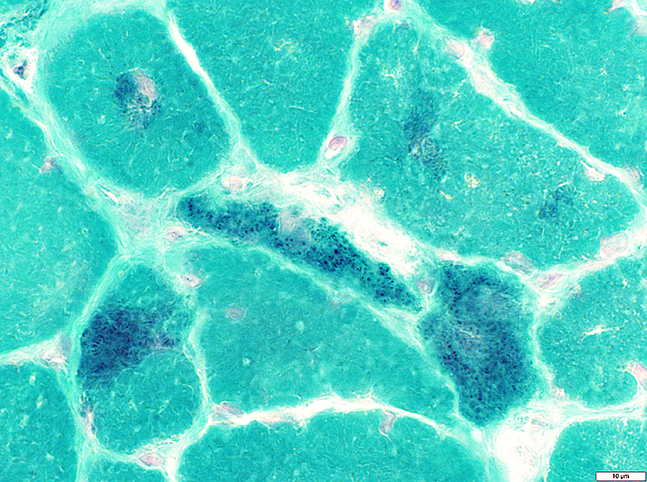 Gomori trichrome stain |
Size: Very small
Distribution in fiber: Diffuse; Often in large of cytoplasm
May be more common in small fibers
 Gomori trichrome stain |
 Gomori trichrome stain |
Size: Very small
Distribution in fiber: Diffuse; Often in large of cytoplasm
May be more common in small fibers
 Gomori trichrome stain |
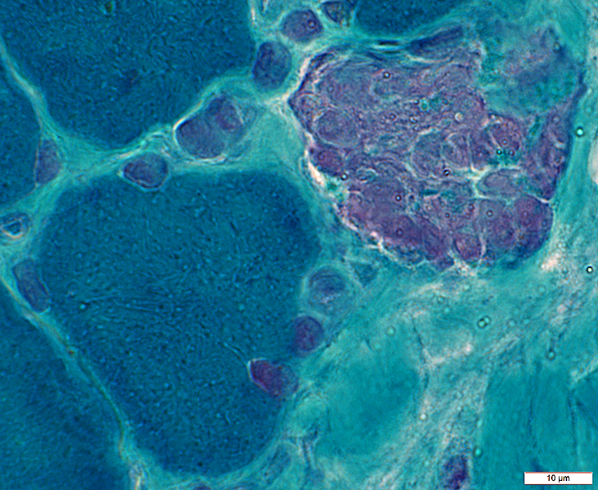
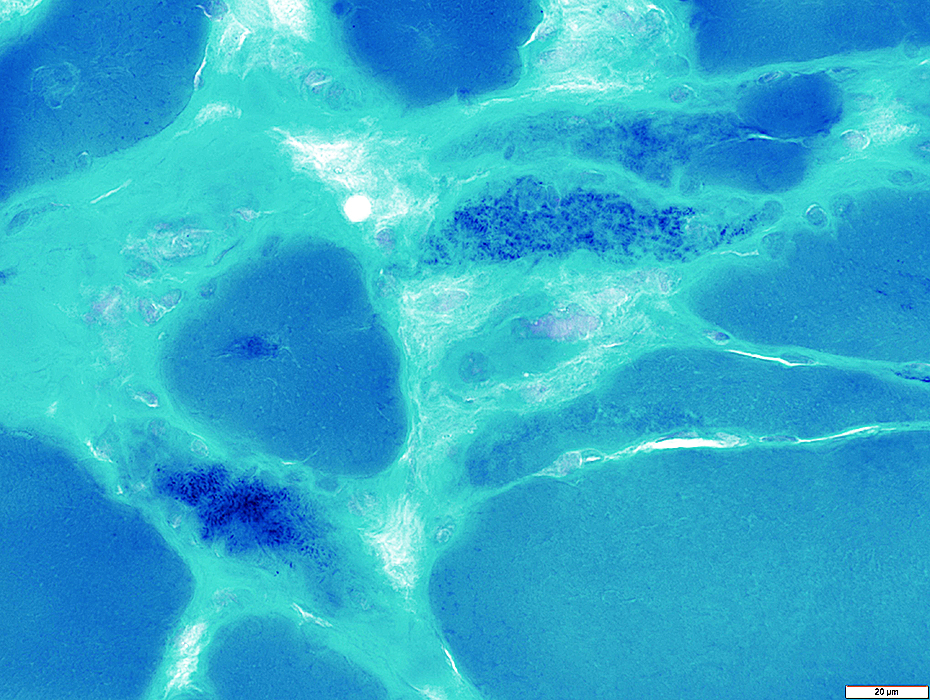
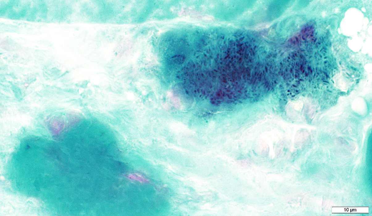
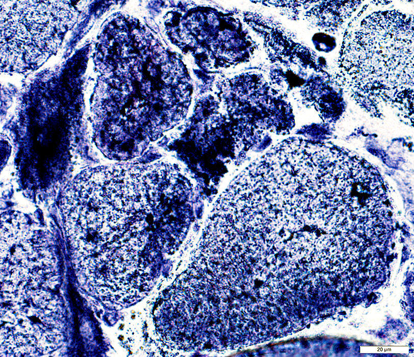
SLONM: Clustered Rod-like Material
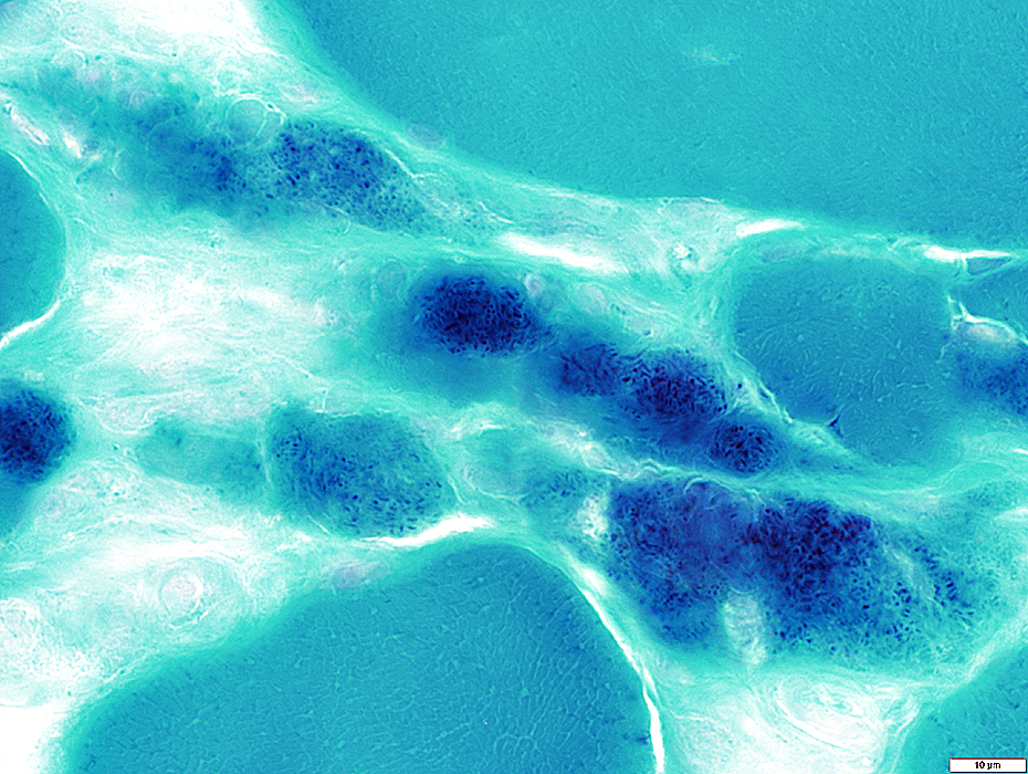 Gomori trichrome stain |
Size: Very small
Distribution in fiber: Diffuse; Often in large of cytoplasm
May be more common in small fibers
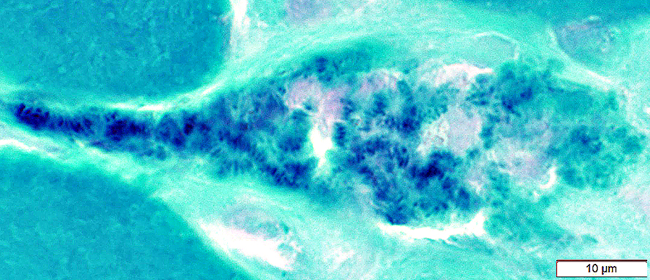 Gomori trichrome stain |
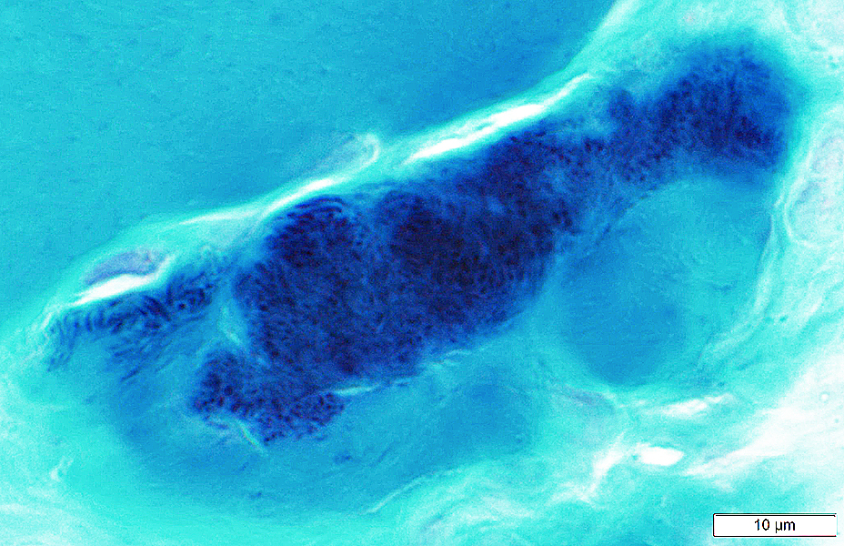 Gomori trichrome stain |
Size: Very small
Distribution in fiber: Diffuse; Often in large of cytoplasm
May be more common in small fibers
 Gomori trichrome stain |
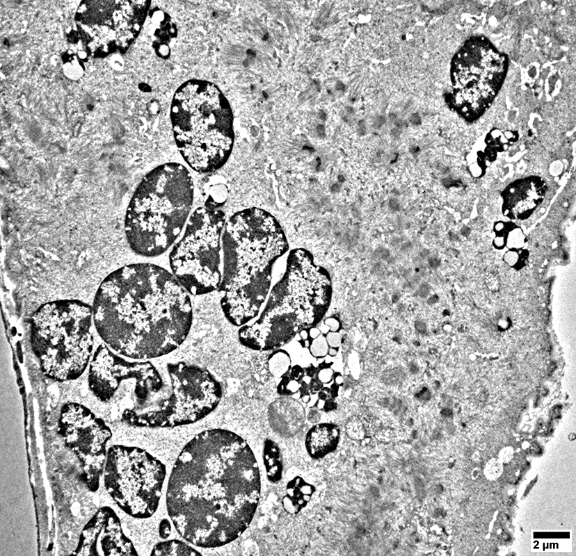
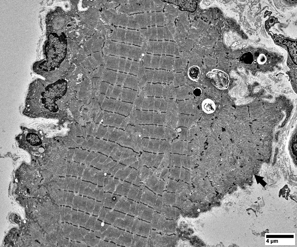
SLONM Rods: Ultrastructure

|
Multiple small rods are present in small muscle fibers

|
Rods
Elongated
Along length of muscle fiber
Ends may be continuous with myofilaments
 From: R Schmidt |

|
Multiple small rods are present in small muscle fibers

|
Rods
May be extensions from Z-bands (Arrow)
Often have fibrillar extensions that are not as common in hereditary rod myopathies
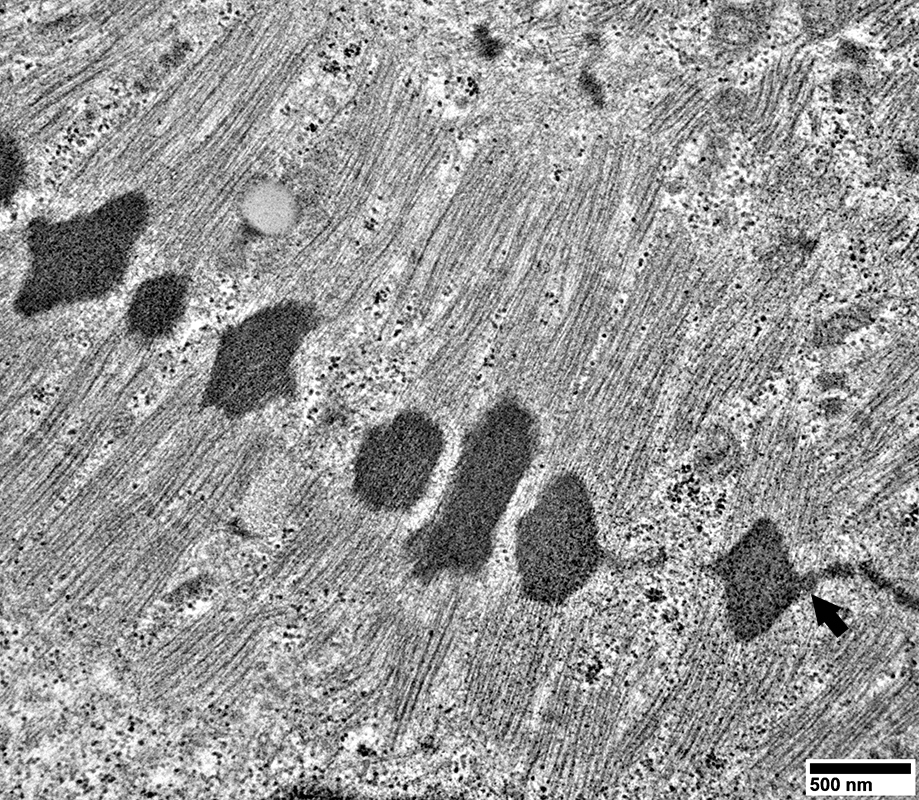 From: R Schmidt |
Rods
Myofilaments extend from ends of some rods
 From: R Schmidt |
SLONM: AMPDA aggregates
 AMPDA stain |
SLONM: Fiber types
Type I predominance
Small fibers mostly 2C
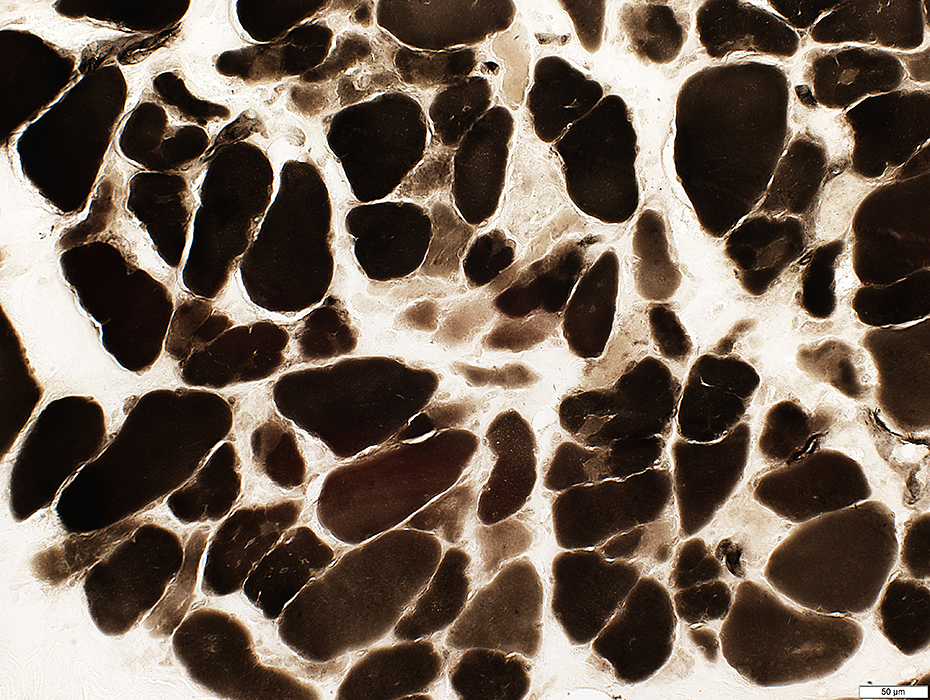 ATPase pH 4.3 stain |

|
Proliferation
Abnormal shapes
Lipid droplets

|
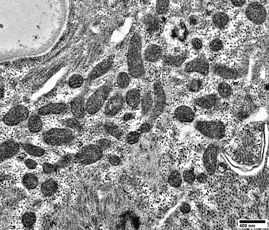 From: R Schmidt |
Ring fiber

|
Return to: Sporadic, Late Onset Nemaline Myopathy (SLONM)
Return to: Neuromuscular Home Page
5/24/2026