RYR3 Myopathy: Nemaline rods 144
All images from: Yalda Nilipour MD
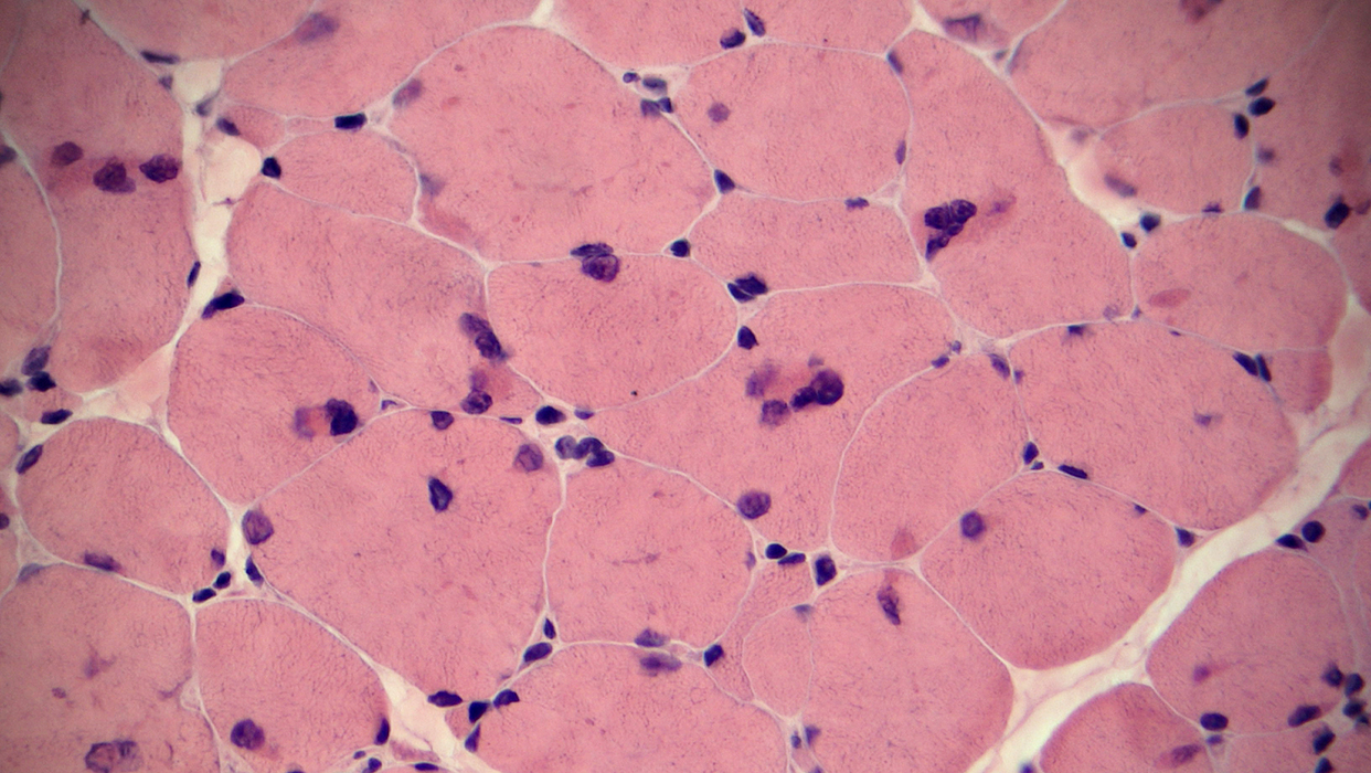
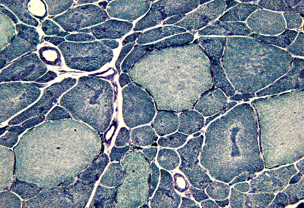
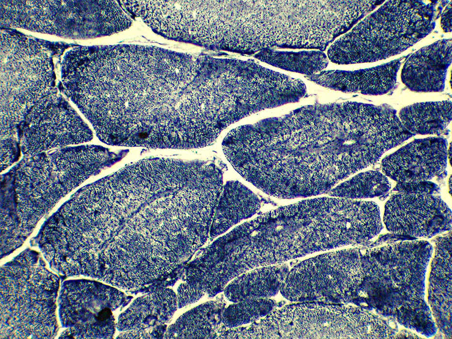
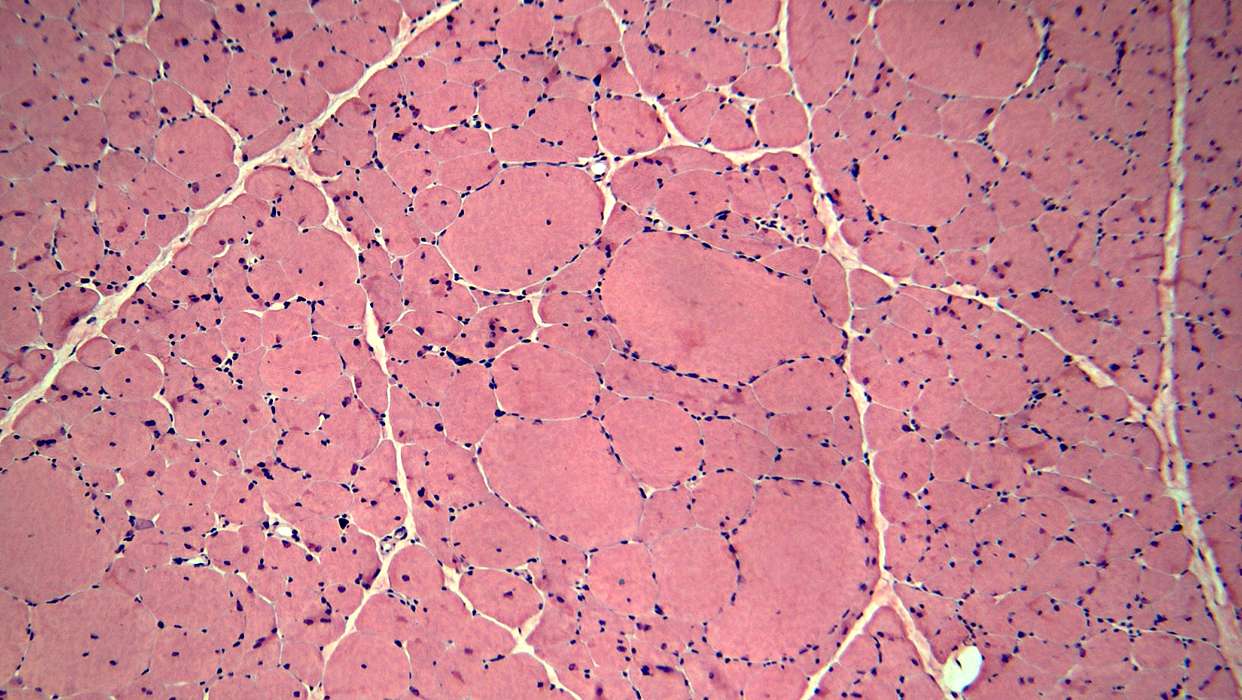 H&E stain |
Type 1 fibers: Small & Intermediate sized
Type 2 fibers: Large
Necrosis & Immaturity
Few muscle fibers (Below)
Myonuclei
Internal in some fibers
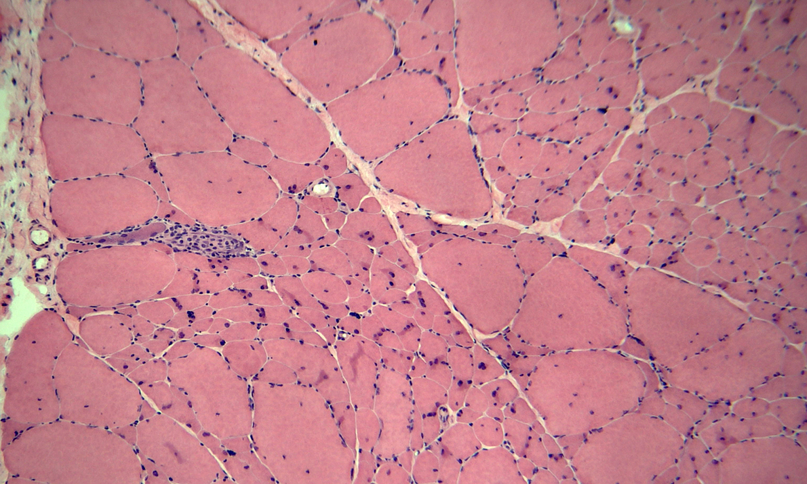 H&E stain |
Type 1 fibers: Small & Intermediate sized
Type 2 fibers: Large
Fiber types
Type 1: Predominant
 NADH stain |
 H&E stain |
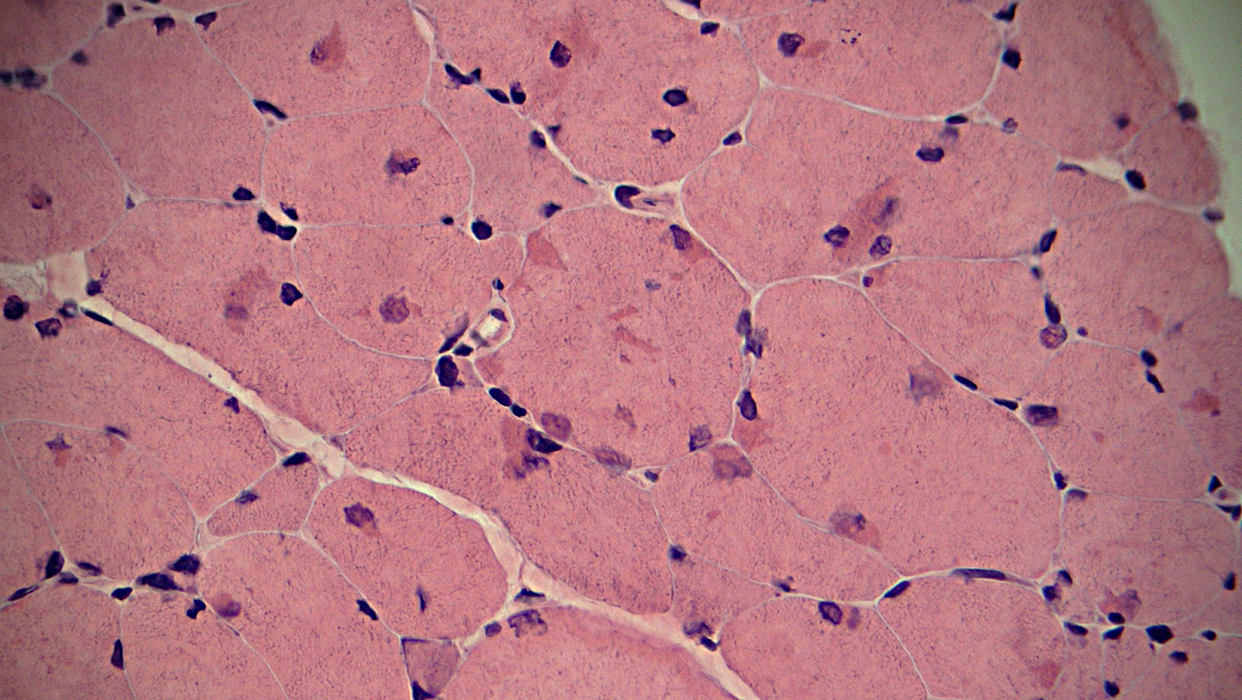
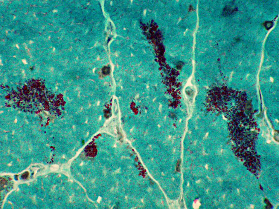
Aggregates
Eosinophilic
Location: Often subsarcolemmal
Myonuclei
Oten near, or within, aggregates
Irregular shapes
May be clustered
 H&E stain |
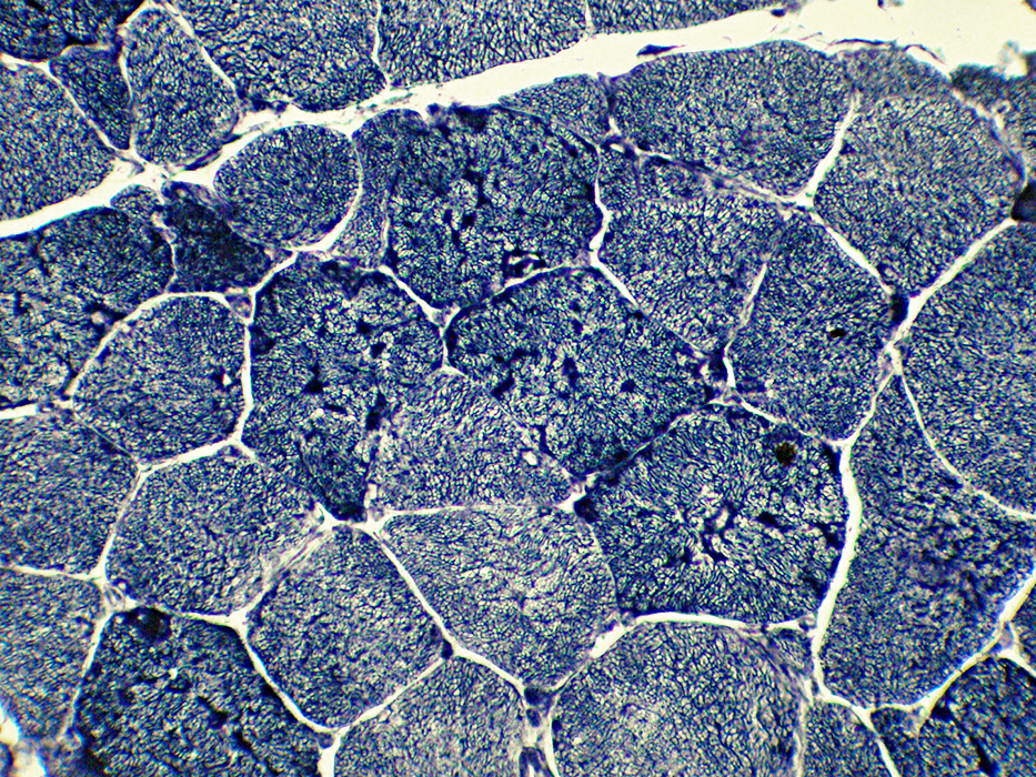
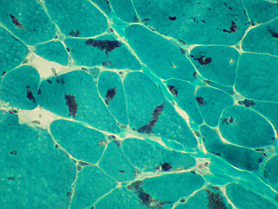 Gomori trichrome stain |
Present in many larger & intermediate-sized muscle fibers
Occur in clusters
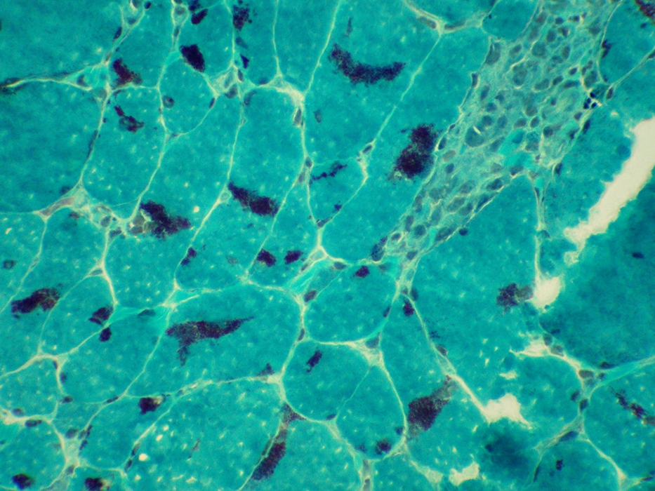 Gomori trichrome stain |
 Gomori trichrome stain |
Present in many larger & intermediate-sized muscle fibers
Occur in clusters
 Gomori trichrome stain |
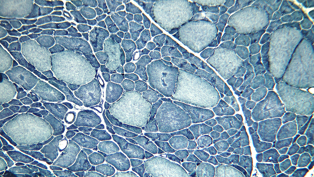
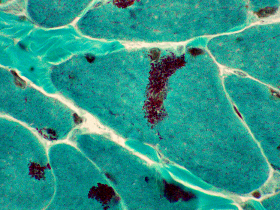
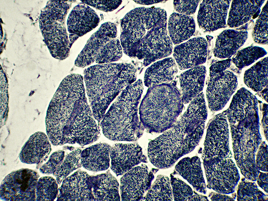
 NADH stain |
Irregular-shaped inclusions in some darker stained muscle fibers
 NADH stain |
 NADH stain |
Irregular in darker stained muscle fibers
 NADH stain |
Return to: Neuromuscular Home Page
8/13/2021