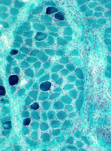
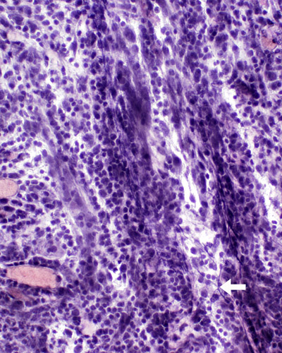
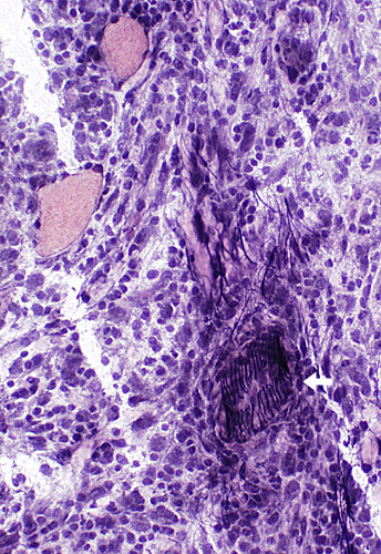
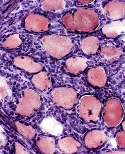
NODULAR MYOSITIS
Case 1
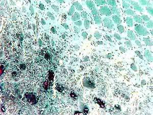 Gomori Trichrome (GT); From S Ringel |
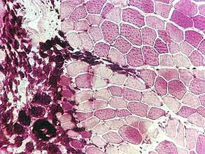 Esterase |
|
|
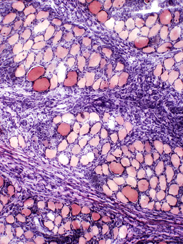
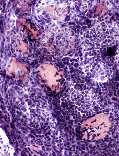
Case 2
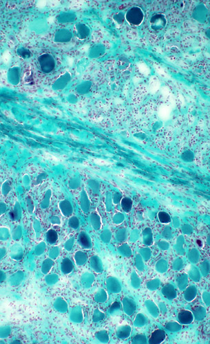 Gomori Trichrome (GT) |
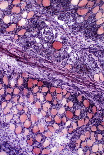 VvG |
|
|
 Gomori Trichrome (GT) |
 VvG |
|
|
 Gomori Trichrome (GT) |
 Gomori Trichrome (GT) |
|
|
 VvG |
 VvG |
|
|
|
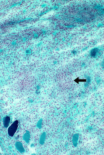
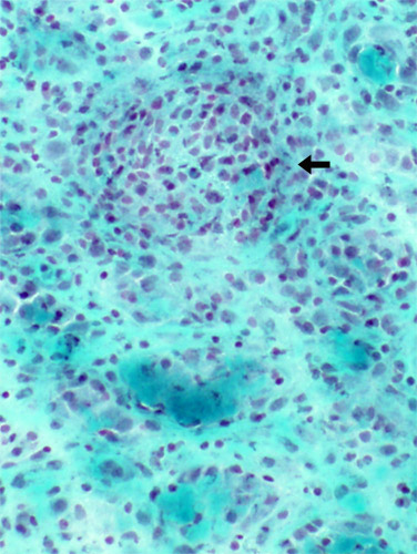 Gomori Trichrome (GT) "Granuloma-like" cellularity |
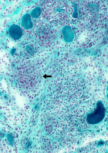
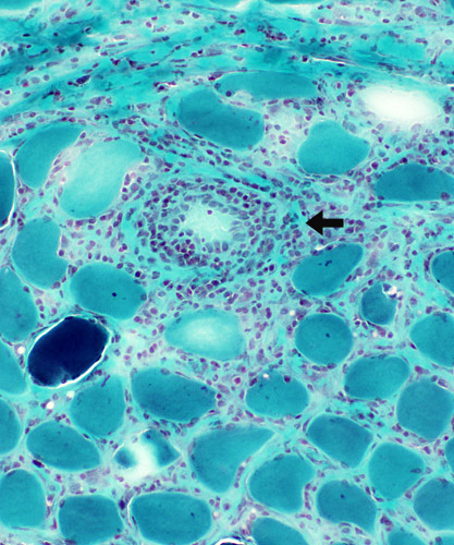 Gomori Trichrome (GT) Cells within the wall of an intermediate-sized vessel |
|
|
|
 VvG |
 VvG |
Muscle fiber pathology
|
|
Return to Neuromuscular Home Page
Return to Inflammatory myopathies
Return to Nodular myositis
8/22/2005